Research Article
Cyclooxygenase-2 Functions as a Downstream Mediator of Lysophosphatidic Acid to Promote Aggressive Behavior in Ovarian Carcinoma Cells 1
2
4
4
Jaime Symowicz, Brian P. Adley, Michelle M.M. Woo, Nelly Auersperg, Laurie G. Hudson, 1,3 and M. Sharon Stack
5
Departments of 1 Cell and Molecular Biology and 2 Pathology; 3 Robert H. Lurie Comprehensive Cancer Center, Northwestern University Feinberg School of Medicine, Chicago, Illinois; 4 Department of Obstetrics and Gynaecology, University of British Columbia, Vancouver, British Columbia, Canada; and 5 College of Pharmacy, University of New Mexico, Albuquerque, New Mexico
Abstract Elevated levels of the bioactive lipid lysophosphatidic acid (LPA) are detectable in the majority of patients with both early- and late-stage ovarian cancer, suggesting that LPA promotes early events in ovarian carcinoma dissemination. LPA contributes to the development, progression, and metastasis of ovarian cancer in part by inducing the expression of genes that contribute to proliferation, survival, or invasion, including cyclooxgenase-2 (COX-2) and matrix metalloproteinase–2 (MMP-2). We have previously shown that LPA promotes proMMP-2 activation and MMP-2–dependent migration and invasion in ovarian cancer cells. The purpose of the current study was to determine whether the effect of LPA on acquisition of the metastatic phenotype in ovarian cancer cells is mediated via a COX-2–dependent mechanism. Immunohistochemical analysis of 173 ovarian tumors showed strong COX-2 immunoreactivity in 63% of tumor specimens, including 50% of borderline tumors. LPA increased COX-2 protein expression in a time- and concentration-dependent manner in two of three immortalized borderline ovarian epithelial cells as well as in four of six ovarian cancer cell lines. This was accomplished by both activation of the Edg/ LPA receptor and LPA-mediated transactivation of the epidermal growth factor receptor, which increased COX-2 expression via the Ras/mitogen-activated protein kinase pathway. COX-2 also played a role in LPA-induced invasion and migration, as treatment with the COX-2 specific inhibitor NS-398 reduced LPA-induced proMMP-2 protein expression and activation and blocked MMP-dependent motility and invasive activity. These data show that COX-2 functions as a downstream mediator of LPA to potentiate aggressive cellular behavior. (Cancer Res 2005; 65(6): 2234–42)
Introduction Ovarian cancer is the leading cause of death from gynecologic disease in the United States. Each year, f24,000 women are newly diagnosed and 14,000 women will die from ovarian cancer. Most women are diagnosed at stage III or IV after the ovarian tumor has spread throughout the abdominal cavity, necessitating an under-
Requests for reprints: M. Sharon Stack, Department of Cell and Molecular Biology, Northwestern University, 303 East Chicago Avenue, Tarry 8-715, Chicago, IL 60611. Phone: 312-908-8216; Fax: 312-503-7912; E-mail:
[email protected]. I2005 American Association for Cancer Research.
Cancer Res 2005; 65: (6). March 15, 2005
standing of the mechanisms supporting ovarian cancer invasion and metastasis (1). Lysophosphatidic acid (LPA) contributes to the development, progression, and metastasis of ovarian cancer and is increased in both the plasma and ascites of ovarian cancer patients, reaching concentrations of 80 Amol/L (2–4). Ovarian tumor cells also produce LPA, thereby maintaining an LPA-rich microenvironment (2, 5–8). Elevated LPA levels are detectable in 98% of patients with ovarian cancer, including 90% of patients with stage I disease, suggesting that LPA promotes early events in ovarian carcinoma dissemination. This is supported by studies demonstrating that treatment of ovarian tumor cells with LPA in vitro results in an enhanced metastatic phenotype, characterized by increased proteolytic activity, stimulation of motility, and more aggressive invasive behavior (9, 8). LPA also induces the expression of additional genes that contribute to proliferation, survival, or metastasis, including c-myc, vascular endothelial growth factor, interleukin-8, and cyclooxgenase-2 (COX-2; refs. 5, 9–14). The main function of COX-2 is to catalyze the rate-limiting step in prostaglandin synthesis from arachidonic acid, generating prostaglandin H2 that is subsequently converted to prostaglandin E2 and other prostaglandins (15). Although not constitutively expressed, COX-2 expression is induced by growth factors, cytokines, and tumor promoters and is inducible in most cells and tissues (16–18). COX-2 contributes to tumorigenesis by changing the levels of pro- and antiapoptotic factors to inhibit apoptosis, by increasing growth factor expression to promote angiogenesis, and by increasing matrix metalloproteinase (MMP) expression to enhance invasiveness (18). Data supporting a role for COX-2 in ovarian physiology and pathobiology are complex, with opposing reports of COX-2 prevalence in normal human ovarian tissue (19–22). COX-2 expression has only been observed in the corpus luteum during menstruation (20), but COX-2 inhibition delays and blocks ovulation in humans (23, 24) and mice genetically deficient in COX-2 also fail to ovulate (25), suggesting COX-2 activity is essential for normal ovarian function. COX-2 induction is thought to be necessary for the rupture of the preovulatory follicle and subsequent release of oocytes during ovulation. It has been speculated that COX-2 or prostaglandins may increase collagenase and proteolytic activity and decrease synthesis of basement membrane components in ovarian granulosa and surface epithelial cells, permitting ovulation (26, 27). In addition, COX-2 expression has been reported in benign, borderline, and malignant ovarian tumors (19, 21, 22, 28, 29) wherein it is associated with chemotherapy resistance and a poor survival rate (30). Ascites from ovarian cancer patients contain elevated levels of prostaglandin E2 compared with nonmalignant ascites or ascites from other carcinomas (19), further supporting a role for COX-2
2234
www.aacrjournals.org
LPA Induction of COX-2 in Ovarian Cancer
in ovarian pathobiology. A specific role for COX-2 in regulating ovarian cancer metastasis has not been reported, although it was recently shown that COX-2 staining is significantly higher in metastatic ovarian tumors (22). The current study was undertaken to test the hypothesis that LPA promotes the metastatic phenotype of ovarian cancer cells via a COX-2–dependent mechanism. Evaluation of human ovarian tumors showed positive COX-2 immunoreactivity in 98% of cases, with 70% displaying moderate to high-level expression, including 50% of borderline ovarian tumors. Treatment of ovarian tumor cells with LPA in vitro induced COX-2 protein expression in a timeand concentration-dependent manner, whereas COX-1 expression was not affected. In addition to signaling via Edg/LPA receptors, LPA-induced transactivation of the epidermal growth factor receptor (EGFR) increased COX-2 expression via the Ras/mitogen-activated protein kinase pathway. Inhibition of COX-2 activity decreased proMMP-2 expression and LPA-induced proMMP-2 activation and reduced MMP-dependent motility and invasion. These data show that COX-2 functions as a downstream mediator of LPA to potentiate aggressive cellular behavior in ovarian carcinoma cells.
Materials and Methods Materials. LPA [1-oleoyl-2-hydroxy-sn-glycero-3-phosphate (sodium salt)] was purchased from Avanti (Alabaster, AL) in solution in chloroform. The chloroform was allowed to evaporate at room temperature and the LPA was reconstituted in PBS (Cellgro, Mediatech, Herndon, VA) at a concentration of 2 mmol/L. COX-2 inhibitor, NS-398, was purchased from Cayman Chemical (Ann Arbor, MI) and suspended in DMSO at a concentration of 50 mmol/L. Pertussis toxin was purchased from Biomol (Plymouth Meeting, PA) and was reconstituted in sterile water at a concentration of 100 ng/AL. AG1478 was purchased from Calbiochem (San Diego, CA) and suspended in DMSO at a concentration of 10 mmol/L. PD58059 was purchased from Calbiochem and suspended in DMSO at a concentration of 50 mmol/L. COX-2 and COX-1 monoclonal antibodies as well as COX-2 (ovine) and COX-1 (ovine) electrophoresis standards were purchased from Cayman Chemical. EGFR antibody and a PhosphoEGF Receptor Antibody Sampler Kit were purchased from Cell Signaling (Beverly, MA) and a mixture of the four phosphoEGFR antibodies (specific to residues Tyr845, Tyr992, Tyr1,045, and Tyr1,068) was used for detection of activated EGFR. Total extracellular signal-regulated kinase 1/2 antibodies were purchased from Santa Cruz Biotechnology (Santa Cruz, CA). Peroxidaseconjugated anti-mouse immunoglobulin G was purchased from Sigma (St. Louis, MO). Anti-rabbit immunoglobulin G horseradish peroxidase– conjugated antibody was purchased from Cell Signaling. Matrigel was purchased from Becton Dickinson (San Jose, CA). Falcon HTS Fluoroblok Insert Systems (8 Am pore size) were purchased from Becton Dickinson. Calcein acetoxymethyl ester was purchased from Molecular Probes (Eugene, OR). A Quantikine Human/Mouse MMP-2 (total) Immunoassay kit was purchased from R&D Systems (Minneapolis, MN). Immunohistochemistry. Immunohistochemical analysis was done retrospectively on tumor tissue microarrays prepared by the Pathology Core Facility of the Robert H. Lurie Comprehensive Cancer Center at Northwestern University assembled from tissue originally taken for routine diagnostic purposes. The microarray tissue specimens included human ovarian carcinomas (77 serous, 45 endometroid, 9 mucinous, and 18 clear cell types) and borderline tumors (16 serous, 6 mucinous, 1 mixed, and 1 clear cell types). Samples were cut 3 to 4 Am thick and deparaffinized. The cores were 1 mm in diameter. The tissue microarray was divided into two blocks, one containing 106 cores and the other containing 87 cores. Antigen retrieval was accomplished by heat induction at 99jC for f45 minutes. Immunohistochemical staining with antibodies to COX-1 (Cayman Chemical; 1:50 dilution), COX-2 (Cayman Chemical; 1:100 dilution), and cytokeratin-7 (DakoCytomation, Carpinteria, CA; 1:200 dilution) was done
www.aacrjournals.org
according to standard procedures. Colon adenocarcinoma was used as a positive control for COX-1 and COX-2. Analysis of tissue sections was done by light microscopy by a pathologist (B.P.A.) without prior knowledge of the clinical variables. Scoring of COX-1 and COX-2 was assigned according to the intensity of the staining and graded 0, 1+ (weak), 2+ (moderate), or 3+ (strong). Statistical analyses were done by the Biostatistics Core Facility of the Robert H. Lurie Comprehensive Cancer Center. Cell Culture. DOV13 cells, provided by Dr. Robert Bast (M.D. Anderson Cancer Center, Houston, TX), were maintained in MEM (Gibco Invitrogen, Carlsbad, CA), 10% fetal bovine serum (Gibco Invitrogen), penicillin/ streptomycin (Cellgro, Mediatech), amphotericin B (Cellgro, Mediatech), nonessential amino acids (Cellgro, Mediatech), sodium pyruvate (Cellgro, Mediatech), and insulin from bovine pancreas (10 mg/L; Sigma) at 37jC in 5% CO2. OVCA433 and OVCA429 cells, provided by Dr. Robert Bast, were maintained in MEM, 10% fetal bovine serum, penicillin/streptomycin, amphotericin B, nonessential amino acids, and sodium pyruvate at 37j in 5% CO2. Immortalized human borderline ovarian epithelial cells (HuIOSBT-1.5, HuIOSBT-2.2, and HuIOSBT-3.3), generated by Dr. Nelly Auersperg (University of British Columbia, Vancouver, BC) using SV40, were maintained in a 1:1 mixture of media 199 (Sigma) and MCDB105 (Sigma), 5% fetal bovine serum, and 50 Ag/mL gentamicin (Sigma). SKOV-3 cells were maintained in McCoy’s media (Cellgro, Mediatech), 10% fetal bovine serum, penicillin/streptomycin, amphotericin B, nonessential amino acids, and sodium pyruvate at 37jC in 5% CO2. OVCAR-3 cells were maintained in RPMI 1640 (American Type Culture Collection, Manassas, VA), 20% fetal bovine serum, penicillin/streptomycin, amphotericin B, nonessential amino acids, sodium pyruvate, and insulin from bovine pancreas (10 mg/L) at 37jC in 5% CO2. CaOV-3 cells were maintained in DMEM (American Type Culture Collection), 10% fetal bovine serum, penicillin/streptomycin, nonessential amino acids, and sodium pyruvate. With the exception of CaOV-3 cells, all ovarian carcinoma cell lines are known to be ascites derived. Western Blot Analysis. Cells were subcultured in six-well plates at 90% to 100% confluence. After 1 day, cells were serum starved overnight in the appropriate medium. In some experiments, cells were pretreated with inhibitor (or equivalent concentrations of DMSO) for 1.5 to 3 hours in serum-free media. Cells were then treated with LPA for time points ranging from 1 to 24 hours before lysis in modified radioimmunoprecipitation assay lysis buffer (50 mmol/L Tris pH 7.5, 150 mmol/L NaCl, 0.1% SDS, 1% Triton X-100, 5 mmol/L EDTA). Protein concentrations of the resulting lysates were determined using the Bio-Rad protein assay (Hercules, CA). Lysates (ranging from 30 to 70 Ag, as indicated) were electrophoresed on an 8% SDS-polyacrylamide gel (31), electroblotted to a polyvinylidene difluoride (PVDF) membrane (32), and blocked in 5% milk/ TBS-T (25 mmol/L Tris pH 7.5, 150 mmol/L NaCl, 0.1% Tween 20) or 3% bovine serum albumin/TBS-T at room temperature for 1 to 3 hours. Blots were incubated overnight with 1:1,000 dilution of the primary antibody. The immunoreactive bands were visualized using peroxidase-conjugated antimouse or rabbit immunoglobulin G (1:5,000 in 3% bovine serum albumin/ TBS-T) and enhanced chemiluminescence. To evaluate loading controls, blots were stripped of primary antibody using a low-pH buffer (400 mmol/L glycine pH 2.5), blocked again in 3% bovine serum albumin/TBS-T, and reprobed with primary antibody. Gelatin Zymography. DOV13 cells were serum starved overnight and pretreated with NS-398 (50-100 Amol/L) or DMSO vehicle control for 2 to 3 hours before incubation with 30 Amol/L LPA for 24 additional hours. Conditioned media (25 AL) was electrophoresed under nonreducing conditions on a 9% SDS-polyacrylamide gel containing f0.1% gelatin (33). The gel was washed for 2 hours in 2.5% Triton X-100, incubated at 37jC in 20 mmol/L Tris (pH 8.3), 10 mmol/L CaCl2, 1 Amol/L ZnCl2 for 65 to 72 hours, and stained with Coomassie blue. Enzyme activity was indicated by zones of gelatin clearance in the gel. Matrix Metalloproteinase–2 ELISA. DOV13 cells were serum starved overnight, and pretreated with NS-398 (50-100 Amol/L) or DMSO vehicle control for 2 to 3 hours before incubation with 30 Amol/L LPA for 24 additional hours. Conditioned media was then analyzed with the Quantikine human/mouse MMP-2 (total) immunoassay kit (R&D Systems)
2235
Cancer Res 2005; 65: (6). March 15, 2005
Cancer Research
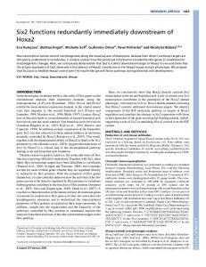
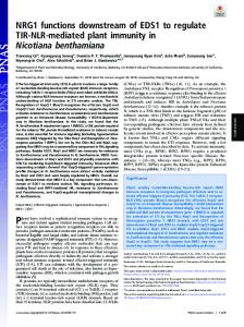
according to Fe´de´ration Internationale des Gynaecologistes et Obstetristes stage or histotype. LPA levels are often elevated in both the plasma and ascites of ovarian cancer patients wherein it has been reported to contribute to ovarian cancer development, progression, and metastasis (2–4). As plasma and ascites LPA levels were not available from the patient population above, the effect of LPA on COX-2 expression was examined in several ovarian cell lines, including immortalized borderline ovarian carcinoma cells (HuIOSBT-1.5, HuIOSBT-2.2, and HuIOSBT-3.3) and several malignant ovarian carcinoma cell lines (OVCA 429, OVCA 433, CaOV-3, OVCAR3, SKOV3, and DOV13). With the exception of the CaOV-3 cells, all ovarian carcinoma cell lines are known to be isolated from the ascites of distinct patients. Cells were treated with 30 Amol/L LPA for 3 hours and the basal and induced levels of COX-2 protein were analyzed by Western blotting relative to a COX-2 standard (Fig. 2). In borderline ovarian carcinoma cells, HuIOSBT-1.5 and HuIOSBT-3.3 cells expressed low basal levels of COX-2 that were greatly increased with LPA treatment, whereas HuIOSBT-2.2 cells expressed high basal levels of COX-2 that were unchanged with the addition of LPA. Differential responses were observed in the six malignant ovarian carcinoma cell lines, with four of the six cell lines responding to LPA treatment by induction of COX-2 (Fig. 2; OVCA429, OVCA433, CaOV-3, and DOV13). Neither SKOV-3 nor OVCAR3 cells expressed detectable constitutive or LPA-inducible COX-2. As shown in a representative example using DOV13 cells, induction of COX-2 was time dependent, reaching a maximum at 2 to 4 hours following LPA treatment (Fig. 3A). Induction was observed at low LPA concentrations (10 Amol/L) and was positively regulated by increasing LPA concentration (Fig. 3B). No constitutive or LPA-inducible COX-1 expression was observed in the DOV13 cells under these conditions (Fig. 3C). LPA transduces signals via the endothelial differentiation gene Edg/LPA subfamily of G protein–coupled receptors leading to changes in adenylate cyclase activity, activation of the Ras-Raf-Erk pathway, and stimulation of phospholipases C and D (5, 14). With the exception of OVCAR3 and SKOV3, expression of Edg/LPA receptor family members has not been characterized in these cell lines. Treatment of cells with LPA in the presence of pertussis toxin decreased COX-2 induction (Fig. 4A, top, lane 3), implicating LPAdependent signaling through Gi protein–coupled receptors in this process (3). Activation of EGFR family members has also been reported to induce COX-2 expression (34, 35). Further, recent studies have implicated LPA in EGFR transactivation via both pertussis toxin–sensitive and –insensitive pathways (36–40). Treatment of DOV13 cells with LPA under serum-free conditions induced phosphorylation of EGFR (Fig. 4A, middle), showing that LPA can transactivate EGFR in DOV13 cells. LPA-mediated
according to the specifications of the manufacturer. The data include normalized values from four separate experiments. In vitro Wound Scratch Assay. DOV13 cells were plated in eight-well plates, cultured to confluence, and serum starved overnight. Two scratch wounds were made in each well using a micropipette tip. The cells were then treated with NS-398 and LPA as indicated. DMSO concentrations remained constant within each experiment. Two points were randomly selected, marked for each scratch, and photographed using a digital camera at 0, 24, and 48 hours. Five relative measurements were taken for each of the four points for each experimental condition using the MetaMorph Imaging System (Universal Imaging Corporation, Downington, PA). These resulting five measurements for each point were averaged and then normalized based on the initial measurement for that point at 0 hour. The four normalized values were then averaged for each experimental condition. The data include results from three separate assays. Matrigel Invasion Assay. Matrigel (50 AL of 0.1 mg/mL) was added to each chamber of the Falcon HTS Fluoroblok Insert System and left to dry overnight. DOV13 cells were serum starved overnight in serum-free MEM, trypsinized, and resuspended in phenol red–free medium at a concentration of 500,000 cells/mL in the presence of NS-398 or DMSO as indicated. Cells (250,000 cells in 500 AL) were then added to the top chamber of the HTS Fluoroblok Insert System with serum-free, phenol red–free MEM (500 AL) in the bottom chamber. After 1 hour, LPA (30 Amol/L) was added and chambers were incubated for 48 hours before labeling with calcein acetoxymethyl ester (100 AL, final concentration 5 Ag/mL) for 30 minutes at 37jC in 5% CO2. Relative invasion was quantified by analysis of the fluorescent signal for the bottom chamber using a Wallac 1420 Victor2 multilabel plate reader (Perkin-Elmer, Shelton, CT). Assays were done in triplicate and analyzed relative to blank wells containing only medium.
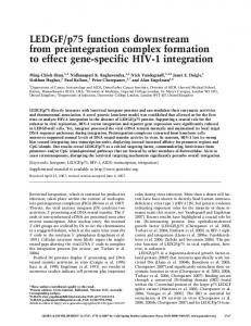
Results Analysis of Cyclooxgenase-2 Expression in Human Ovarian Cells and Tissues. Samples from 173 patients were examined for COX-2, COX-1, and cytokeratin-7 immunoreactivity. Of these samples, 77 (45%) were serous carcinoma, 45 (26%) were endometroid carcinoma, 18 (10%) were clear cell carcinomas, 9 (5%) were mucinous carcinomas, and 24 (14%) were borderline tumors. The vast majority of ovarian tumors (98%) displayed positive COX-2 immunoreactivity. COX-2 expression was high (3+ or 2+) in 63% of patients compared with 39% with high COX-1 staining (Table 1). A representative example of a serous ovarian tumor with intense COX-2 immunoreactivity is shown relative to COX-1 and keratin 7 in Fig. 1A-D. Of note, 8 of 9 (89%) mucinous tumors displayed high COX-2 expression (Fig. 1E-H). COX-2 expression was also elevated in borderline ovarian tumors (50%) relative to 29% displaying high COX-1 staining (Fig. 1I-L; Table 1). Significant differences in high (3+ or 2+) COX-2 expression between borderline and other tumors were not observed. No significant COX-2 staining was observed in the stromal compartment. Strong COX-2 positivity (3+ or 2+) was not differentially distributed
Table 1. Immunohistochemical expression of COX-1 and COX-2 in borderline and malignant ovarian epithelial tumors Histotype
COX-2 3+
Serous (77) Endometroid (45) Clear Cell (18) Mucinous (9) Borderline (24)
30 25 7 5 6
COX-1 2+
(39%) (56%) (39%) (56%) (25%)
25 11 3 3 6
1+ (32%) (24%) (17%) (33%) (25%)
Cancer Res 2005; 65: (6). March 15, 2005
19 9 7 1 12
0 (25%) (20%) (39%) (11%) (50%)
3 0 1 0 0
3+ (4%) (0%) (5%) (0%) (0%)
2236
10 6 0 2 3
2+ (13%) (13%) (0%) (22%) (12%)
35 6 1 1 4
1+ (45%) (13%) (5%) (11%) (17%)
20 15 3 0 7
0 (26%) (34%) (17%) (0%) (29%)
12 18 14 6 10
(16%) (40%) (78%) (67%) (42%)
www.aacrjournals.org
LPA Induction of COX-2 in Ovarian Cancer
Figure 1. Immunohistochemical expression of COX-1, COX-2, and cytokeratin-7 in ovarian tumor samples. Samples were stained with antibodies to COX-2 (1:200; A, E , I), COX-1 (1:50; B, F , J ), cytokeratin-7 (CK7 , 1:200; C, G , K ) or H&E (H + E; D , H , L) as detailed in Materials and Methods. A -D, serous carcinoma; E -H, mucinous carcinoma; I -L, borderline tumor.
transactivation of EGFR was pertussis toxin insensitive (Fig. 4A, middle, lane 3). Cotreatment with LPA and the EGFR-specific tyrosine kinase inhibitor AG1478 also abrogated the ability of LPA to induce COX-2 expression (Fig. 4A, top, lane 4). Similar results
were obtained following inhibition of the Ras/mitogen-activated protein kinase pathway using the mitogen-activated protein/ extracellular signal-regulated kinase kinase inhibitor PD98059 (Fig. 4B). Combined treatment of DOV13 cells with LPA, pertussis
Figure 2. Effect of LPA on COX-2 expression in ovarian cells. Cells were cultured as indicated in Materials and Methods, serum starved overnight, and cultured in the presence or absence of 30 Amol/L LPA for 3 hours. Cell lysates [50 Ag for all cell lines except OVCAR3 (30 Ag)] were electrophoresed on an 8% SDS-polyacrylamide gel, electroblotted to PVDF membrane, and probed with anti-COX-2 (1:1,000 dilution), followed by peroxidase-conjugated secondary antibody (1:5,000) and enhanced chemiluminescence detection. A COX-2 standard (50 ng) was included as a control.
www.aacrjournals.org
2237
Cancer Res 2005; 65: (6). March 15, 2005
Cancer Research
3-5), implicating COX-2 as a mediator of LPA-induced MMP-2 activation. At higher concentrations (100 Amol/L), down-regulation of proMMP-2 protein expression was also observed (Fig. 5, lane 5) although no effect on cell viability was evident (data not shown). This was confirmed by an MMP-2 ELISA, showing a decrease in total MMP-2 (pro and active) expression in the presence of NS398 (Fig. 5B, *P < 0.05, #P < 0.005), indicating that COX-2 modulates pericellular proteolytic potential via regulation of both proMMP-2 expression and activation. No change in either membrane type 1 MMP (MMP-14) or tissue inhibitor of metalloproteinase-2 was observed (data not shown). LPA has been previously reported to potentiate the motility and invasiveness of ovarian cancer cells (9, 44, 45). Similarly, COX-2 activity is necessary for enhanced migration and
Figure 3. LPA induces COX-2 expression in a time- and concentrationdependent manner. A, DOV13 cells were cultured in the presence of 30 Amol/L LPA for time points indicated. Lysates (65 Ag) were electrophoresed on an 8% SDS-polyacrylamide gel, electroblotted to PVDF membrane, and immunoblotted with anti-COX-2 (1:1,000), followed by peroxidase-conjugated secondary antibody (1:5,000) and enhanced chemiluminescence detection. A COX-2 standard (50 ng) was included as a control. B, DOV13 cells were cultured for 3 hours in the presence of increasing concentrations of LPA as indicated. Lysates (80 Ag) were electrophoresed on an 8% SDS-polyacrylamide gel, electroblotted to PVDF membrane, and immunoblotted with anti-COX-2 (1:1,000), followed by peroxidase-conjugated secondary antibody (1:5,000) and enhanced chemiluminescence detection. A COX-2 standard (50 ng) was included as a control (not shown). C, DOV13 cells were cultured for 3 hours in the presence of increasing concentrations of LPA, as indicated. Lysates (65 Ag) were electrophoresed on an 8% SDS-polyacrylamide gel, electroblotted to PVDF membrane, and immunoblotted with anti-COX-1 (1:1,000), followed by peroxidase-conjugated secondary antibody (1:5,000) and enhanced chemiluminescence detection. A COX-1 standard (50 ng) was included as a control.
toxin, and AG1478, resulted in a greater reduction in COX-2 protein induction relative to treatment with each inhibitor individually (Fig. 4A, top, lane 5), suggesting that distinct signaling pathways promote COX-2 expression. To provide further support for this hypothesis, DOV13 cells were treated with LPA and EGF. Both EGF (Fig. 4C, lane 2) and LPA (Fig. 4C, lane 3) individually induced COX-2 protein expression, whereas cotreatment with both EGF and LPA further enhanced COX-2 expression (Fig. 4C, lane 4). Together these data support a major role for LPA-mediated EGFR transactivation in COX-2 induction and suggest both Edg/LPA receptors and EGFR contribute to optimal LPA-mediated COX-2 induction DOV13 cells. Functional Effect of Cyclooxgenase-2 Inhibition on Lysophosphatidic Acid–Induced Aggressive Behavior. Separate studies have implicated either LPA or COX-2 in expression and activation of proMMP-2 (9, 41–43). To evaluate whether COX-2 activity is necessary for LPA-induced proMMP-2 activation, DOV13 cells were pretreated with increasing concentrations of the specific COX-2 inhibitor NS-398 followed by LPA for an additional 24 hours, and changes in proMMP-2 processing were evaluated via gelatin zymography. As previously reported using LPA concentrations as low as 2.5 to 5 Amol/L (9), LPA stimulates activation of proMMP-2, as indicated by the appearance of a lower molecular weight band representative of the activated form of the enzyme (Fig. 5, lane 2, arrow). Treatment with NS-398 blocked the ability of LPA to induce proMMP-2 activation in a dose-dependent manner (Fig. 5A, lanes
Cancer Res 2005; 65: (6). March 15, 2005
Figure 4. LPA induces COX-2 expression via Edg/LPA receptor and EGFR transactivation. A, DOV13 cells were pretreated with pertussis toxin (PTX , 100 ng/mL) in the presence or absence of the EGFR tyrosine kinase inhibitor AG1478 (10 Amol/L) for 2 hours before addition of LPA (30 Amol/L) for 3 hours. Lysates (50 Ag) were electrophoresed on an 8% SDS-polyacrylamide gel, electroblotted to PVDF membrane, and immunoblotted with anti-COX-2 (1:1,000), anti-phosphoEGFR mixture (1:1,000) or anti-total EGFR (1:1,000) as indicated, followed by peroxidase-conjugated secondary antibody (1:5,000) and enhanced chemiluminescence detection. OVCA433 cells treated with EGF (20 ng/mL) served as a positive control for EGFR phosphorylation (middle ). B, DOV13 cells were pretreated with the mitogen-activated protein/extracellular signal-regulated kinase kinase inhibitor PD98059 (50 Amol/L) before the addition of LPA (30 Amol/L) for 3 hours. Lysates (75 Ag) were electrophoresed on an 8% SDS-polyacrylamide gel, electroblotted to PVDF membrane, and immunoblotted with anti-COX-2 (1:1,000), followed by peroxidase-conjugated secondary antibody (1:5,000) and enhanced chemiluminescence detection. A COX-2 standard (50 ng) was included as a control (not shown). C, DOV13 cells were treated with EGF (30 ng/mL) and/or LPA (30 Amol/L) for 4 hours. Lysates (70 Ag) were electrophoresed on an 8% SDS-polyacrylamide gel, electroblotted to PVDF membrane, and immunoblotted with anti-COX-2 (1:1,000), followed by peroxidase-conjugated secondary antibody (1:5,000) and enhanced chemiluminescence detection. A COX-2 standard (50 ng) was included as a control (not shown). The blot was stripped and reprobed with anti–total extracellular signal-regulated kinase 1/2 (ERK 1/2 ; 1:2,000) to verify equal loading.
2238
www.aacrjournals.org
LPA Induction of COX-2 in Ovarian Cancer
Figure 5. COX-2 inhibitor NS-398 decreases LPA-induced proMMP-2 activation and proMMP-2 expression. A, DOV13 cells were pretreated with NS-398 as indicated for 3 hours before culture in the presence of LPA (30 Amol/L) for 24 hours. Conditioned media was analyzed by gelatin zymography. Arrow, migration position of active MMP-2. B, conditioned media was also analyzed using ELISA to detect total MMP-2 expression. The data include results from four separate experiments (#, P < 0.005; *, P < 0.05).
spreading of cancer cells and endothelial cells (46–49), and overexpression of COX-2 enhances invasiveness of colon carcinoma cells (41). To determine whether the LPA-induced increase in motility and invasiveness in ovarian cancer cells requires COX-2 activity, an artificial wound was created in confluent cultures of DOV13 cells and the effect of COX-2 inhibition on LPA-stimulated wound closure was evaluated. Inhibition of COX-2 activity blocked LPA-stimulated wound closure in a dose-dependent manner at both 24 and 48 hours (Fig. 6A and B, P < 0.05). We have previously reported that LPA stimulates the MMP-dependent invasive activity of DOV13 cells (9). To evaluate the requirement for COX-2 activity in this process, cells were seeded into HTS Fluoroblok inserts overlaid with Matrigel and incubated for 48 hours before labeling followed by quantification of the invasive cells. Inhibition of COX-2 activity abrogated the stimulatory effect of LPA on ovarian cancer cell invasive activity (Fig. 6C, P < 0.005).
Discussion In ovarian cancer patients, LPA concentrations are elevated in the ascites and range from 1 to 80 Amol/L, providing an LPA-rich microenvironment for ovarian tumors (2, 4–8). LPA promotes the proliferation, survival, and metastasis of ovarian cancer by inducing the expression of key regulatory genes (5, 11–13). Proteinase regulation is also modulated by LPA in ovarian cancer cells, leading to LPA-dependent changes in motility and invasive behavior (9, 8). The current data show that LPA also induces COX-2 expression in premalignant and malignant ovarian epithelium, indicating a role for COX-2 as a downstream mediator of LPA. This
www.aacrjournals.org
is supported by analysis of human ovarian tumors, the majority of which exhibit strong COX-2 immunoreactivity (refs. 19, 21, 28, 29, 50 and current study). Whereas data regarding COX-2 expression in the normal ovary suggest a functional link to ovulation (19–21, 25), COX-2 expression has been observed in benign, borderline, and malignant ovarian tumors (19, 21, 22, 28, 29). Our data are in agreement with the published results. Whereas the majority of studies report lack of correlation between COX-2 immunoreactivity and tumor stage, grade, or histologic type, COX-2 positivity has been proposed as an independent prognostic indicator (19, 29, 30). Although our data also show a lack of correlation between COX-2 immunoreactivity and tumor stage, it is interesting to note that eight of nine cases with a mucinous histotype were strongly COX-2 positive (2+ or 3+), as previously reported for two mucinous ovarian tumors (30). The current data support a role for LPA in the induction of COX-2 expression in ovarian carcinoma cells and tumors. This is consistent with the observation that expression of both LPA and COX-2 is detectable in ovarian cancer patients with early-stage disease (3, 19, 29, 30, 51). The magnitude of LPA-induced COX-2 expression varied among the immortalized borderline and malignant ovarian carcinoma cell lines. These cell lines are derived from distinct patients, accounting for their varying physiologic characteristics, and likely differentially express members of the Edg/LPA receptor family (8). COX-2 induction was blocked by pertussis toxin, implicating LPA signaling through Gi protein–coupled receptors in the Edg/LPA receptor family (5). Our data also further support a mechanism in which LPA transactivates EGFR and show that EGFR tyrosine kinase activity is also necessary for maximal LPA-induced COX-2 expression. EGFR family members (EGFR, ErbB2, and ErbB3) are frequently overexpressed in ovarian tumors (50) and EGFR overexpression is associated with a more invasive and malignant phenotype in ovarian cancer cells (52–54). In addition, EGFR signaling mediates COX-2 induction in other cancer cell lines (35, 55–57). Crosstalk between EGFR and Gi protein–coupled receptors can promote EGFR transactivation in the absence of EGF (58). LPA has been previously reported to transactivate EGFR in several different cells types, including head and neck squamous carcinoma cell lines (37), PC12 cells (38), keratinocytes (59), COS-7 cells (59), and Rat-1 fibroblasts (40). In addition, ErbB-2 was recently reported to associate with a specific sequence in the COX-2 promoter to increase COX-2 gene expression (60). Together these data support a mechanism wherein both LPA-induced transactivation of EGFR and activation of the Edg/LPA receptor result in up-regulated COX-2 expression in ovarian tumors. It should be noted that in two recent studies, COX-2 immunoreactivity did not correlate with EGFR expression in ovarian tumors (29, 50). However, the presence of LPA in the ascites or serum of these patients may lead to amplification of EGFR signaling without altering EGFR expression status. Analysis of a potential relationship between EGFR activation (i.e., phosphorylation) and COX-2 expression has not been reported. LPA-induced expression of COX-2 may contribute to ovarian cancer progression via multiple mechanisms. COX-2 expression was recently correlated with tumor angiogenesis in patients with high-grade, advanced stage serous ovarian carcinoma (29) and other reported functions of COX-2 include inhibition of apoptosis and promotion of proliferation and angiogenesis (18). Treatment
2239
Cancer Res 2005; 65: (6). March 15, 2005
Cancer Research
with COX-2 inhibitors such as NS-398 may block these pathways through COX-2–dependent and –independent mechanisms (61–63). The current data show that inhibition of COX-2 activity decreases proMMP-2 expression and LPA-induced proMMP-2 activation and subsequently inhibits LPA-induced motility and invasive activity. In support of this observation, COX-2-overexpressing colon carcinoma cells exhibit enhanced proMMP-2 activation and invasiveness that is blocked by treatment with a COX inhibitor (41) and COX-2 inhibition in lung and prostate cancer cells leads to decreased MMP-2 expression (42, 43). Currently very little is known about the mechanisms by which COX-2 or prostaglandins increase MMP activity and cell invasiveness, but it has been previously shown that the COX-2 inhibitor NS-398 decreases the transcription of MMP-2, reducing both its expression and activity (43). Our data showing that NS-398 treatment decreases expression of proMMP-2 suggest that MMP-2 activity is decreased in a similar manner in ovarian carcinoma. A slight increase in COX-2 protein levels was observed following longterm treatment with high concentrations of NS-398 (data not shown), however, LPA-induced COX-2 expression clearly predominates in this system. Further, the decrease in pro- and active MMP-2 following NS-398 treatment for 24 hours indicates that COX-2 activity remains inhibited by NS-398 at this time point. Altered COX-2 protein in response to NS-398 has been observed in colorectal cancer cell lines at 72 and 96 hours (64, 65) and in pancreatic cancer cell lines at 48 hours (66). Many reports examining the clinical benefits of COX-2 inhibitors and nonsteroidal anti-inflammatory drugs in ovarian
cancer have addressed the role of these compounds in chemoprevention (67–70), but their therapeutic efficacy at modulating progression is yet to be determined. It is possible that COX-2 inhibitors may have a detrimental effect when administered with other chemotherapeutic agents, as shown in an in vitro study showing reduced apoptotic effects of paclitaxel on two ovarian cancer cell lines cotreated with NS-398 (71). However, COX-2 inhibitor therapy has been promising in the treatment of other cancers and recent data suggest that celecoxib may actually improve the preoperative response to paclitaxel and carboplatin in patients with non–small cell lung cancer (72). Another recent study suggests that rofecoxib, a specific COX-2 inhibitor, may negatively regulate angiogenesis in human colorectal cancer liver metastases (73). At this time, many clinical trials investigating the efficacy of celecoxib in breast, cervical, pancreatic, non–small-celllung, colon, and prostate cancer are under way (www.cancer.gov). Based on the results of the current study and other published data, it is reasonable to speculate that COX-2 inhibitor therapy may also prove efficacious for ovarian cancer patients. However, many questions regarding the therapeutic use of COX-2 inhibitors remain, such as the stage(s) of tumor development when treatment will be most effective and the combination of therapies that can be administered with COX-2 inhibitors for the greatest benefit. COX-2 overexpression has been observed in ovaries experiencing early preneoplastic changes, leading to speculation that COX-2 may mimic ovulation by promoting the loss of the basement membrane of the ovarian surface epithelium, increasing the risk of ovarian tumorigenicity (70, 74); therefore, COX-2
Figure 6. COX-2 inhibitor NS-398 decreases LPA-induced motility and invasion. A and B, scratch wounds were introduced into confluent cultures of DOV13 cells as indicated in Materials and Methods before treatment with NS-398 in the presence or absence of LPA, as indicated. At preselected points, cultures were photographed using a digital camera and the relative scratch width determined using the MetaMorph Imaging System. The data include results from three separate assays. Representative images are shown for the 48-hour time point in A and quantitative data are shown for the 24-hour time point in B (*, P < 0.05). C, DOV13 cells were added to the Matrigel-coated top chamber of the HTS Fluoroblok Insert System and preincubated for 1 hour in the presence or absence of NS-398 as indicated. LPA (30 Amol/L) was added and cells were allowed to invade for 48 hours before staining with calcein acetoxymethyl ester and quantitation of fluorescence as indicated in Materials and Methods. Data represent the average of three separate experiments (**, P < 0.005).
Cancer Res 2005; 65: (6). March 15, 2005
2240
www.aacrjournals.org
LPA Induction of COX-2 in Ovarian Cancer
inhibitors may be more beneficial in the early stages of cancer or as chemopreventive agents. In addition, preclinical data using the Min mouse model of colon cancer have shown that combination therapy comprised of both a COX-2 and an MMP inhibitor is more efficacious than either agent alone (75), suggesting combination therapy may be more beneficial in treating stage III and IV ovarian cancer. Based on our results, future development of molecular diagnostic techniques that allow individual characterization of multiple variables, such as the presence and concentration of LPA in serum or ascites, the expression and activity of EGFR and COX-2, and the presence of active proteinases such as MMP-2, may allow for the development
References 1. Fishman DA, Bozorgi K. The scientific basis of early detection of epithelial ovarian cancer: the national ovarian cancer early detection program (NOCEDP). Stack MS, Fishman DA, editors. Cancer Treatment and Research: Ovarian Cancer. Boston: Kluwer Academic Publishers; 2001. p. 3–28. 2. Fang X, Gaudette D, Furui T, et al. Lysophospholipid growth factors in the initiation, progression, metastases, and management of ovarian cancer. Ann NY Acad Sci 2000;905:188–208. 3. Xu Y, Gaudette DC, Boynton JD, et al. Characterization of an ovarian cancer activating factor in ascites from ovarian cancer patients. Clin Cancer Res 1995;1: 1223–32. 4. Westermann AM, Havik E, Postma FR, et al. Malignant effusions contain lysophosphatidic acid (LPA)-like activity. Ann Oncol 1998;9:437–42. 5. Fang X, Schummer M, Mao M, et al. Lysophosphatidic acid is a bioactive mediator in ovarian cancer. Biochim Biophys Acta 2002;1582:257–64. 6. Xu Y, Shen Z, Wiper DW, et al. Lysophosphatidic acid as a potential biomarker for ovarian and other gynecologic cancers. JAMA 1998;280:719–72. 7. Xiao YJ, Schwartz B, Washington M, et al. Electrospray ionization mass spectrometry analysis of lysophospholipids in human ascitic fluids: comparison of the lysophospholipid contents in malignant vs. nonmalignant ascitic fluids. Anal Biochem 2001;290: 302–13. 8. Mills GB, Eder A, Fang X, et al. Critical role of lysophospholipids in the pathophysiology, diagnosis, and management of ovarian cancer. Stack MS, Fishman DA, editors. Cancer Treatment and Research: Ovarian Cancer. Boston: Kluwer Academic Publishers; 2001. p. 259–84. 9. Fishman DA, Liu Y, Ellerbroek SM, Stack MS. Lysophosphatidic acid promotes matrix metalloproteinase (MMP) activation and MMP-dependent invasion in ovarian cancer cells. Cancer Res 2001;61:3194–9. 10. Reiser CO, Lanz T, Hoffman F, Hofer G, Rupprecht HD, Goppelt-Struebe M. Lysophosphatidic acid-mediated signal-transduction pathways involved in the induction of the early-response gene prostaglandin G/H synthase-2 and Egr-1: a critical role for the mitogen-activated protein kinase p38 and for Rho proteins. Biochem J 1998;330:1107–14. 11. Moolenar WH, Kruijer W, Tilly BC, Verlaan I, Bierman AJ, de Laat SW. Growth factor-like action of phosphatidic acid. Nature 1986;323:171–3. 12. Hu YL, Tee MK, Goetzl EJ, et al. Lysophosphatidic acid induction of vascular endothelial growth factor expression in human ovarian cancer cells. J Natl Cancer Inst 2001;93:762–8. 13. Schwartz BM, Hong G, Morrison BH, et al. Lysophospholipids increase interleukin-8 expression in ovarian cancer cells. Gynecol Oncol 2001;81:291–300. 14. Mills GB, Moolenaar WH. The emerging role of lysophosphatidic acid in cancer. Nat Rev Cancer 2003; 3:582–91.
www.aacrjournals.org
of more effective ovarian cancer patient-targeted combination therapies.
Acknowledgments Received 8/3/2004; revised 12/6/2004; accepted 1/11/2005. Grant support: NIH training grant T32CA09560 (J. Symowicz) and research grants RO1 CA86984 (M.S. Stack) and RO1 CA90492 (L.G. Hudson). The costs of publication of this article were defrayed in part by the payment of page charges. This article must therefore be hereby marked advertisement in accordance with 18 U.S.C. Section 1734 solely to indicate this fact. We thank Dr. Richard Bell (Department of Surgery, Northwestern University) for the use of the multilabel plate reader and Dr. Alfred Rademaker, director of the Biostatistics Core Facility of the Robert H. Lurie Comprehensive Cancer Center, Northwestern University, for statistical analyses.
15. Cao Y, Prescott SM. Many actions of cyclooxygenase2 in cellular dynamics and in cancer. J Cell Physiol 2002; 190:279–86. 16. Dubois RN, Abramson SB, Crofford L, et al. Cyclooxygenase in biology and disease. FASEB J 1998;12: 1063–73. 17. Katori M, Majima M. Cyclooxygenase-2: its rich diversity of roles and possible application of its selective inhibitors. Inflamm Res 2000;49:367–92. 18. Dempke W, Rie C, Grothey A, Schmoll H-J. Cyclooxygenase-2: a novel target for cancer chemotherapy. J Cancer Res Clin Oncol 2001;127:411–7. 19. Denkert C, Kobel M, Pest S, et al. Expression of cyclooxygenase 2 is an independent prognostic factor in human ovarian carcinoma. Am J Pathol 2002;160: 893–903. 20. Matsumoto Y, Ishiko O, Deguchi M, Nakagawa E, Ogita S. Cyclooxygenase-2 expression in normal ovaries and epithelial ovarian neoplasms. Int J Mol Med 2001;8: 31–6. 21. Landen CN, Mathur SP, Richardson MS, Creasman WT. Expression of cyclooxygenase-2 in cervical, endometrial, and ovarian malignancies. Am J Obstet Gynecol 2003;188:1174–6. 22. Li S, Miner K, Fannin R, Barrett JC, Davis BJ. Cyclooxygenase-1 and 2 in normal and malignant ovarian epithelium. Gynecol Oncol 2004;92:622–7. 23. Norman RJ. Reproductive consequences of COX-2 inhibition. Lancet 2001;358:1287–8. 24. Pall M, Friden BE, Brannstrom M. Induction of delayed follicular rupture in the human by the selective COX-2 inhibitor rofecoxib: a randomized double-blind study. Hum Reprod 2001;16:1323–8. 25. Lim H, Paria BC, Das SK, et al. Multiple female reproductive failures in cyclooxygenase 2-deficient mice. Cell 1997;91:197–208. 26. Tsafriri A. Ovulation as a tissue remodeling process. Proteolysis and cumulus expansion. Adv Exp Med Biol 1995;377:121–40. 27. Butler TA, Zhu C, Mueller RA, Fuller GC, Lemaire WJ, Woessner JF Jr. Inhibition of ovulation in the perfused rat ovary by the synthetic collagenase inhibitor SC44463. Biol Reprod 1991;44:1183–8. 28. Klimp AH, Hollema H, Kempinga C, van der Zee AGJ, de Vries EGE, Daemen T. Expression of cyclooxygenase2 and inducible nitric oxide synthase in human ovarian tumors and tumor-associated macrophages. Cancer Res 2001;61:7305–9. 29. Ali-Fehmi R, Che M, Khalifen I, et al. The effect of cyclooxygenase-2 expression on tumor vascularity in advanced stage ovarian serous carcinoma. Cancer 2003; 98:1423–9. 30. Ferrandina G, Lauriola L, Zannoni GF, et al. Increased cyclooxygenase (COX-2) expression is associated with chemotherapy resistance and outcome in ovarian cancer patients. Ann Oncol 2002;13: 1205–11. 31. Laemmli UK. Cleavage of structural proteins during the assembly of the head of bacteriophage T4. Nature (London) 1970;227:680–5.
2241
32. Matsudaira P. Sequence from picomole quantities of proteins electroblotted onto polyvinylidene difluoride membranes. J Biol Chem 1987;262:10035–8. 33. Heussen C, Dowdle EB. Electrophoretic analysis of plasminogen activators in polyacrylamide gels containing sodium dodecyl sulfate and copolymerized substrates. Anal Biochem 1980;102:196–202. 34. Coffey RJ, Hawkey CJ, Damstrup L, et al. Epidermal growth factor receptor activation induces nuclear targeting of cyclooxygenase-2, basolateral release of prostaglandins, and mitogenesis in polarizing colon cancer cells. Proc Natl Acad Sci U S A 1997;94:657–62. 35. Vadlamudi R, Mandal M, Adam L, Steinbach G, Mendelsohn J, Kumar R. Regulation of cyclooxygenase-2 pathway by HER2 receptor. Oncogene 1999;18:305–14. 36. Laffargue M, Raynal P, Yart A, et al. An epidermal growth factor receptor/Gab1 signaling pathway is required for activation of phosphoinositide 3-kinase by lysophosphatidic acid. J Biol Chem 1999;274: 32835–41. 37. Gshwind A, Prenzel N, Ullrich A. Lysophosphatidic acid-induced squamous cell carcinoma cell proliferation and motility involves epidermal growth receptor signal transactivation. Cancer Res 2002;62: 6329–36. 38. Kim SN, Park JC, Lee EB, Kim SS, Yoo YS. Characteristics of epidermal growth factor receptor function in lysophosphatidic acid signaling in PC12 cells. J Cell Biochem 2000;76:386–93. 39. Daub H, Wallasch C, Lankenau A, Herrlich A, Ullrich A. Signal Characteristics of G protein–transactivated EGF receptor. EMBO J 1997;16:7032–44. 40. Daub H, Weiss FU, Wallasch C, Ullrich A. Role of transactivation of the EGF receptor in signalling by Gprotein-coupled receptors. Nature 1996;379:557–60. 41. Tsujii M, Kawano S, DuBois RN. Cyclooxygenase-2 expression in human colon cancer cells increases metastatic potential. Proc Natl Acad Sci U S A 1997;94:3336–40. 42. Attiga FA, Fernandez PM, Manyak MJ, Patierno SR. Effect of the cyclooxygenase-2-selective inhibitor NS398 on the secretion of matrix metalloproteinases (MMP2 and MMP9) and tissue inhibitors of metalloproteinase (TIMP-1 and TIMP-2) from human prostate tumor cells. Proc Am Assoc Cancer Res 2000;41:131–2. 43. Pan M-R, Chuang L-Y, Hung W-C. Non-steroidal antiinflammatory drugs inhibit matrix metalloproteinase-2 expression via repression of transcription in lung cancer cells. FEBS Lett 2001;508:365–8. 44. Pustilnik TB, Estrella V, Wiener JR, et al. Lysophosphatidic acid induces urokinase secretion by ovarian cancer cells. Clin Cancer Res 1999;5:3704–10. 45. Sawada K, Morishige K, Tahara M, et al. Alendronate inhibits lysophosphatidic acid-induced migration of human ovarian cancer cells by attenuating the activation of Rho. Cancer Res 2002;62:6015–20. 46. Timoshenko AV, Xu G, Chakrabarti S, Lala PK, Charraborty C. Role of prostaglandin E2 receptors in migration of murine and human breast cancer cells. Exp Cell Res 2003;289:265–74.
Cancer Res 2005; 65: (6). March 15, 2005
Cancer Research 47. Eibl G, Bruemmer D, Okada Y, et al. PGE(2) is generated by specific COX-2 activity and increases VEGF production in COX-2-expressing human pancreatic cancer cells. Biochem Biophys Res Commun 2003;306:88–897. 48. Dormond O, Foletti A, Paroz C, Ruegg C. NSAIDs inhibit aVh3 integrin-mediated and Cdc42/Rac-dependent endothelial cell spreading, migration, and angiogenesis. Nat Med 2001;7:1041–7. 49. Dormond O, Bezzi M, Mariotti A, Ruegg C. Prostaglandin E2 promotes integrin aVh3-dependent endothelial adhesion, rac-activation, and spreading through camp/PKA-dependent signaling. J Biol Chem 2002;277: 45838–46. 50. Ferrandina G, Ranelletti FO, Lauriola L, et al. Cyclooxygenase-2 (COX-2), epidermal growth factor receptor (EGFR), and Her-2/neu expression in ovarian cancer. Gynecol Oncol 2002;85:305–10. 51. Sutphen R, Xu Y, Wilbanks GD, et al. Lysophospholipids are potential biomarkers of ovarian cancer. Cancer Epidemiol Biomarkers Prev 2004;13: 1185–91. 52. Alper O, Bergmann-Leitner ES, Bennett TA, Hacker NF, Stromberg K, Stetler-Stevenson WG. Epidermal growth factor receptor signaling and the invasive phenotype of ovarian carcinoma cells. J Natl Cancer Inst 2001;93:1375–84. 53. Brader KR, Wolf JK, Chakrabarty S, Price JE. Epidermal growth factor (EGFR) antisense transfection reduces the expression of EGFR and suppresses the malignant phenotype of a human ovarian cancer cell line. Oncol Rep 1998;5:1269–74. 54. Alper O, DeSantis ML, Stromberg K, Hacker NF, Cho-Chung YS, Salomon DS. Antisense suppression of epidermal growth factor receptor expression alters cellular proliferation, cell adhesion, and tumorigenicity in ovarian cancer cells. Int J Cancer 2000;88:566–74. 55. Sheng H, Shao J, DuBois RN. K-Ras-mediated increase in cyclooxygenase 2 mRNA stability involves activation of the protein kinase B. Cancer Res 2001; 61:2670–5.
Cancer Res 2005; 65: (6). March 15, 2005
56. Ohno R, Yoshinaga K, Fujita T, et al. Depth of invasion parallels increased cyclooxygenase 2 levels in patients with gastric carcinoma. Cancer 2001;91: 1876–81. 57. Kulkarni S, Rader JS, Zhang F, et al. Cyclooxygenase-2 is overexpressed in human cervical cancer. Clin Cancer Res 2001;7:429–34. 58. Gschwind A, Zwick E, Prenzel N, Leserer M, Ullrich A. Cell communication networks: epidermal growth factor receptor transactivation as the paradigm for interreceptor signal transmission. Oncogene 2001;20: 1594–600. 59. Daub H, Wallasch C, Lankenau A, Herrlich A, Ullrich A. Signal characteristics of G protein–transactivated EGF receptor. EMBO J 1997;16:7032–44. 60. Wang S-C, Lien H-C, Xia W, et al. Binding at and transactivation of the COX-2 promoter by nuclear tyrosine kinase receptor ErbB-2. Cancer Cell 2004; 6:251–61. 61. Rodriguez-Burford C, Barnes MN, Oelschlager DK, et al. Effects of nonsteroidal anti-inflammatory agents (NSAIDs) on ovarian carcinoma cell lines: preclinical evaluation of NSAIDs as chemopreventive agents. Clin Cancer Res 2002;8:202–9. 62. Denkert C, Furstenberg A, Daniel PT, et al. Induction of G0/G1 cell cycle arrest in ovarian carcinoma cells by the anti-inflammatory drug NS398, but not COX-2-specific RNA interference. Oncogene 2003;22:8653–61. 63. Grosch S, Tegeder I, Niederberger E, Brautigam L, Geisslinger G. COX-2 independent induction of cell cycle arrest and apoptosis in colon cancer cells by the selective COX-2 inhibitor celecoxib. FASEB J 2001;15: 2742–4. 64. Elder DJE, Halton DE, Crew TE, Paraskeva C. Apoptosis induction and cyclooxygenase-2 regulation in human colorectal adenoma and carcinoma cell lines by the cyclooxygenase-2-selective non-steroidal anti-inflammatory drug NS-398. Int J Cancer 2000;86: 553–60. 65. Elder DJE, Halton DE, Playle LC, Paraskeva C. The MEK/ERK pathway mediates COX-2 selective NSAID-
2242
induced apoptosis and induced COX-2 protein expression in colorectal carcinoma cells. Int J Cancer 2002;99: 323–7. 66. Molina M, Sitja-Arnau M, Lemoine MG, Frazier ML, Sinicrope FA. Increased cyclooxygenase-2 expression in human pancreatic carcinoma and cell lines: growth inhibition by nonsteroidal anti-inflammatory drugs. Cancer Res 1999;59:4356–62. 67. Mills GB. Mechanisms underlying chemoprevention of ovarian cancer. Clin Cancer Res 2002;8: 7–10. 68. Akhmendkhanov AA, Toniolo P, Zeleniuch-Jacquotte A, Kato I, Koenig KL, Shore RE. Aspirin and risk of epithelial cancer. Prev Med 2001;33:682–7. 69. Tavani A, Gallus S, La Vecchia C, Conti E, Montella M, Franceschi S. Aspirin and ovarian cancer: an Italian case-control study. Ann Oncol 2001;11:1171–3. 70. Smith ER, Daly MB, Xu X-X. A mechanism for COX-2 inhibitor anti-inflammatory activity in chemoprevention of epithelial cancers. Cancer Epidemiol Biomarkers Prev 2004;13:144–5. 71. Munkarah AR, Genhai Z, Morris R, et al. Inhibition of paclitaxel-induced apoptosis by the specific COX-2 inhibitor, NS398, in epithelial ovarian cancer cells. Gynecol Oncol 2003;88:429–33. 72. Altorki NK, Keresztes RS, Port JL, et al. Celecoxib, a selective cyclo-oxygenase-2 inhibitor, enhances the response to preoperative paclitaxel and carboplatin in early-stage non–small-cell lung cancer. J Clin Oncol 2003;21:2645–50. 73. Fenwick SW, Toogood GJ, Lodge JP, Hull MA. The effect of selective cyclooxygenase-2 inhibitor rofecoxib on human colorectal cancer liver metastases. Gastroenterology 2003;125:716–29. 74. Roland IH, Yang W-L, Yang D-H, et al. Loss of surface and cyst epithelial basement membranes and preneoplastic morphologic changes in prophylatic oophorectomies. Cancer 2003;98:2607–23. 75. Wagenaar-Miller RA, Hanley G, Shattuck-Brandt R, et al. Cooperative effects of matrix metalloproteinase and cyclooxygenase-2 inhibition on intestinal adenoma reduction. Br J Cancer 2003;88:1445–52.
www.aacrjournals.org