Vol. 263, No. 8, Issue of March 15, pp. 4029-4032,1988 Printed in U.S.A.
THEJOURNALOP BIOLOGICAL CHEMISTRY 0 1988 hy The American Society for Biochemistry and Molecular Biology, Inc.
Decrease in Transforming Growth Factor-@ Binding and Action during Differentiation in Muscle Cells* (Received for publication, September 25, 1987)
Daina Z. EwtonS, Gwendolyn Spizzp, Eric N. Olson§V, andJames R. Floriniz From the $Biology Department, Syracuse University, Syracuse, New York 13210 and the $Department of Biochemistry and Molecular Biology, University of Texas, M. D. Anderson Hospital and Tumor Institute, Houston, Texas 77030
We report here the effects of differentiation on the vation of creatine kinase, and acetylcholine receptors, inbinding and action of transforming growth factor-@ creases inproteins of the contractile apparatusandtheir (TGF-@) in threelines of myogenic cells. In two lines corresponding mRNAs, transition from @, y to a-actinare all (L6-A1 andC2)which irreversibly differentiate by blocked. All muscle cells tested were prevented from differfusing to form postmitotic myotubes, there is a virtual entiating by TGF-@; thisincluded primary cultures from rat, disappearance of TGF-@ binding sites as differentiation chick, and quail muscle as well as cells of the L6, C-2, and occurs. Analysesof the binding curves bythe method BC,H1 lines. Increased synthesis of extracellular proteins of Scatchard indicates that thereis little or no change including fibronectin and at least two collagens (5) and tranin affinity but a substantial decrease in the number ofsient induction of the c-myc and c-fos proto-oncogenes (8) binding sites. In L6-A1 cells, responsiveness to TGF-8 were also reported in myoblasts exposed to
[email protected] in parallel to the loss of receptors. The decreases in TGF-@ binding activity and with differentia- logical changes, as well as inhibition of differentiation, were tion are not paralleled by similar changes in another observed in all cases, and all three groups found the inhibition growth factor,insulin-like growth factor-I, whichex- of differentiation by TGF-@to be reversible. No inhibition of processes not associated with differentiation was reported, hibits little change in binding and only a modest derecently been found to have effects similar to crease in activity as L6-A1 myoblasts differentiate to and TGF-@ has those of IGF-I on amino acid uptake.’In view ofthe frequently formmyotubes. In athird cell line (BC,Hl), which stated belief that mitogens inhibit myogenesis, it is striking exhibits reversible differentiation without fusion, there is little or no change in TGF-@ binding as the that TGF-@exerted neither a positive nor a negative effect on cell proliferation in any muscle cell types examined. cells differentiate. Comparisonswithreporteddecreases in binding of fibroblast and epidermal growth We report here our observations that TGF-#3binding defactors indicates that there are substantial differences creases dramatically as myoblasts fuse to form postmitotic in growth factor binding and actions as muscle cells myotubes, both in rat L6 and in mouse C2 cells. We found a differentiate, but it is not possible to make the simple parallel loss of responsiveness to TGF-@ as the number of generalization that differentiation is accompanied by receptors decreased. However, there was a much smaller dea decrease in binding of all growth factors. crease with differentiation of the nonfusing BC3H1 muscle cell line, and Massague et al. ( 5 ) reported no appreciable loss of TGF-#3binding during fusion of L6E9 cells, suggesting that The remarkably varied actions of transforming growth fac- the decrease in TGF-@binding observed in L6-A1 and C2 tor-@ are now being recognized in a number of laboratories cells represents a consequence, rather than a cause, of differstudying a variety of systems (for reviews, see Refs. 1 and 2). entiation. Nevertheless, this is one of several cases of subIt has recently been reported in three independent studies(3- stantial decreases in growth factor receptors on muscle cell 5) that TGF-@’is a potent inhibitor of myoblast differentia- surfaces that occurs as the cells differentiate, indicating that tionin several cell lines. An earlierreport with partially there are major changes in responsiveness to exogenous horpurified TGF-@ secreted by Buffalo rat liver cells demon- mones and growth factors of terminally differentiated myostrated that this growth factor also inhibited differentiation tubes compared to myoblasts. in primary muscle cell cultures (6). From these observations, EXPERIMENTALPROCEDURES as well as othersmade with crude preparations of TGF-@(7), a reasonably coherent picture can be drawn. All measured Materials-Tissue culture medium components were purchased aspects of myoblast differentiation are inhibited; fusion, ele- fromGibco Laboratories. Disuccinimidyl suberate was purchased
* This work wassupported by National Institutes of Health Grants HL11551 and AGO5557 (to J. R. F.) and by grants from the American Heart Association (to E. N. 0.)and Muscular Dystrophy Association (to J. R. F.). The costs of publication of this article were defrayed in part by the payment of page charges. This article must therefore be hereby marked “advertisement” in accordance with 18 U.S.C. Section 1734 solely to indicate this fact. li Established Investigator of the American Heart Association. The abbreviations used are: TGF-@:transforming growth factor@; AIB, a-amino isobutyric acid; DMEM, Dulbecco’s modified Eagle’s medium; EGTA, [ethylenebis(oxyethylenenitrilo)]tetraacetic acid; IGF-I, insulin-like growth factor-I; SDS,sodium dodecyl sulfate; PBS, phosphate-buffered saline (0.9% NaCl in 0.05 M NaPOI, pH 7.4); Hepes, 4-(2-hydroxyethyl)-l-piperazineethanesulfonic acid.
from Pierce Chemical Co. TGF-@(purified to homogeneity by analysis on SDS-polyacrylamide gel electrophoresis as well as by amino acid analysis) was a generous gift of Anita Roberts and Michael Sporn, National Institutes of Health, as was “51-TGF-p. The IGF-I used in these studies was the N-methionine analog produced by recombinant DNA technology; it was a gift from Dr. B. D. Burleigh, IMC, Inc. Insulin and other biochemical reagents were purchased from Sigma. Cell Cultures-Cells of the L6-A1 clone of myoblasts (9) were maintained in Dulbecco’s modified Eagle’s medium containing 10% horse serum and 2% chick embryo extract as detailed earlier (6). To stimulate myotube formation, cultures were transferred to DMEM containing 0.3 pM insulin and 1% horse serum. Florini, J. R., and Ewton, D. Z. (1988) J. Cell Physwl., submitted for publication.
4029
4030
and
Myogenesis
The mouse muscle cell lines, C2 and BC3H1, were cultured in DMEM with 20% fetal bovine serum as described previously (4, 10). To initiate differentiation, cultureswere transferred to medium with 10% horse serum (C2 cells) or 0.5% fetal bovine serum (BC3H1cells). Nonfusedcells in culturesof C2 myotubes were eliminated by addition of DMEM with 20% fetal bovine serum and 0.1 mM cytosine arabinoside after 48-h incubation in 10% horse serum. Measurements of TGF-P Actions-Effects of TGF-/3on amino acid uptake were measured as detailed by Ewton et al. (11).In essence, AIB uptake was determined during a 5-minincubation done 5 (IGFI) or 7 (TGF-8) h after addition of the growth factor to cells that had been plated overnight in 5% horse serum and thenwashed twice with serum-free DMEM. Binding Assays and AffinityLabeling-L6-A1 myoblasts were plated a t lo5 cells/2 cm2 well (24-well plates) in growth medium and incubated for 24 h a t 37 “C. They were washed two times with binding buffer (DMEM containing 0.025 M Hepes, 0.1% bovine serum albumin, pH 7.4) a t room temperature. “‘I-TGF-/3 (50,000 cpm/well) was added in the absence or presence of unlabeled TGF-8 (100 ng/ml). The cells were incubated for 2 hat room temperature. The radioactive medium was removed and cells washed four times with cold PBS containing 0.1% bovine serum albumin. The cell monolayers were solubilized in prewarmed solubilization buffer (0.02 M Hepes, 10% glycerol, 1%Triton X-100) for 30 min at 37 “C. The cell lysate was triturated thoroughly and counted in a Beckman Model4000 ycounter. For quantitation of TGF-/3 receptors on C2 and BC3H1 cells, cultures were plated on Falcon 35-mm dishes in DMEM with 20% fetal bovine serum for 24 h prior to initial binding assays. Binding of 1251-TGF-p to monolayers was measured as described in the preceding paragraph. For affinity labeling studies, confluent L6 myoblasts or myotubes were washed with binding buffer and then incubated 2 h a t 37 “C in binding buffer. “’I-TGF-/3 (50,000 cpm/well) was added in the absence or presence of unlabeled TGF-8 (125 ng/ml) in binding buffer and incubated for 3 h a t 15 ‘C. The solution was removed by aspiration, and cells were washed with cold PBS. Next, 0.1 mM disuccinimidyl suberate (in 0.12 M NaCI, 5 mM KCI, 1.2 mM MgS04, and 8 mM glucose in 0.025 M Hepes, pH 7.4) was added and cells incubated for an additional 15 min. The reaction was quenched by the addition of 3 volumes of cold 10 mM Tris-1 mM EDTA. After 5 min,the buffer was aspirated, and cells were treated with 2.0% SDS in electrophoresis sample buffer containing 100 mM dithiothreitol. The cells were scraped from the surface and the lysate transferred to a 1.5-ml microfuge tube and boiled for 3 min. The samples were subjected to electrophoresis on 5% polyacrylamide gels according to the method of Laemmli (12). The gels were stained with Coomassie Blue, destained, dried, and autoradiographed with Kodak X-Omat film with DuPont Cronex Lightning Plus enhancing screens. Identical gel patterns were obtained when the cells were solubilized in Hepes/Triton X-lOO/glycerol plus phenylmethylsulfonyl fluoride (10 yglml).
TGF-p Binding
1
2 3 4 Days after Insulin Addition
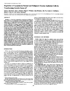
FIG. 1. Time course of differentiation and loss of TGF-8 receptors in L6 cells. Cultures were prepared as described under “Experimental Procedures.” A t zero time, the monolayers were washed and incubated with DMEM containing 1%horse serum and 0.3 p M insulin to stimulate differentiation. A t the indicated times, parallel cultures were removed for creatine kinase ( C K ) analysis and for measurement of TGF-/3binding as specified under “Experimental Procedures.” Results are means of triplicate determinations.
125
-
I -TGF-I3
+
205 k-
116 k-
97 k-
RESULTS
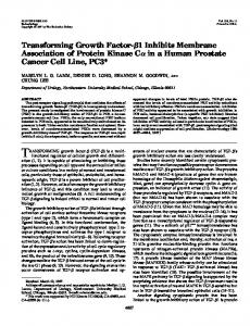
Effects of Differentiation on Binding of TGF-/3 in Muscle Cells-We have measured the binding of TGF-/3 before and after differentiation in three cell lines, L6, C2, and BC3H1. The first two fuse to form multinucleated myotubes as part of their differentiation program, whereas the last exhibits many of the biochemical aspects of myogenesis when transferred to medium lacking mitogens but does not fuse or irreversibly withdraw from the cell cycle. As indicated in Fig. 1, L6 cells show a very substantial loss of TGF-/3 receptors as they differentiate (quantitated here as elevation of creatine kinase activity, which is closely paralleled by percent nuclei, in myotubes). It shouldbenoted that the fall in TGF-/3 receptors precedes the elevation of creatine kinase; by day 2, TGF-/3 binding had decreased to less than half of that found initially, whereas there was only a very small increase in creatine kinase activity at thattime. The disappearance of TGF-/3 receptors upon differentiation is also demonstrated by affinity labeling of the cell surface receptors with lZ5I-labeledTGF-/3 (Fig. 2). As reported earlier by Massague et al. (5), L6 myoblasts do not possess the large (>250 kDa) TGF-P receptors found on many other cell types,
66 k-
MYOBLAST
MYOTUBE
FIG. 2. Affinity labeling of TGF-8 receptors in L6 myoblasts and myotubes. L6 myoblast and myotube cultures were prepared, labeled in the absence (-) or presence (+) of unlabeled TGF-8 (125 ng/ml) and analyzed as described under “Experimental Procedures.”
and it appears that the smaller 85- and 65-kDa receptors mediate the actions exerted by TGF-/3 on L6 myoblasts. The loss of TGF-/3 binding does not occur in all kinds of muscle cells. Fig. 3 presents a comparison of three cell lines at various stages of differentiation; L6 cells about 80% fused
Myogenesis and TGF-p Binding
TGF-beta Bound (fmollmg Protein)
FIG. 3. Effects of differentiation on binding of TGF-,9 in three muscle celllines. Binding of TGF-8to the cultures of myogenic cell lines was determined before and after extensive differentiation as described under “Experimental Procedures.” TABLEI Properties of TGF-8 receptors on muscle cell lines The constants presented here werecalculated from the data plotted in Fig. 3. Cell line
State
L6-Al
Myoblasts 15 Myotubes Myoblasts Myotubes 20 Undifferentiated Differentiated
c2 BCsH1
5
0
Kd
Binding cauacitv
p~
fmollmg protein
19 12 8 8 10 14
58 16 3 10
160
c
c
0
0
140 120
190
TGF-O (pgiml)
FIG. 4. Effects of TGF-8 on amino acid uptake by L6 myoblasts. AIB uptake was measured 7 h after addition of the indicated concentrations of TGF-p to the cell cultures, which were preparedas described under “Experimental Procedures.”
(visual estimate), C2 cells virtually completely fused, and BC3H1 cells at aquiescentsubconfluent state that corresponds to maximum differentiation. Because BC3H1 cells differentiate inthe absence of fusion, it was of special interest to compare levels of TGF-(3 receptors in the undifferentiated and differentiated states and thereby determine whether or not the decline in receptors associated with terminal differentiation was coupled to differentiation or to fusion. Binding data were analyzed by the method of Scatchard (13). This plot yields a slope proportional to the affinity andan x
intercept corresponding to the number of binding sites. As shown in Fig. 3, L6 and C2 myoblasts exhibit a decline in TGF-(3 receptors following fusion, whereas BC3H1 cells lose no more than half of their TGF-(3receptors as they differentiate. The decrease in binding in L6-A1 and C2 myoblasts apparently resulted from a decrease in number, not alessened affinity of the receptors (Table I). The dissociation constants found here are in the range (1-60 PM) recently reported by Wakefield et al. (14) in their survey of properties of TGF-(3 receptors in a wide range of cell types. In the L6-Al cells, for which cell counts were also done, the number of TGF-P receptors/cell was18,000; the results with these cells fit exactly on the log-log plot of receptor affinity uersus number of TGF-(3 receptors/cell for 34 cell types analyzed by Wakefield et al. (14). In other experiments (data not shown) there was no detectable loss of receptors in differentiated BC3H1cells. Olson et al. (4) have reported that these cells remain responsive to TGF-(3after differentiation, so the retention of receptors after differentiation was not unexpected. Thus, it appears that the loss of receptors is more closely associated with fusion than with the central controls of differentiation. Effects of Differentiation on Responsiveness to TGF-@-If the binding measured here represented true receptors for TGF-(3, then the loss of binding should be accompanied by a loss of responsiveness of the cells. To investigate this point, we measured the effects of TGF-(3 on amino acid uptake in myoblasts and myotubes. Earlier, Boerner et al. (15) had shown thatTGF-P stimulated AIB uptakein normal rat kidney cells, and we have also demonstrated stimulation of amino acid uptake by TGF-(3 in L6-Al.’ As shown in Fig. 4, the stimulation of AIB uptake by TGF-(3 isalmost completely lost in myotubes; the small remaining response can be attributed to a few undifferentiated myoblasts remaining in the cultures. ComparisontoChanges in IGF Receptors-The loss of growth factor receptors is not a universal result of myoblast differentiation. As shown in Fig. 5, binding of IGF-I per microgram DNA is not decreased as L6 myoblasts fuse to form myotubes, although there is some decrease in sensitivity to the hormone. (Similar decreases in responsiveness to rIGF11 and insulin were observed, which is to be expected if they both act through the type I IGFreceptor, as hasbeen reported by Ewton et al. (ll).)Given the substantial metabolic differences between myoblasts and differentiated myotubes, it seems likely that the displacement of the response curves in myotubes can best be explained by differences in interactions of intracellular mediators of the IGF effects, as has recently been considered in detail by Loeb and Strickland (16). DISCUSSION
The results presented here demonstrate that binding of TGF-P decreases dramatically as myoblasts fuse to form
25 I - IGF - I BINDING
FIG. 5. Effects of differentiation on responses to IGF-I and its binding to receptors on L6 myoblasts and myotubes. Actions ofIGF-Iwere 3 measured as detailed under “Experimen- 5 tal Procedures.” and specific binding of the hormonewas measured as described by Ewton et al. (11).
4031
PROTEOLYSIS
AIB UPTAKE
6oo 400
150
200 0.1
UNLABELED IGF - I (ngml)
1
10
100
0
1
IGF - I CONCENTRATION (ngiml)
10
100
4032
Myogenesis and TGF-8 Binding
postmitotic myotubes, and thisdecrease is paralleled by a loss of responsiveness to TGF-(3. We point out that this parallel loss in receptors and responsiveness is one of the relatively few indications that thespecific and saturable growth factorbinding protein onthe cell membrane represents a true receptor; in this case, its presence or absence is clearly correlated with an activity evoked by the growth factor. As Loeb and Strickland (16) have pointed out,exact coincidence of binding and activity concentration curves is not a valid criterion for a receptor role of a binding protein, because this occurs only when the affinity of an intracellular mediator for its receptor is identical to thatof the growth factor and itsreceptor. Thus, this absolute relationship of loss of activity (Fig. 4) and loss of receptors (Fig. 1)is a more persuasive indication of involvement of these cell surface receptors in thestimulation of AIB uptake. Previous studies demonstrated that differentiated BC3H1 cells, which do notfuse, remained sensitive to theTGF-(3 (4). Similarly, C2 cells maintained in mitogen-free medium containing EGTA expressed muscle mRNAs and proteins in the absence of fusion; under these conditions, TGF-(3 receptors remain at levels similar to those in proliferating undifferentiated myoblasts? The loss of responsiveness of the differentiation program in myotubes to TGF-(3 is probably due in large part todisappearance of the TGF-(3 receptors from the cell surface. However, a basal level of TGF-P receptors was detected in fully differentiated C2 myotubes (Fig. 3B) so alterations in the intracellular signal transduction pathway utilized by TGF-fi may also accompany myoblast fusion. The loss of TGF-(3receptors reported here parallels the loss of epidermal growth factor (17) and fibroblast growth factor4 receptors in another line of mouse cells. Hauschka4 has suggested that thismight be a mechanism by which differentiated muscle cells become postmitotic, i.e. unresponsive to mitogens. However, we have found (Fig. 5) that postmitotic L6 myotubes do not lose receptors for IGF-I (a potent mitogen for L6 myoblasts ( l l ) ) , andtheycontinue to show other responses to thehormone. Furthermore, we (3,4) and others (5) have demonstrated that TGF-(3 is not mitogenic in any muscle cells tested. Taken together, these observations demonstrate that the loss of growth factor receptors cannot be viewedsolely as a mechanism by which myotubes become postmitotic. It is not surprising that BC3H1 cells, which differentiate reversibly, show little or no loss in TGF-P binding as they differentiate; they remain responsive to the TGF-(3 even in the differentiated state. It is more difficult to understand the substantial difference from our results reported by Massague et al. (5)in a study of the L6E9 clone; they found no loss of receptors or decrease in responsiveness of the myotubes to TGF-P. It seems unlikely that the differences result from simple technical problems; both laboratories have extensive experience with peptide hormone receptors, and in both cases, the cells showed parallel changes (or lack of them) in responsiveness to TGF-(3. It seems probable that the differences in loss ofTGF-(3receptors with differentiation reflect differences in the L6 lines studied. It has been at least 15 years since the L6E9 and L6-A1 clones were separated, and the repeated reclonings done since that time may well have ledto variants that differed in a property notclosely related to thebasis for clone selection, i.e. relatively rapid differentiation. We have found an analogous difference between L6 clones in comparJ . S. Hu, G. Spizz, and E. N. Olson, unpublished results.
ison of effects of differentiation on responsiveness to insulin; Beguinot et al. (18) found that differentiation was accompanied by a substantial increase in responsiveness to insulin, while Ewton et al. (11)observed a slight decrease in sensitivity of myotubes to insulin (similar to the loss in sensitivity to IGF-I shown in Fig. 5C). The appearance of acetylcholine receptors accompanying differentiation of muscle cells has long been usedas amarker of myogenic differentiation (19). De Vroede et al. (20) and Beguinot et al. (18) have shown that insulin receptors (and responsiveness) increase dramatically in somemusclecell lines as theydifferentiate. Hauschka’s group (see Footnote 4 ) has demonstrated strikingdecreases in epidermal growth factor and fibroblast growth factor receptors in differentiating mouse muscle cells.We now report similar decreases in TGF(3 receptors in two rodent muscle celllines, with little decrease in another line. Taken together, these observations indicate that myogenic differentiation is a process that involves a number of specific, individually regulated changes in important membrane components as well as simple fusion to form multinucleated myotubes. Acknowledgments-We thank Drs. Anita Roberts and Michael Sporn for highly purified TGF-8 and lZ61-labeledTGF-8 and Dr. B. Daniel Burleigh for recombinant DNA-produced IGF-I. The technical assistance of Mary Ellen Perry, LiIie Welych, and Cathleen Jenney is gratefully acknowledged. REFERENCES 1. Moses, H. L.,Childs, C. B., Halper, J., Shipley, G. D., and Tucker, R. F. (1984) in Control of Cell Growth and Proliferation (Veneziale, C.M., ed) pp. 147-167, Van Nostrand Reinhold, New York 2. Sporn, M. B., Roberts, A. B., Wakefield, L. M., and Assoian, R. K. (1986) Science 33,532-534 3. Florini, J. R., Roberts, A. B., Ewton, D. Z., Falen, S. L., Flanders, K. C., and Sporn, M.B. (1986) J. Biol.Chem. 2 6 1 , 1650916513 4. Olson, E. N., Sternberg, E., Hu, J. S., Spizz, G., and Wilcox, C. (1986) J. Biol. Chem. 1 0 3 , 1799-1805 5. Massague, J., Cheifetz, S., Endo, T., and Nadal-Ginard, B. (1986) Proc. Natl. Acad. Sci. U. S. A. 8 3 , 8206-8210 6. Florini, J. R., Ewton, D. Z., Evinger-Hodges, M. J., Falen, S. L., Lau, R. L., Regan, J. F., and Vertel, B. M. (1984) In Vitro 2 0 , 942-958 7. Evinger-Hodges, M. J., Ewton, D. Z., Seifert, S. C., and Florini, J. R. (1982) J. Cell Biol. 93,395-401 8. Spizz, G., Hu, J.-S., and Olson, E. N. (1987) Deu. Biol. 123,500507 9. Ewton, D. Z., Erwin, B. G., Pegg, A. E., and Florini, J. R. (1984) J. Cell. Physiol. 1 2 0 , 263-270 10. Olson, E. N., Caldwell, K. L., Gordon, J. I., and Glaser, L. (1983) J. Biol. Chem. 2 5 8 , 2644-2652 11. Ewton, D. Z., Falen, S. L., and Florini, J. R. (1987) Endocrinology 120,115-124 12. Laemmli, U. K. (1970) Nature 227,680-685 13. Scatchard, G. (1949) Ann. N. Y. Acad. Sci. 51,660-872 14. Wakefield, L. M., Smith, D. M., Masui, T., Harris, C. C., and Sporn, M. B. (1987) J. Biol. Chem. 105,965-975 15. Boerner, P., Resnick, R. J., and Racker, E. (1985) Proc. Natl. Acad. Sci. U. S. A. 82,1350-1353 16. Loeb, J. N., and Strickland, S. (1987) Mol. Endocrinol. 1, 75-82 17. Lim, R. W., and Hauschka, S. D. (1984) J. Biol. Chem. 9 8 , 739747 18. Beguinot, F., Kahn, C. R., Moses, A. C., and Smith, R.J. (1986) Endocrinology 18,446-455 19. Fambrough, D.M. (1979) Physiol. Reu. 5 9 , 165-227 20. De Vroede, M. A., Romanus, J. A., Standaert, M. L., Pollett, R. J., Nissley, S. P., and Rechler, M.M. (1984) Endocrinology 114,1917-1929
B. B. Olwin and S. D. Hauschka, personal communication.