The Journal of Immunology
Dectin-1 Expression and Function Are Enhanced on Alternatively Activated and GM-CSF-Treated Macrophages and Are Negatively Regulated by IL-10, Dexamethasone, and Lipopolysaccharide1 Janet A. Willment,* Hsi-Hsen Lin,* Delyth M. Reid,† Philip R. Taylor,* David L. Williams,‡ Simon Y. C. Wong,† Siamon Gordon,* and Gordon D. Brown2* Dectin-1 is the major macrophage receptor for -glucans and generates a proinflammatory response through the recognition of these carbohydrates on fungal pathogens. We have examined the effects of cytokines and other agents on the expression and functions of dectin-1 in both resident and elicited murine peritoneal macrophages (M). Dectin-1 expression was found to be highly up-regulated by GM-CSF and by the cytokines that induce alternative macrophage activation, IL-4 and IL-13. In contrast, IL-10, LPS, and dexamethasone, but not IFN-␥, down-regulated the expression of this receptor. Modulation of dectin-1 receptor levels correlated with the ability of these macrophages to bind zymosan and significantly affected the contribution of this receptor to the resultant proinflammatory response, as measured by the production of TNF-␣, although some M-specific differences were observed. These results correlate with the known effects of these cytokines and other agents on the ability of the immune system to recognize and respond to fungal pathogens. The Journal of Immunology, 2003, 171: 4569 – 4573.

-Glucans are structural components of fungal cell walls with well-characterized immunostimulatory properties and as such have been widely used to study the effector functions of leukocytes as well as inflammatory processes in vivo. We identified dectin-1 as a major receptor involved in the recognition of these carbohydrates, including both soluble and particulate forms, such as the -glucan-rich Saccharomyces cerevisiae cell wall fraction, zymosan (1, 2). Dectin-1 is a type II transmembrane receptor containing a single extracellular C-type lectin-like domain and an immunoreceptor tyrosine-based activation motif in its cytoplasmic tail (3). This receptor is predominantly expressed on cells of the monocyte/macrophage (M)3 and neutrophil lineages as well as on dendritic cells and a minor subpopulation of splenic T cells (4). We have recently shown that dectin-1 mediates the biological effects of -glucans through cooperation with the Tolllike receptor pathway at the cell surface (5). In addition to these exogenous ligands, dectin-1 can also recognize selected T cells, stimulating their proliferation through unidentified mechanisms (3). The successful control of invading microbes is critically dependent on the type of immune response mounted, which is orches-
*Sir William Dunn School of Pathology, University of Oxford, Oxford, United Kingdom; †Edward Jenner Institute for Vaccine Research, Compton, Berkshire, United Kingdom; and ‡Department of Surgery, James H. Quillen College of Medicine, Johnson City, TN 37614 Received for publication June 5, 2003. Accepted for publication August 21, 2003. The costs of publication of this article were defrayed in part by the payment of page charges. This article must therefore be hereby marked advertisement in accordance with 18 U.S.C. Section 1734 solely to indicate this fact. 1 This work was supported by the Wellcome Trust, the Arthritis Research Campaign, the Medical Research Council, and the Histiocytosis Research Foundation. 2 Address correspondence and reprint requests to Dr. Gordon D. Brown, Sir William Dunn School of Pathology, Oxford University, South Parks Road, Oxford, U.K. OX1 3RE. E-mail address:
[email protected]
Abbreviations used in this paper: M, macrophage; res-M, resident macrophage; thio-M, thioglycolate-elicited macrophage.
3
Copyright © 2003 by The American Association of Immunologists, Inc.
trated mostly through the actions of cytokines. Since dectin-1 is likely to be centrally involved in the innate response to fungal pathogens (5), we examined the effects of cytokines and other biological response modifiers on the expression and functions of this receptor. We show here that these agents significantly affect the expression of dectin-1 in primary M, altering their ability to recognize and respond to fungal particles.
Materials and Methods Cells and media Resident peritoneal M (res-M) and thioglycolate-elicited M (thioM), generated using standard procedures, were harvested from 6- to 10wk-old BALB/c mice (Sir William Dunn School of Pathology, Oxford, U.K.). Animals were kept and handled according to institutional guidelines. The cells were washed and maintained in RPMI 1640 (Life Technologies, Gaithersburg, MD) supplemented with 10% FCS, 100 U/ml penicillin, 0.1 mg/ml streptomycin, 2 mM L-glutamine, and 20 mM HEPES. For cytokine treatment, M were plated at 5 ⫻ 105 cells/well in six-well plates and allowed to adhere for 2 h at 37°C in 5% CO2. After thorough washing to remove nonadherent cells, fresh medium was added, supplemented with the appropriate cytokines (R&D Systems, Abingdon, U.K.) and other agents at the following concentrations: IL-4, IL-10, and GM-CSF at 20 ng/ml; TNF-␣, M-CSF, human TGF-, IL-13, and Escherichia coli LPS (Sigma-Aldrich, St. Louis, MO) at 10 ng/ml; IFN-␥ at 200 U/ml; and dexamethasone (Sigma-Aldrich) at 0.5 M. M were then incubated for an additional 4 or 24 h, as indicated, at 37°C in 5% CO2.
Dectin-1 mRNA expression Northern blots of total RNA extracted from res-M treated with the various cytokines indicated were prepared as previously described (6). Blots were probed with 32P-radiolabeled, full-length dectin-1 cDNA (4).
FACS analysis FACS was performed according to conventional protocols at 4°C in the presence of 2 mM NaN3, as previously described (4). Nonspecific binding sites on cells were blocked with 5% heat-inactivated rabbit serum, 0.5% BSA, 5 mM EDTA, and 4 g/ml 2.4G2 (anti-Fc␥RII and -III; BD PharMingen, San Diego, CA) before the addition of primary Abs. Surfaceexpressed dectin-1 was detected using 10 g/ml of biotin-labeled 2A11 mAb (2) and was compared with an irrelevant IgG2b isotype control. The biotinylated Abs were detected using streptavidin-allophycocyanin (BD 0022-1767/03/$02.00
4570
REGULATION OF DECTIN-1 EXPRESSION AND FUNCTION
PharMingen). Cells were fixed with 1% formaldehyde in PBS before analysis. Fold changes in surface staining were determined using the following equation: receptor surface expression after treatment (mean fluorescence of receptor specific mAb – mean fluorescence of isotype control)/receptor expression of untreated cells (mean fluorescence of receptor specific mAb – mean fluorescence of isotype control). All FACS experiments were repeated independently at least three times. The activity of the cytokines was confirmed by examining their effects on the surface expression of F4/80, scavenger receptor A, CR3, or MHC class II in cultured thio-M (data not shown).
incubated for an additional 6 h at 37°C in fresh medium containing the various treatments. Levels of TNF-␣ released into the supernatants were measured using the OptEIA murine TNF-␣ ELISA kit (BD PharMingen) as described by the manufacturer. The amount of TNF-␣ was normalized to the untreated control value, and the data presented represent pooled values from at least three independent experiments.
Fluorescent zymosan binding assays
Results and Discussion
The fluorescence-based binding assays using FITC-labeled zymosan were performed at 4°C, as described previously (2). Briefly, M were plated at 3 ⫻ 105 cells/well in 24-well plates, allowed to adhere for 2 h, washed three times, and then incubated for 24 h with and without treatment. The cells were cooled to 4°C and washed three times with prechilled culture medium. Where required, laminarin (Sigma-Aldrich; 100 g/ml) was added 20 min before the addition of FITC-labeled zymosan (Molecular Probes, Eugene, OR; 25 particles/cell), and the cells were incubated for an additional 1 h. After removal of unbound zymosan by extensive washing, the cells were lysed in 3% Triton X-100. FITC in lysates was quantified using a Titer-Tek Fluoroskan II (Labsystems Group, Basingstoke, U.K.), and the amount of fluorescence was normalized to the untreated control value. Each experiment was performed in triplicate, and the data shown represent pooled values from at least two independent experiments.
Dectin-1 expression is up-regulated on IL-4/IL-13- and GM-CSF-treated peritoneal M
TNF-␣ assays M were harvested, plated at 2 ⫻ 105 cells/well in 24-well plates, and treated for 24 h with the appropriate agents. At the start of the assay the cells were cooled to 4°C and washed three times with prechilled culture medium. When appropriate, glucan phosphate (7) was added to 100 g/ml, and the cells were incubated for 20 min at 4°C to allow inhibition of dectin-1 (2). After the addition of zymosan, the cells were incubated at 37°C in 5% CO2 for 30 min, washed to remove unbound particles, and then
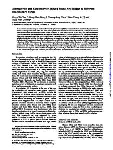
FIGURE 1. Dectin-1 expression on res-M is modulated by cytokines and other agents. A, Representative FACS profiles of 24-h cultured untreated (filled histograms) and treated (unfilled histograms) M, demonstrating the effects of IL-4, GM-CSF, and LPS on dectin-1 expression. M were stained with the anti-dectin-1 Ab 2A11 (dark gray) or isotype control (light gray). The changes in dectin-1 receptor levels at 4 h (B) and 24 h (C) were quantified as described in Materials and Methods. The data were generated from at least three independent experiments and were normalized to the untreated control value. Error bars indicate the SEM. D, Northern blot analysis demonstrating that the agents tested affect the levels of dectin-1 mRNA. The 28S rRNA band is shown as a loading control.
Statistical analysis Statistics were calculated using paired t tests in GraphPad PRISM (version 2.0; GraphPad, Berkeley, CA).
As dectin-1 is involved in the innate recognition and response to -glucans and fungal pathogens (5), we investigated whether the expression and function of this receptor could be modulated by exogenous agents. We initially performed these analyses in resM, in which up-regulation of this receptor after 24 h of in vitro culture had been previously noted (4). Since we observed that upregulation of dectin-1 could occur within 4 h of culture (data not shown), the effects of cytokines and other agents were examined at both early (4 h) and late (24 h) time points. Using a FACS-based approach with the anti-dectin-1 mAb 2A11 (2), we found that surface expression of dectin-1 was markedly influenced by cytokines and biological response modifiers, and that this regulation was reflected in the levels of mRNA present in the cell (Fig. 1, A–D). Strikingly, dectin-1 appeared to be highly up-regulated within 4 h by IL-4 and IL-13. These cytokines are central to the induction of Th2-type responses and have been shown to induce alternative activation of M, characterized by cell surface and phenotypic
The Journal of Immunology changes, including elevated levels of certain pattern recognition receptors, such as the mannose receptor (8). The effects of these cytokines on dectin-1 expression as well as the high levels of dectin-1 on alveolar macrophages (4), which are considered similar to alternatively activated M (9), are consistent with this phenotype. Interestingly, IFN-␥, a Th1-type cytokine, had little direct effect on the expression of dectin-1. The binding of IL-4 to its heterodimeric receptor leads to STAT6 activation, dimerization, nuclear translocation, and binding to specific elements of various IL-4/IL-13-responsive genes (10). As the IL-4-mediated regulation of dectin-1 is rapid and affected the levels of mRNA (Fig. 1D), STAT6 is likely to be mediating this effect, as has been shown for other IL-4-induced M genes (11). Indeed, at least one STAT6 consensus region was present in the putative promoter region of dectin-1 (data not shown). Furthermore, the ability of IFN-␥ to antagonize the IL-4-mediated induction in res-M (Fig. 1C) after 24 h is consistent with the ability of IFN-␥ to inhibit STAT6 activation through the induction of SOCS-1 (12). This mechanism, however, has yet to investigated further in the context of the dectin-1 promoter. Furthermore, IFN-␥ did not inhibit IL-4-mediated induction of dectin-1 in elicited M (see below), suggesting that additional regulatory mechanisms may be involved. GM-CSF and, to a lesser extent, TGF- also up-regulated the expression of dectin-1, but this effect was only detectable after 24 h of culture (Fig. 1C). Although GM-CSF has been shown to be cooperative with IL-4 in inducing the expression of other lectinlike receptors, such as DC-SIGN (13), the combination of these two cytokines was not additive for dectin-1 expression at either time point. Dexamethasone, IL-10, and LPS negatively regulate dectin-1 expression We also identified dexamethasone, IL-10, and LPS as negative regulators of dectin-1 expression, whose effects were only apparent after 24 h of culture (Fig. 1C). The inhibitory effect of dexamethasone probably occurs at the transcriptional level (14); this is consistent with previously characterized effects of this glucocorticoid on -glucan receptor activity (15). IL-10 is produced by M
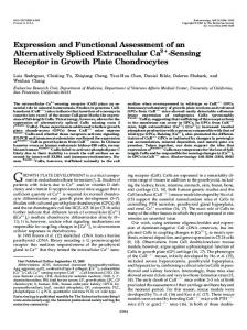
FIGURE 2. Modulation of dectin-1 expression on 24-h treated thio-M. Although similar, the modulation of receptor levels is quantitatively less than that obtained with res-M. The data were generated from at least three independent experiments, and the error bars indicate the SEM. Note that thio-M respond differently from res-M (Fig. 1) to IL-4 plus IFN-␥ and to TGF-.
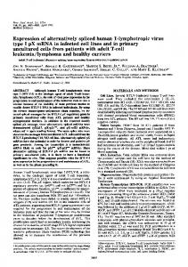
4571 after stimulation by zymosan and other agents, and modulates the resultant inflammatory response through STAT3-dependent mechanisms (16). Thus, the negative effect of IL-10 on dectin-1 expression may represent a negative feedback mechanism, as previously suggested (17). LPS has been shown to negatively regulate the expression of several M receptors, including the mannose receptor (18), but the significance of this effect on dectin-1 is unclear. No changes in the surface expression of dectin-1 were observed at either time point in cells cultured in M-CSF or TNF-␣. Dectin-1 regulation on elicited macrophages As we have shown significant quantitative differences between the levels of dectin-1 surface expression in resident and elicited M (4), we also examined the effects of these cytokines in thio-M (Fig. 2). Although most of the cytokines had similar effects on dectin-1 expression, following 24 h of treatment the magnitude of the response in thio-M was less than that observed in res-M. This may be related to the lower initial levels of surface-expressed dectin-1 in res-M (4), giving rise to a more pronounced effect after cytokine treatment. The inability of TGF- to affect dectin-1 expression and the lack of antagonism of IFN-␥ on the IL-4-mediated regulation of dectin-1, were the most notable differences in this M population. Functional consequences of modulation of dectin-1 expression in primary macrophages We next examined the effect of cytokine modulation on the ability of these primary cells to recognize the -glucan-rich particle, zymosan (19). Using laminarin to specifically inhibit dectin-1 (1), we found that the cytokine-induced effects on the levels of surfaceexpressed dectin-1 after 24 h closely correlated with the ability of the primary cells to recognize this fungal-derived particle (Fig. 3). In untreated res-M, approximately half the zymosan binding capacity was due to dectin-1 (Fig. 3A), consistent with our previous findings that these cells possess an additional, mannan-inhibitable, receptor(s) for zymosan (4). Notably, only the dectin-1mediated component of zymosan recognition was significantly modulated by the cytokine treatments. In thio-M, almost all the zymosan binding activity was due to dectin-1 (Fig. 3B), as we have previously described (2). IL-4, GM-CSF, IFN-␥, and dexamethasone have been shown to have significant effects on the immune response to fungal infections (20 –23). As dectin-1 is involved in mediating a proinflammatory response to these pathogens (5), we examined what effect these agents would have on this response. In untreated peritoneal M, zymosan-stimulated TNF-␣ production could be inhibited when the cells were pretreated with the specific dectin-1 antagonist, glucan phosphate (Fig. 4) (5). In contrast to thio-M (Fig. 4B), however, addition of glucan phosphate to res-M (Fig. 4A) only partially inhibited TNF-␣ production, suggesting the presence of additional signaling mechanisms in these cells. Differences between these M populations were also observed in the levels of TNF-␣ produced in response to zymosan after cytokine treatment, which was consistent with previous descriptions of these M populations (24). IL-4-treated M are generally thought to act as suppressor cells, limiting the Th1-mediated inflammatory response and promoting wound healing. A number of studies, however, have demonstrated that IL-4 pretreatment can also induce a proinflammatory response, leading to protective Th1 development (25, 26). Indeed, the IL-4 priming mechanism is critical for control of Candida albicans infection (20). IL-4 pretreatment appeared to have opposite effects on the inflammatory response depending on the M population
4572
FIGURE 3. Zymosan recognition by peritoneal M correlates with the expression of dectin-1. Cytokines, LPS, and dexamethasone affect the ability of res-M (A) and thio-M (B) to bind zymosan, correlating with the levels of dectin-1 on these cells (compare with Figs. 1 and 2). The contribution of dectin-1 to this activity was confirmed using the dectin-1-specific inhibitor laminarin (lam) (1). In res-M, the contribution of the non-glucan receptor(s) was largely unaffected by cytokine treatment, as similar levels of residual zymosan binding were obtained after laminarin treatment. The percent binding of FITC-labeled zymosan by treated cells is expressed relative to untreated M, pooled from two independent experiments, and the error bars indicate the SEM.
examined, with the response being enhanced in res-M, but suppressed in thio-M (Fig. 4). In res-M, the levels of increased TNF-␣ produced corresponded to the increase in dectin-1 on the surface of these cells (Fig. 1), and the inhibition of this response by glucan phosphate confirmed that this effect was being mediated by dectin-1 (5). Thus, these data suggest a mechanism in specific M populations by which IL-4-mediated receptor up-regulation, such as dectin-1, can lead to a proinflammatory, Th1, response. GM-CSF is known to prime neutrophils and M for an enhanced proinflammatory response to microbial stimuli, including zymosan, and has been suggested for use as an adjunct therapy for the treatment of systemic fungal infections (27). Although up-regulation of dectin-1 by GM-CSF leads to a proportionate dectin-1mediated increase in the ability of these M to bind zymosan (Fig. 3), a disproportionate increase in the level of TNF-␣ was observed in both M populations examined (Fig. 4). Dectin-1 had a major contribution to this activity, as determined by glucan phosphate inhibition (⬃50 and⬃75% inhibition in res-M and thio-M, respectively). The exaggerated response observed with GM-CSF results in part from the up-regulation of signaling components, including TLR-2 (28), although the exact mechanisms are still unclear. IFN-␥ has been shown to be critical in the generation of protective Th1 responses for the control of fungal infections (29, 30). Although IFN-␥ did not greatly alter the expression of dectin-1 (Figs. 1 and 2), we examined the effect of this cytokine on the proinflammatory response to zymosan (Fig. 4). Consistent with its
REGULATION OF DECTIN-1 EXPRESSION AND FUNCTION
FIGURE 4. IL-4, GM-CSF, IFN-␥, and dexamethasone treatments influence the proinflammatory response in peritoneal M. The effects of these agents on the production of TNF-␣ in response to zymosan by res-M (A) and thio-M (B) were determined as described in Materials and Methods. The contribution of dectin-1 to this response was measured using glucan phosphate (GluP) (5). The partial inhibition by glucan phosphate in untreated res-M, indicates the presence of an additional signaling component(s) in these cells. The histograms show the percentage of TNF-␣ produced relative to that in untreated cells. No TNF-␣ was detected when the cells were treated with these agents alone in the absence of zymosan (data not shown). The data shown are the pooled average of at least three independent experiments, normalized to the untreated control value, and error bars indicate the SEM. ⴱ, p ⬍ 0.05; n.s., not significantly different, p ⬎ 0.05.
ability to prime classical macrophage activation (31), pretreatment with IFN-␥ greatly enhanced the production of TNF-␣ in response to zymosan in both M populations. Although dectin-1 made a major contribution to this response in thio-M, as determined by glucan phosphate inhibition, the response appeared to be largely dectin-1 independent in res-M. Thus, the data suggest that IFN-␥ can prime M for enhanced responses to fungal products, but that this involves different mechanisms in each M population. Immunosuppression induced by corticosteroids, such as dexamethasone, is known to strongly predispose patients to fungal infections (22). Dexamethasone significantly down-regulates dectin-1 expression on peritoneal M (Figs. 1 and 2) and abolishes the TNF-␣ response to zymosan (Fig. 4). This effect could be a contributing factor to fungal susceptibility, but the ability of dexamethasone to repress the proinflammatory responses is more complex than receptor regulation, as it also represses NF-B through induction of IB, inhibiting the production of TNF-␣ (32). Although we have focused on the effects of cytokines on the recognition and response of M to yeast particles, it should be noted that the modulation of dectin-1 expression may also impact on its interactions with T cells. Indeed, IL-4-induced, alternatively activated M have been implicated in the inhibition of T cell proliferation, via cell-to-cell contact, and suppression of the mitogenactivated CD4⫹ T cell secretory response (9). The role of dectin-1,
The Journal of Immunology if any, in these interactions remains unclear, however, as both human and murine dectin-1 have been shown to stimulate, not inhibit, the proliferation of T cells (3, 33). In conclusion, we have shown that the expression of dectin-1 on primary M can be modulated by cytokines and other agents and that this affects the ability of these cells to recognize and respond to fungal particles. The effects of these agents on dectin-1 are consistent with their known effects on the host response to fungal infection, and this suggests that variations in the expression of this receptor may influence the ability of the immune system to respond to these pathogens in vivo.
4573
14. 15. 16.
17.
18.
Acknowledgments
19.
We thank the staff of our animal facility for the care of the animals used in this study.
20.
References 1. Brown, G. D., and S. Gordon. 2001. Immune recognition: a new receptor for -glucans. Nature 413:36. 2. Brown, G. D., P. R. Taylor, D. M. Reid, J. A. Willment, D. L. Williams, L. Martinez-Pomares, S. Y. C. Wong, and S. Gordon. 2002. Dectin-1 is a major -glucan receptor on macrophages. J. Exp. Med. 296:407. 3. Ariizumi, K., G. L. Shen, S. Shikano, S. Xu, R. Ritter III, T. Kumamoto, D. Edelbaum, A. Morita, P. R. Bergstresser, and A. Takashima. 2000. Identification of a novel, dendritic cell-associated molecule, dectin-1, by subtractive cDNA cloning. J. Biol. Chem. 275:20157. 4. Taylor, P. R., G. D. Brown, D. M. Reid, J. A. Willment, L. Martinez-Pomares, S. Gordon, and S. Y. C. Wong. 2002. The -glucan receptor, dectin-1, is predominantly expressed on the surface of cells of the monocyte/macrophage and neutrophil lineages. J. Immunol. 269:3876. 5. Brown, G. D., J. Herre, D. L. Williams, J. A. Willment, A. S. J. Marshall, and S. Gordon. 2003. Dectin-1 mediates the biological effects of -glucan. J. Exp. Med. 197:1119. 6. Stacey, M., G. W. Chang, S. L. Sanos, L. R. Chittenden, L. Stubbs, S. Gordon, and H. H. Lin. 2002. EMR4, a novel epidermal growth factor (EGF)-TM7 molecule up-regulated in activated mouse macrophages, binds to a putative cellular ligand on B lymphoma cell line A20. J. Biol. Chem. 277:29283. 7. Muller, A., P. J. Rice, H. E. Ensley, P. S. Coogan, J. H. Kalbfleish, J. L. Kelley, E. J. Love, C. A. Portera, T. Ha, I. W. Browder, et al. 1996. Receptor binding and internalization of a water-soluble (133)--D-glucan biologic response modifier in two monocyte/macrophage cell lines. J. Immunol. 156:3418. 8. Gordon, S. 2003. Alternative macrophage activation. Nat. Rev. Immunol. 3:23. 9. Goerdt, S., and C. E. Orfanos. 1999. Other functions, other genes: alternative activation of antigen-presenting cells. Immunity 10:137. 10. Nelms, K., A. D. Keegan, J. Zamorano, J. J. Ryan, and W. E. Paul. 1999. The IL-4 receptor: signaling mechanisms and biologic functions. Annu. Rev. Immunol. 17:701. 11. Welch, J. S., L. Escoubet-Lozach, D. B. Sykes, K. Liddiard, D. R. Greaves, and C. K. Glass. 2002. TH2 cytokines and allergic challenge induce Ym1 expression in macrophages by a STAT6-dependent mechanism. J. Biol. Chem. 277:42821. 12. Dickensheets, H. L., C. Venkataraman, U. Schindler, and R. P. Donnelly. 1999. Interferons inhibit activation of STAT6 by interleukin 4 in human monocytes by inducing SOCS-1 gene expression. Proc. Natl. Acad. Sci. USA 96:10800. 13. Relloso, M., A. Puig-Kroger, O. M. Pello, J. L. Rodriguez-Fernandez, G. de la Rosa, N. Longo, J. Navarro, M. A. Munoz-Fernandez, P. Sanchez-Mateos, and A. L. Corbi.
21. 22. 23.
24.
25.
26.
27.
28.
29. 30.
31. 32.
33.
2002. DC-SIGN (CD209) expression is IL-4 dependent and is negatively regulated by IFN, TGF-, and anti-inflammatory agents. J. Immunol. 168:2634. Adcock, I. M. 2001. Glucocorticoid-regulated transcription factors. Pulm. Pharmacol. Ther. 14:211. Goldman, R. 1988. Characteristics of the -glucan receptor of murine macrophages. Exp. Cell Res. 174:481. Lang, R., D. Patel, J. J. Morris, R. L. Rutschman, and P. J. Murray. 2002. Shaping gene expression in activated and resting primary macrophages by IL-10. J. Immunol. 169:2253. Ajuebor, M. N., A. M. Das, L. Virag, R. J. Flower, C. Szabo, and M. Perretti. 1999. Role of resident peritoneal macrophages and mast cells in chemokine production and neutrophil migration in acute inflammation: evidence for an inhibitory loop involving endogenous IL-10. J. Immunol. 162:1685. Shepherd, V. L., R. Abdolrasulnia, M. Garrett, and H. B. Cowan. 1990. Downregulation of mannose receptor activity in macrophages after treatment with lipopolysaccharide and phorbol esters. J. Immunol. 145:1530. Di Carlo, F. J., and J. V. Fiore. 1958. On the composition of zymosan. Science 127:756. Mencacci, A., G. Del Sero, E. Cenci, C. F. d’Ostiani, A. Bacci, C. Montagnoli, M. Kopf, and L. Romani. 1998. Endogenous interleukin 4 is required for development of protective CD4⫹ T helper type 1 cell responses to Candida albicans. J. Exp. Med. 187:307. Armitage, J. O. 1998. Emerging applications of recombinant human granulocytemacrophage colony-stimulating factor. Blood 92:4491. Maertens, J., M. Vrebos, and M. Boogaerts. 2001. Assessing risk factors for systemic fungal infections. Eur. J. Cancer Care 10:56. Kaposzta, R., P. Tree, L. Marodi, and S. Gordon. 1998. Characteristics of invasive candidiasis in ␥ interferon- and interleukin-4-deficient mice: role of macrophages in host defense against Candida albicans. Infect. Immun. 66:1708. Stein, M., and S. Gordon. 1991. Regulation of tumor necrosis factor (TNF) release by murine peritoneal macrophages: role of cell stimulation and specific phagocytic plasma membrane receptors. Eur. J. Immunol. 21:431. Major, J., J. E. Fletcher, and T. A. Hamilton. 2002. IL-4 pretreatment selectively enhances cytokine and chemokine production in lipopolysaccharide-stimulated mouse peritoneal macrophages. J. Immunol. 168:2456. D’Andrea, A., X. Ma, M. Aste-Amezaga, C. Paganin, and G. Trinchieri. 1995. Stimulatory and inhibitory effects of interleukin (IL)-4 and IL-13 on the production of cytokines by human peripheral blood mononuclear cells: priming for IL-12 and tumor necrosis factor ␣ production. J. Exp. Med. 181:537. Hubel, K., D. C. Dale, and W. C. Liles. 2002. Therapeutic use of cytokines to modulate phagocyte function for the treatment of infectious diseases: current status of granulocyte colony-stimulating factor, granulocyte-macrophage colonystimulating factor, macrophage colony-stimulating factor, and interferon-␥. J. Infect. Dis. 185:1490. Kurt-Jones, E. A., L. Mandell, C. Whitney, A. Padgett, K. Gosselin, P. E. Newburger, and R. W. Finberg. 2002. Role of toll-like receptor 2 (TLR2) in neutrophil activation: GM-CSF enhances TLR2 expression and TLR2-mediated interleukin 8 responses in neutrophils. Blood 100:1860. Allendoerfer, R., and G. S. Deepe, Jr. 1997. Intrapulmonary response to Histoplasma capsulatum in ␥ interferon knockout mice. Infect. Immun. 65:2564. Cenci, E., A. Mencacci, G. Del Sero, C. F. d’Ostiani, P. Mosci, A. Bacci, C. Montagnoli, M. Kopf, and L. Romani. 1998. IFN-␥ is required for IL-12 responsiveness in mice with Candida albicans infection. J. Immunol. 161:3543. Mosser, D. M. 2003. The many faces of macrophage activation. J. Leukocyte Biol. 73:209. Auphan, N., J. A. DiDonato, C. Rosette, A. Helmberg, and M. Karin. 1995. Immunosuppression by glucocorticoids: inhibition of NF-B activity through induction of IB synthesis. Science 270:286. Grunebach, F., M. M. Weck, J. Reichert, and P. Brossart. 2002. Molecular and functional characterization of human dectin-1. Exp. Hematol. 30:1309.