ISSN 1607-6729, Doklady Biochemistry and Biophysics, 2018, Vol. 480, pp. 173–176. © Pleiades Publishing, Ltd., 2018. Original Russian Text © A.P. Puzyr, S.E. Medvedeva, A.E. Burov, Yu.P. Zernov, V.S. Bondar, 2018, published in Doklady Akademii Nauk, 2018, Vol. 480, No. 6, pp. 743–746.
BIOCHEMISTRY, BIOPHYSICS, AND MOLECULAR BIOLOGY
Detection of Hispidin by a Luminescent System from Basidiomycete Armillaria borealis A. P. Puzyra, *, S. E. Medvedevaa, A. E. Burova, b, Yu. P. Zernovc, and V. S. Bondara Presented by Academician A.G. Degermendzhi January 15, 2018 Received January 18, 2018
Abstract—In in vitro experiments, the possibility of using a luminescent system extracted from the luminous fungus Armillaria borealis has been shown to detect and determine the concentration of hispidin. A linear dependence of the luminescent response on the content of hispidin in solutions in the concentration range of 5.4 × 10⁻⁵– 1.4 × 10⁻² μM was detected. The stability of the enzyme system and the high sensitivity of the bioluminescent reaction allows carrying out multiple measurements with the analyte detection limit of 1.3 × 10⁻¹¹ g. The obtained results show the prospects of creating a rapid bioluminescent method for the analysis of medical substances or extracts from various biological objects for the presence of hispidin. DOI: 10.1134/S1607672918030146
One of the important directions in modern biotechnology is associated with the identification of new natural sources of raw materials for obtaining target products that are in demand in medicine, biology, pharmaceutical industry, etc. In particular, in China, Japan, Korea, and other countries in the Asia-Pacific region, studies on the pharmacological activity and practical use of compounds synthesized by basidiomycetes are actively performed [1–4]. These compounds include hispidin, the metabolite produced by a number of basidiomycetes that exhibits antioxidant and antitumor activity [5–8]. The properties of hispidin are usually studied using its commercial preparations or extracts from various fungal species [6, 8–11]. The determination of the presence and concentration of hispidin in fungal extracts is a fairly complex and multistage procedure. The method that is most commonly used in laboratories is based on recording the retention time on columns during HPLC; it requires specialized equipment and is highly timeconsuming [12]. a Institute
of Biophysics, Krasnoyarsk Research Center, Siberian Branch, Russian Academy of Sciences, Krasnoyarsk, 660036 Russia b Institute of Computational Technologies, Siberian Branch, Russian Academy of Sciences, Krasnoyarsk, 660049 Russia c Voevodsky Institute of Chemical Kinetics and Combustion, Siberian Branch, Russian Academy of Sciences, Novosibirsk, 630090 Russia *e-mail:
[email protected]
Earlier [13, 14], we obtained extracts from three species of luminous basidiomycetes (Neonothopanus nambi, Armillaria borealis, and Mycena citricolor), containing fungal luminescent systems exhibiting enzymatic activity in vitro. With the use of the extracts from the fungus N. nimbi, it was shown [15] that hispidin is at least one of the precursors (preluciferin) of the substrate of the light emission reaction in higher fungi. This served as a basis for testing the applicability of luminescent systems of luminous basidiomycetes in creating new rapid methods for testing hispidin in extracts from various natural objects in order to identify the potential sources of this compound. It is commonly known that bioluminescent assay methods are much more sensitive as compared to, for example, spectral methods (spectrophotometry and colorimetry), which are used in biological and medical studies. In this paper, we present the results of experiments on the determination of hispidin in solutions in vitro using the luminescent system isolated from the basidiomycete Armillaria borealis. In the study, we used the culture of the higher luminous fungus A. borealis IBSO 2328 (Collection of Microorganisms CCIBSO 836, Institute of Biophysics, Krasnoyarsk Research Center, Siberian Branch, Russian Academy of Sciences). The mycelium was obtained by submerged culturing of the fungus. The procedures of growing the mycelium, isolation of the luminescent system from the biomass, and obtaining freeze-dried samples that retain enzymatic activity for a long time were described in our earlier study [14]. Before studying, the freeze-dried samples of the luminescent system were dissolved in deionized water
173
174
PUZYR et al.
Luminescence, arb. units 2.0×10⁵ 1
1.5×10⁵ 7
1.0×10⁵ 6 5 0.5×10
5
4 2 0
3
50
100
150
200 Time, min
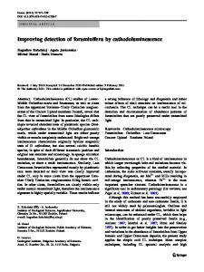
Fig. 1. Amplitude and kinetics of light signals detected when using the luminescent system from the basidiomycete A. borealis for testing: (1) after the addition of NADPH, (2–7) after subsequent periodic additions of hispidin solutions of increasing concentrations.
(Milli-Q system, Millipore, United States), and the resulting solutions were used in experiments. To test the luminescent activity, we used NADPH (Serva, Germany) and hispidin (Sigma-Aldrich, United States). The initial hispidin solution with a concentration of 20 mM was prepared in methanol (Sigma-Aldrich) and stored at –20°C. For experiments, samples with different hispidin concentrations were prepared by successive dilutions of the stock solution with deionized water. During the measurements of luminescence, the amplitude and dynamics of the light signal were recorded with a Glomax® 20/20 luminometer (Promega BioSystems Sunnyvale, Inc., United States) in the mode of one measurement per second. The intensity of light emission of the samples was expressed in arbitrary luminescent units. The reaction was initiated by adding 5 μL of 10 mM NADPH and 5 μL of a hispidin solution with a known concentration to 100 μL of the luminescent system sample. When studying the in vitro properties of the luminescent enzyme system isolated from the A. borealis mycelium biomass, we found that a light signal was recorded after the addition of NADPH (Fig. 1, peak 1). This effect was observed in the case when the enzyme system contained the endogenous substrate of the luminescent reaction. When the light signal intensity decreased to a certain steady-state level, the reaction
mixture was supplemented with the test solutions of hispidin as an exogenous substrate of the luminescent reaction. The results of consistent measurements of six hispidin samples with increasing analyte concentration using the same enzyme system sample are shown in Fig. 1 (peaks 2–7). The possibility of continuous recording of light signals at successive multiple additions of hispidin can be explained by the high thermostability of the enzymes of the luminescent system used, which retained their activity at room temperature for 6–8 h. In our opinion, the presence of the endogenous substrate in the samples of the luminescent system of A. borealis is a positive factor. On the one hand, the recording of a light signal after the addition of NADPH (Fig. 1, peak 1) is an indication of the functional activity of the luminescent system enzymes. At the same time, the presence of the endogenous substrate was not an obstacle to using this system for further testing of hispidin (Fig. 1, peaks 2–7). One of the most important characteristics of any analytical system is the lower limit of quantitation of the analyte. In our experiments (Fig. 2), for the luminescent system from the fungus A. borealis, this parameter was 5.4 × 10⁻⁵ μM (1.3 × 10⁻¹¹ g) of hispidin. This figure shows that, at this hispidin concentration, we recorded a detectable light emission level. Possibly, the sensitivity of the luminescent method for hispidin determination can be increased by optimizing the test
DOKLADY BIOCHEMISTRY AND BIOPHYSICS
Vol. 480
2018
DETECTION OF HISPIDIN BY A LUMINESCENT SYSTEM
175
Luminescence, arb. units 8000
7000 2 6000
5000 1
4000 Background of the device 3000
0
500
1000
1500
2000 Time, s
Fig. 2. Light signals detected using the luminescent system from A. borealis at hispidin concentrations in the reaction mixture of (1) 5.4 × 10⁻⁵ μM and (2) 1.1 × 10⁻⁴ μM.
Luminescence, arb. units 2.5×10⁸
2.0×10⁸
1.5×10⁸
1.0×10
4×10⁷
8
3×10⁷ 2×10⁷
0.5×10⁸ 1×10⁷
0
0
0.002
0.004
0.006
0.0005
0.008
0.0010
0.0015
0.010 0.012 0.014 Concentration, µM
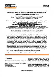
Fig. 3. Luminescence intensity of the light-emitting system from the fungus A. borealis depending on the hispidin concentration in the reaction mixture. The inset shows this dependence for small concentrations of the analyte. DOKLADY BIOCHEMISTRY AND BIOPHYSICS
Vol. 480
2018
176
PUZYR et al.
conditions, as evidenced by the results of preliminary experiments. We have found (Fig. 3) that, in the studied range of hispidin concentrations (5.4 × 10⁻⁵–1.4 × 10⁻² μM), a linear dependence of the recorded level of light emission on the hispidin concentration was observed (correlation coefficient R2 > 0.99). This relationship was retained after sequential additions of hispidin solutions at both increasing and decreasing concentrations. The obtained dependence can be used as a calibration curve to quantify the presence of hispidin in extracts from natural objects (e.g., higher fungi). The comparative studies performed at the next stage of this work showed that the spectral method of hispidin determination is much less sensitive than the luminescent testing method. When measuring the optical density of solutions with different concentrations of the analyte at a wavelength of 357 nm using a UV-1800 spectrophotometer (Shimadzu, Japan), we found that its threshold concentration that can be detected spectrophotometrically is approximately 1 μM (2.5 × 10⁻⁷ g). Thus, we have experimentally demonstrated the applicability of the luminescent system from the luminous basidiomycete A. borealis for detecting and determining the concentration of hispidin. The stability of the enzymes of the light-emitting system and the high sensitivity of the luminescent reaction allow multiple measurements of the analyte with a detection limit of 1.3 × 10⁻¹¹ g. This creates prerequisites for the development of a new highly sensitive rapid method of hispidin analysis in medicines and extracts from various biological sources. Further research will be focused on optimizing the testing method and increasing its sensitivity.
5. Lim, J.H., Lee, Y.M., Park, S.R., Kim, D.H., and Lim, B.O., Anticancer activity of hispidin via reactive oxygen species-mediated apoptosis in colon cancer cells, Anticancer Res., 2014, vol. 34, pp. 4087–4093. 6. Li, N., Zhao, L., Ng, T.B., Wong, J.H., Yan, Y., Shi, Z., and Liu, F., Separation and purification of the antioxidant compound hispidin from mushrooms by molecularly imprinted polymer, Appl. Microbiol. Biotechnol., 2015, vol. 99, pp. 7569–7577. 7. Lv, L.X., Zhou, Z.X., Zhou, Z.Z., Zhang, L.J., Yan, R., Zhao, Z., Yang, L.Y., Bian, X.Y., Jiang, H.Y., Li, Y.D., Sun, Y.S., Xu, Q.Q., Hu, G.L., Guan, W.J., and Li, Y.Q., Hispidin induces autophagic and necrotic death in SGC-7901 gastric cancer cells through lysosomal membrane permeabilization by inhibiting tubulin polymerization, Oncotarget, 2017, vol. 8, pp. 26992– 27006. 8. Lin, W.C., Deng, J.S., Huang, S.S., Wu, S.H., Lin, H.Y., and Huang, G.J., Evaluation of antioxidant, antiinflammatory and anti-proliferative activities of ethanol extracts from different varieties of sanghuang species, RSC Adv., 2017, vol. 7, pp. 7780–7788. 9. Anouar, H., Ali, ShahS.A., Hassan, N.B., Moussaoui, N.El., Ahmad, R., Zulkefeli, M., and Weber, J.-F.F., Antioxidant activity of hispidin oligomers from medicinal fungi: a DFT study, Molecules, 2014, vol. 19, pp. 3489–3507. 10. Tu, P.T.B. and Tawata, S., Anti-obesity effects of hispidin and Alpinia zerumbet bioactives in 3T3-L1 adipocytes, Molecules, 2014, vol. 19, pp. 1656–1667. 11. Shao, H.J., Jeong, J.B., Kima, K.-J., and Leea, S.-H., Anti-inflammatory activity of mushroom-derived hispidin through blocking of activation, J. Sci. Food Agric., 2015, vol. 95, pp. 2482–2486. 12. Lee, I.K., Cho, S.M., Seok, S.J., and Yun, B.S., Chemical constituents of Gymnopilus spectabilis and their antioxidant activity, Mycobiology, 2008, vol. 36, pp. 55–59.
1. Wu, T. and Xu, B., Antidiabetic and antioxidant activities of eight medicinal mushroom species from china, Int. J. Med. Mushrooms, 2015, vol. 17, pp. 129–140.
13. Bondar, V.S., Puzyr, A.P., Purtov, K.V., Petunin, A.I., Burov, A.E., Rodicheva, E.K., Medvedeva, S.E., Shpak, B.A., Tyaglik, A.B., Shimomura, O., and Gitelson, J.I., Isolation of luminescent system from the luminescent fungus Neonothopanus nimbi, Dokl. Biochem. Biophys., 2014, vol. 455, pp. 56–58.
2. Kalaras, M.D., Richie, J.P., Calcagnotto, A., and Beelman, R.B., Mushrooms: a rich source of the antioxidants ergothioneine and glutathione, Food Chem., 2017, vol. 233, pp. 429–433.
14. Puzyr, A.P., Medvedeva, S.E., Artemenko, K.S., and Bondar, V.S., Luminescence of cold extracts from mycelium of luminous basidiomycetes during longterm storage, Curr. Res. Environ. Appl. Mycol., 2017, vol. 7, pp. 227–235.
REFERENCES
3. Prasad, S., Rathore, H., Sharma, S., and Yadav, A.S., Medicinal mushrooms as a source of novel functional food, Int. J. Food Sci. Nutr. Diet., 2015, vol. 4, pp. 221– 225. 4. Greeshma, P., Ravikumar, K.S., Neethu, M.N., Pandey, M., Zuhara, K.F., and Janardhanan, K.K., Antioxidant, anti-inflammatory, and antitumor activities of cultured mycelia and fruiting bodies of the elm oyster mushroom, Hypsizygus ulmarius (agaricomycetes), Int. J. Med. Mushrooms, 2016, vol. 18, pp. 235–244.
15. Purtov, K.V., Petushkov, V.N., Baranov, M.S., Mineev, K.S., Rodionova, N.S., Kaskova, Z.M., Tsarkova, A.S., Petunin, A.I., Bondar, V.S., Rodicheva, E.K., Medvedeva, S.E., Oba, Yuichi., Oba, Yumiko., Arseniev, A.S., Lukyanov, S., Gitelson, J.I., and Yampolsky, I.V., The chemical basis of fungal bioluminescence, Angew. Chem., Int. Ed. Engl., 2015, vol. 54, pp. 8124–8128.
Translated by M. Batrukova
DOKLADY BIOCHEMISTRY AND BIOPHYSICS
Vol. 480
2018