n ing atio inu c nt Edu Co ical ed M
CLINICAL PODIATRY
Developing a Comprehensive Diagnostic and Treatment Plan for Charcot Neuroarthropathy—Pt.1 Successful outcomes for this insidious condition are dependent on a proper work-up. BY BRENT BERNSTEIN, DPM, HUOI LAM, DPM AND JOHN MOTKO, RN
Goals and Objectives 1) The practitioner will be able to discuss the main theoretical causes of neuroarthropathy. 2) The practitioner will be able to list the diagnostic options for neuroarthropathy and identify the gold standard option and implement them in clinical practice. 3) The practitioner will be able to classify a case of neuroarthropathy by both disease stage and anatomic location based on radiographic and clinical examination. 4) The practitioner will be able to formulate a conservative plan for treatment of a case of neuroarthropathy and identify the gold standard offloading technique.
Welcome to Podiatry Management’s CME Instructional program. Our journal has been approved as a sponsor of Continuing Medical Education by the Council on Podiatric Medical Education. You may enroll: 1) on a per issue basis (at $22.00 per topic) or 2) per year, for the special rate of $169 (you save $51). You may submit the answer sheet, along with the other information requested, via mail, fax, or phone. You can also take this and other exams on the Internet at www.podiatrym.com/cme. If you correctly answer seventy (70%) of the questions correctly, you will receive a certificate attesting to your earned credits. You will also receive a record of any incorrectly answered questions. If you score less than 70%, you can retake the test at no additional cost. A list of states currently honoring CPME approved credits is listed on pg. 188. Other than those entities currently accepting CPME-approved credit, Podiatry Management cannot guarantee that these CME credits will be acceptable by any state licensing agency, hospital, managed care organization or other entity. PM will, however, use its best efforts to ensure the widest acceptance of this program possible. This instructional CME program is designed to supplement, NOT replace, existing CME seminars. The goal of this program is to advance the knowledge of practicing podiatrists. We will endeavor to publish high quality manuscripts by noted authors and researchers. If you have any questions or comments about this program, you can write or call us at: Podiatry Management, P.O. Box 490, East Islip, NY 11730, (631) 563-1604 or e-mail us at
[email protected]. Following this article, an answer sheet and full set of instructions are provided (pg. 188).—Editor What Is Charcot Foot? Jean Martin Charcot first published a description of neuropathic arthropathy in 1868, although William Musgrave first noted arthropathy in syphilitic patients in 1703.1-3 Synonyms for the disorder are numerous, including Charcot’s foot, www.podiatrym.com
Charcot’s joint, Charcot’s fracture, neuropathic osteoarthropathy, and neuroarthropathy, to name a few.(I moved this from the last sentence up) There are a number of precipitating factors that can contribute to the development of charcot neuroarthropathy; however, it usually begins with a
neuropathic foot that typically suffers from some type of trauma, sometimes very minor or unremembered. This incident is followed by an acute inflammatory stage with progressive fragmentation of bone and joints, disorganization, and finally collapse of Continued on page 178 FEBRUARY 2013 | PODIATRY MANAGEMENT | 177
M C ed on ica ti l E nui du ng ca tio n
CLINICAL PODIATRY
NEUROARTHROPATHY
the foot and ankle if weight bearing continues during this stage. The collapse is accentuated by the continued pull of the conjoined triceps tendon. The final stage is characterized by slow resolution of the inflammation with permanent, and sometimes bizarre, deformities remaining that occurred during the prior phase. These can lead to a nonfunctional and many times chronically ulcerated foot. The incidence of neurarthropathy in the insensate diabetic population varies in the literature but at specialty centers where the index of suspicion is most accurate, rates can be as high as 13%.4 Once neuroarthropathy is di-
agnosed, the incidence of contralateral involvement goes up to 30% either due to some inherent predilection for the process or due to increased pressures on the initially uninvolved extremity. A retrospective analysis comparing mortality and major amputation between Charcot patients and simple diabetic foot ulcer patients was published in 2004. This small study did not show a significant difference in mortality Figure 1: Slide Showing Shards of Bone Ground into the between the two groups, alSynovium though the amputation rate trend was higher in the Charcot group.5
TABLE 1
Early Signs and Symptoms of Charcot Neuroarthropathy Dull, Deep, Unilateral Pain Despite Neuropathy Crepitus Sudden Change in Foot Shape Unilateral Edema Unilateral Erythema Unilateral Warmth Small Fleck Fractures on Plain Radiograph
What Causes Charcot Foot? The two classically opposed theories are the German neurotraumatic theory and the French neurovascular theory. The Germans (via Virchow and VolkFigure 2: Infrared Dermal Thermometry mann) believed that the insensitive joints are subject to repetiMore recently, clinicians have accepttive microtrauma and finaled a confluence of these two theories. ly deterioration. The Additionally, researchers have investiFrench (via Charcot) begated the possibility of a pre-existing lieved that deficiencies in “diabetic osteoporosis” in setting the the trophic centers of the stage for neuroarthropathy.8-13 Jeff-
TABLE 2
Differential Diagnosis Acute Traumatic Fracture or Dislocation
Superficial Thrombophlebitis
Stress Fracture
Cellulitis
Bone Tumor
Necrotizing Fasciitis
Gout
Abscess
Pseudogout
Osteomyelitis
Degenerative Joint Disease
Septic Arthritis
Plantar Fibroma
Inflammatory Arthritis
Deep Vein Thrombosis
Reflex Sympathetic Dystrophy
178 | FEBRUARY 2013 | PODIATRY MANAGEMENT
Researchers have investigated the possibility of a pre-existing “diabetic osteoporosis” in setting the stage for neuroarthropathy.8-13 spine led to a vasodilatory “washing out” of the bony substance of the extremity. Both theories have been bolstered by animal experiments.6-7
coate has also implicated the possibility of a cycle of pro-inflammatory cytokine release that allows the vicious cycle of inflammation and osteopenia to continue.14 Lastly, important work has also added the theory of the glycosylated diabetic Achilles tendon to the understanding of the overall picContinued on page 179 www.podiatrym.com
NEUROARTHROPATHY ture of the Charcot foot.15-16 Regardless of which theory one chooses to believe in, the common feature in the affected limb is neuropathy secondary
n ing atio inu c nt Edu Co ical ed M
CLINICAL PODIATRY
from fresh fractures. (Table 6) We feel that this is important because, although these fractures may be treated with a similar protocol as in non-neuropathic fractures initially, they more often than not can trigger a full-blown neuroarthropathy.47-48 We also identify distal absorptive osteopathy that we see in the phalanges. Lastly, we differentiate between medial and lateral midtarsus disease as the treatment differs for each.
cording to both disease stage and anatomic location. There are many anatomic systems available to practitioners.44-46 Our anatomic classifica-
Neuroarthropathy can be classified according to both the stage of disease process and the anatomical location. tion system is an expanded version of the Sanders system that we have modified to capture cases that we feel need to be treated very differently. In addition to the five classic locations described by Sanders, et al., we separate cases of classic neuroarthropathy
How Is Charcot Diagnosed? Diagnosing an early Charcot’s neuro-arthropathic fracture is primarily dependent on clinical assessment Classification Systems and necessitates a high index of suspiNeuroarthropathy can be classicion by the treating physician. A thorfied according to both the stage of ough history and physical should be disease process and the anatomical obtained from the neurolocation. We have not conpathic patient, who will sidered the classic Eichenoften seek medical attention holz and the later Sella and TABLE 3 due to increased edema, Barrette radiographic stagwarmth and mild pain in a ing systems useful in clinipreviously insensate limb. cal practice due to the lack Radiographs should be obof correlation with clinical tained and in early stage findings and thus do not (Stage 0) will demonstrate use them.40-41 Instead, we Diabetes Hemochromatosis subtle radiographic changes. use a four-stage system This coupled with the cliniwhich is a combination of Alcoholism Antiretroviral Therapy cal presentation can be easily the Armstrong and Lavery Spina bifida Lyme Disease mistaken for infection. pragmatic acute-to-chronic (Table 1) After quickly ruling system with the addition of Myelomeningocele Hansen’s Disease out a short list of differential the “pre-Charcot” stage disSyringomyelia Amyloidosis diagnoses such as cellulitis, cussed by Yu, et al.42-43 gout, and etc., (Table 2) one Our system includes a Syphilis Steroid Use should immediately assume post-surgical stage to identiPernicious Anemia Spinal Cord Compression that a neuropathic arthropafy those patients who have Charcot Marie Tooth Syndrome Multiple Sclerosis thy is present and prophylacpost-surgical inflammation tically immobilize and offbut are surgically stabilized. load the extremity while (Table 5) The inactive Charawaiting definitive testing. cot patients are split into TABLE 4 two distinctive groups: Both triple-phase bone techthose with pathology such nitium scans and magnetic as pain, significant deformiresonance imaging are useful ty; and the non-pathologic to show activity out of progroup who are ready for portion to the subtle clinical Tartrate-resitant acid phosphotase is a glycosylated monomeric shoeing. We believe our signs of inflammation to asmetalloenzyme that is used as a bone resorption marker. system captures those presist in diagnosis.17-18 Recently, Osteocalcin is a protein secreted by osteoblast that is used as a clinical cases of neuthe role of positron emission biochemical marker for bone formation. roarthropathy that can be tomography scans have been “nipped in the bud” and investigated, showing that it Bone alkaline phosphate is a hydrolase enzyme responsible for also allows for clear cut dehas a potential role in accuremoving phosphate groups from many types of molecules and is cision-making regarding rately distinguishing charused as a biochemical marker for bone formation. treatment in those patients cot’s neuroarthropathy by rewith full-blown cases. liably differentiating it from Urinary excretion of deoxypridinoline crosslinks is a biochemical Patients enrolled in the osteomyelitis. In this study marker for bone resorption. program are classified acContinued on page 180 to diabetes or any disease complicated by nerve damage.75
Neuropathies Associated with Charcot Joints
Common Bone Markers
www.podiatrym.com
FEBRUARY 2013 | PODIATRY MANAGEMENT | 179
M C ed on ica ti l E nui du ng ca tio n
CLINICAL PODIATRY
NEUROARTHROPATHY
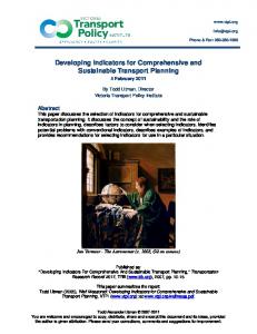
PET scans was 100% sensitive and 93.8% accurate in diagnosing charcot foot.76 All diabetic foot wounds should be carefully explored with a blunt sterile probe.19 A presumptive diagnosis of osteomyelitis can be made when skeletal structures are exposed, which is bolstered by an elevated erythrocyte sedimentation rate and C-reactive protein.20-21 A follow-up bone biopsy and culture will allow appropriate antibiotic treatment of the causative organism and is the gold standard in subtle cases. In cases when the possibility of osteomyelitis versus Figure 3: Technique for Long Leg Reconstructive Plain neuroarthropathy exists, then the Radiographs synovium should be evaluated as bone infection and are followed conwell. (Figure 1) Shards of bone comitantly with our infectious disease ground into the synovium is indicaspecialists. Additionally, patients ditive of Charcot. Horowicz wrote the agnosed with neuroarthropathy definitive paper on this—and every should be fully evaluated for the unclinician treating Charcot should be derlying cause of sensorium loss. familiar with his paper.22 It has been
shown to be a positive indicator of an underlying pathologic condition of the plantar foot.23-25 Skin temperatures of the affected foot and contralateral foot are measured after allowing the skin temperature to equilibrate to room temperature for ten minutes after removing cast, brace or shoes. Measurements with a hand-held infrared dermal thermometric probe are taken over the medial and lateral arch, medial and lateral malleoli, the dorsum of the foot and the tibial crest, with care to avoid direct sunlight on the extremities which can raise surface temperatures falsely. This technique has been well described in the literature.26-32 Markers of Bone Metabolism Researchers are on the hunt for markers of bone resorption and for-
It has been our experience that while many authors reference this original paper, this gold standard diagnostic test is underutilized in actual clinical practice. our experience that while many authors reference this original paper, this gold standard diagnostic test is underutilized in actual clinical practice. Patients who are diagnosed with both osteomyelitis and Charcot have serial erythrocyte sedimentation rates drawn to monitor the treatment of the
We’ve included a list of possible causes of neuroarthropathy in Table 3, although the list is not exhaustive.
Once Diagnosed, How Is Charcot Foot Monitored? Historically, radiographs were monitored as the means to assess the slow consolidation of neuroarthropathy. A more objective and the current standard for serial monitoring is the quantification of TABLE 5 inflammatory activity in a neuroarthropathic joint through the use of dermal infrared thermometry (Figure 2). On a week-to-week basis, temperatures are compared between the affected and unaffectStage 0—Pre-Charcot ed foot (the control) to judge the Stage 1a—Active Charcot efficacy of treatment and readiness of the foot for surgery or Stage 1b—Post-Surgical Charcot shoeing. A temperature change of Stage 2a—Non-Pathologic, Inactive Charcot more than two degrees Celsius compared to the surrounding skin Stage 2b—Pathologic, Inactive Charcot or contralateral site has been
Modified Charcot Staging System
180 | FEBRUARY 2013 | PODIATRY MANAGEMENT
Figure 4: Long Leg Calcaneal Axial View
mation in serum and urine that would allow clinicians to identify Charcot neuroarthropathy at an earlier stage.33 Numerous markers of bone metabolism have been evaluated. Selby, et al. measured urinary deoxypridinoline (bone resorption marker) and bone specific alkaline phosphate (bone formation marker) in patients with acute Charcot neuroarthropathy and non-Charcot patients with diabetes. The authors Continued on page 181 www.podiatrym.com
NEUROARTHROPATHY
n ing atio inu c nt Edu Co ical ed M
CLINICAL PODIATRY
are lucky in that our hospital has a special relationship with General Electric and the latest in CT scanners and digital imaging are available to our patients. Three-dimensional modeling gives our surgeons the ability to visualize these complex, multiplanar corrections prior to making the incisions. (Figures 5a & b)
Figure 5a: Three Dimensional CT Scan with Extrinsic Tendon Reconstruction
Figure 5b: Three Dimensional CT Scan with Hardware Highlighting
found an increase in both markers in Charcot patients, indicating an ongoing remodeling process of bone resorption and formation.34 Ulianova, et al. found, however, that while both processes increase in neuroarthropathy, only resorption increases in osteomyelitis, while other authors found that bone turnover markers are not useful in discrimination between the two entities.35-36 We have recently presented data from our own facility that calls into question the usefulness of the markers listed in Table 4.37 We attempted to correlate abnormal values of the four markers with abnormal infrared pedal temperatures. While trends were present for some of the markers, the correlation was not statistically significant. This, in conjunction with the previously mentioned study showing changes in bone markers in the presence of osteomyelitis, have lead us to consider markers “not ready for prime time.” They are currently utilized at our facility as a second-line diagnostic for decision-making in borderline cases, unusual cases or bilateral cases that make thermometry problematic. We plan on correlation of additional markers with thermometry readings in the future.
tients. The usefulness of these views in surgical planning is indispensable and our radiology department was very helpful in modifying their techniques when we brought the articles to them that outlined the techniques (Figures 3 & 4).38-39 In addition, at St. Luke’s, we are able to take advantage of the hospital’s relationship with General Electric technology to obtain some of the highest resolution computerized tomograms available. We
Radiology All patients who enter our program undergo a plain radiograph series. In addition to standard views, we obtain the long leg calcaneal axial and hindfoot alignment views on all pawww.podiatrym.com
Pressure Mapping and Force Measurement A computerized gait analysis platform is maintained at the center and all patients are evaluated with an initial barefoot static and dynamic image. Any patient who undergoes a surgical intervention is re-evaluated when healed to evaluate the efficacy of the procedure. In-shoe images can also be obtained to assist our pedorthist in shoe and brace modifications (Figure 6). Conservative Treatment Options:
TABLE 6
Immobilization and Off-Loading As long as patients do not have a deep infection requiring immediate debridement, all patients initially begin conservative treatment with the goal of ulceration healing and conversion of the Charcot process from the active phase to the inactive phase, while maintaining the bony architecture of the foot and ankle. The keystone of treatment, just as when treating diabetic foot ulcerations, is immobilization and off-loading. This can be accomplished using a wheelchair, which can create problems with compliance in homes without adequate wheelchair access. Crutches provide another option, but are not easily used by older or obese patents. A Roll-a-bout (Roll-A-Bout Corporation, Frederica, DE, USA) is another product that is maybe easier for older or obese patients and comes
Modified Sanders Anatomic Classification System Level I Forefoot (IPJ, MPJ) A. Absorptive Distal Osteopathy B. Insidious C. Traumatic—Metatarsals Level II Lisfranc’s Joint Level III Midtarsal Joints A. Medial Column B. Lateral Column Level IV Ankle and Subtalar Joints A. Insidious B. Traumatic Ankle Fractures Level V Calcaneal Insufficiency Fracture * Modifications to the Sanders System in Italics
Continued on page 182 FEBRUARY 2013 | PODIATRY MANAGEMENT | 181
M C ed on ica ti l E nui du ng ca tio n
CLINICAL PODIATRY
NEUROARTHROPATHY
Safe application of a total conin different models that tact cast requires simple traincan accommodate paing in either doctoral or posttients up to 400 pounds and doctoral programs, or attenas tall as 6’10”. dance at a casting lab, or The gold standard, howproctoring by a specialist faever, is correct application of miliar with the technique. the classic rigid total contact We are amazed at the cast as described by Brand.4951 amount of dogma surround(Figure 7) While clinicians ing the technique. The literahave made several modificaFigure 7: Rigid Total ture has many references by tions to the original techContact Cast clinicians who note connique over the years, such straints such as application as use of synthetic casting time, material costs, and risks inhermaterial rather than plaster, the basic ent in the technique. It has been our device remains the same.52-53 The cast experience that clinicians make the reduces edema, applies external stabimajority of these statements with eilization of fracture fragments, prether limited clinical contact with patients, or have never actually applied
wounds receive debridement and application of a slow-release antimicrobial dressing at each cast change. Of course, there will be patients who cannot tolerate the cast for reasons such as active infection, overwhelming drainage, obesity, and claustrophobia, necessitating alternative offloading devices and techniques. (Table 7) Once the foot is stable with evidence of healing on follow up xrays or MRI, the patient transitions into a removable cast walker or Charcot Restraint Orthotic Walker (CROW) (Figures 8 & 9).55 Patients progress into extra-depth footgear with custom inserts after one month as long as dermal thermometric measurements remain within two degrees
Once formal casting begins, we will always evaluate each patient for adjunctive therapy to shorten the disease process. a total contact cast. This single example of “diabetic foot dogma” has limited availability of the best treatment for patients, and possibly cost thousands of diabetic amputations. To this vents further injury, enforces compliday, however, we see patients with ance; and most importantly, negates tight Achilles tendons and inflamed, the damaging effect of the triceps edematous Charcot feet being ambusurae while the foot is in the weaklated in diabetic shoes, Unna boots, ened status of active-phase Charcot. or non-custom walking boots. This virtually TABLE 7 guarantees gross deformity and failure. At our program, a total contact cast is applied Non-Removable Cast Brace (“Instant Total Contact Cast”) in approximately 15 minutes. The Removable Cast Brace cost of materials Patellar Tendon Bearing Brace (“PTB”) is well under the third-party reimCharcot Restraining Orthotic Walker (“CROW”) bursement. The Crutches efficient application of total conWheelchair tact casts has Bedrest been discussed by other authors as Knee-Bearing Scooter (“Roll-A-Bout”) well.54 Patients Hands-free crutch device (“iWALKFree”) with open
Figure 6: Example of Plantar Pressure Mapping to Assess Orthotic Un-Loading of Ulceration
Celsius. Patients requiring tri-plane control will be prescribed ankle foot orthoses to be used within the footgear or a custom-molded high-top shoe with extended shanks, rigid counters, and supra-malleolar bracing. (Figure 10) Adjunctive Medical Treatments In addition to mandatory off-loading, many times we will initiate treatContinued on page 183
Alternative to the Rigid Total Contact Cast
182 | FEBRUARY 2013 | PODIATRY MANAGEMENT
Figure 8: Charcot Restraining Orthotic Walker www.podiatrym.com
NEUROARTHROPATHY
n ing atio inu c nt Edu Co ical ed M
CLINICAL PODIATRY
ed foot will be treated bisphosphonate therapy. The ment in patients with with salmon calcitonin majority of our patients are now an extreme amount of preferentially at the prescribed calcitonin-salmon spray inflammation with a present time.62-63 Due to daily, alternating nostrils each day. short course of rest, ice, Researchers have shown this treatthe slow clearance of elevation, compression ment to be beneficial in driving down these drugs from bone, (Jones boot, Unna’s the markers of bone metabolism.64 these drugs are not boot, or pneumatic given if surgery is compression), and bedCalcitonin-salmon nasal spray has being contemplated. rest. Once formal castbeen used to increase vertebral bone ing begins, we will always evaluate each patient for adjunctive Harshorne reported therapy to shorten the the use of electrical energy to directly disease process. After evaluating the bone Figure 9: Cast Walker Immobilizastimulate bone healing in 1841.65 markers and the patien- tion with External Bone Growth t’s renal status, augStimulator mentation treatment with bisphosphonates (oral or intraThe dental literature is being followed mass in post-menopausal osteoporovenous), salmon calcitonin therapy, closely. We still utilize bone oral and sis and to decrease the incidence of or non-invasive bone stimulation will intravenous bisphosphonates in those vertebral fractures. be selected in an effort to attenuate patients with limited deformity in the The treatment adverse reactions the bone destruction. Overall, the reacute phase of Charcot when the goal are mild to moderate in severity search behind these adjunctive treatis to arrest the process without any (mostly local nasal complaints). ments is far from concrete and many reconstruction. We’ve noted an anecdotal decrease in questions remain. Our position, howWe initially used oral bisphosphopain in the involved extremity when ever, has been that due to the severe nates such as Fosamax, Boniva, and utilizing this therapy. It can be used morbidity associated with neuActonel exclusively in the acute phase at any time of the day without regard roarthropathy, adjunctive treatment of CN. We have used IV Pamidronate to food restrictions or supplementashould be offered as long as the risks sparingly. We now require a full dention. The previous research with calare low. tal examination prior to considering citonin-salmon spray did not, however, drive down the skin temBisphosphonates perature faster than the conBisphosphonates are trol, and questions remain. potent inhibitors of osteoWe hope to add to the data TABLE 8 clast activation. There have being gathered on this adbeen trials showing signifijunct. Of important note, cant reduction in sympnone of the above mentioned toms and bone turnover drugs have been approved by Charcot Neuro-arthropathy disease process markers compared to conthe Food and Drug AdminisTreatment options trol groups. They have also tration for use in charcot been shown to normalize arthropathy The Necessity of Off-loading skin temperature differenCompliance with treatment plan tial between affected and Electrical Bone Stimulation non-affected feet.56-60 HowHarshorne reported the Diagnostics use of electrical energy to diever, it has been shown Blood glucose control (HBgA1c) rectly stimulate bone healing that bisphosphonates decrease bone remodeling in 1841.65 Yasuda, et al.66 studHand controls for car and are contraindicated in ied electrical fields and bone Residential Modifications (Ramps, Grab Bars, etc.) patients with renal insuffiformation in the early 1950’s. ciency.61 They were able to demonWeight Management and Conditioning strate the development of subDue to recent questions Dilated Eye Examinations regarding bisphosphonateperiosteal callus in bones Dental Examination if Bisphosphonates Are Planned associated osteonecrosis in under mechanical stress. The patients undergoing dental callus was formed as a result Smoking Cessation surgery, we have taken the of the electrical potentials inPain Management approach that any patient duced by the mechanical who may possibly need stress (piezoelectricity). ElecDepression Management bone surgery on the affectContinued on page 184
Patient Education
www.podiatrym.com
FEBRUARY 2013 | PODIATRY MANAGEMENT | 183
M C ed on ica ti l E nui du ng ca tio n
CLINICAL PODIATRY
NEUROARTHROPATHY
tronegative potentials are generated in areas of compression and electropositive potentials are generated in areas of tension. They were then able to show that passing 10 µA of continuous current along the bone could result in similar callus formation. Increased osteoblastic (bone formation) activity would be seen on the concave side of the bone, which has an electronegative potential. This is why the cathode negatively-charged electrode is placed at the site of nonunion or at the fracture site.67 The amount of current is dose specific; currents less than 5 µA do not cause bone formation, currents of 5 to 20 µA produce progressively increasing amounts of bone formation, currents over 20 µA produce necrosis.68-69 Current can be delivered to the bone by either direct current or by intermittent pulsed electromagnetic fields. Direct current devices are surgically implanted where as intermittent pulsed electromagnetic field devices are noninvasive and deliver current by means of two opposing coils of wires mounted on a cast or skin. The coils face each other at 180 de-
grees.68-69 Research has shown some benefit in the use of stimulation of bone healing through electrostimulation, magnetic fields, and low intensity ultrasound when treating neuroarthropathy.70-74 We typically utilize this adjunct when traumatic fractures that morph into neuroarthropathy, long bone fractures, or high risk fusions exist in the clinical Figure 10: Molded Foot and Ankle Orthosis picture. We utilize
blind, with poor dentition, in chronic renal failure, has glycosylated hemoglobin of 10, and must drive himself to and from doctors’ visits is doomed to failure and complications. At the initial enrollment of these patients, we look at many factors that will enhance the treatment of the neuropathic joint as well as decrease overall morbidity in the
One of the main reasons that we have excellent salvage rates and functional extremities is due to the “whole picture” approach that we take with these patients.
TABLE 9
Risk Factors for Neuroarthropathy Clinical Signs of Peripheral Neuropathy:75 Insensate to Monofilament Decreased Vibratory Sensation Decreased Deep Tendon Reflexes Patient History of: Retinopathy Nephropathy Previous Foot Ulcer Neuro-arthropathy Activities/Events including:112 Use of Ladders or Digging Tools (i.e., Shovels) Obesity Lifting Heavy Objects Sudden Change in Activity Level Impact Sports/Activities (Jogging, Stair-Climber, Dance) Osteopenia Foot or Ankle Surgery of any kind Traumatic Fractures of the Foot or Ankle Minor Trauma (sprains, contusions, etc.)
184 | FEBRUARY 2013 | PODIATRY MANAGEMENT
pulsed electromagnetic field stimulators (EBI Medical Inc, Parsippany, NJ, USA) to speed up bone formation. Direct current devices have been used with success in a few surgical patients with high risk tibiocalcaneal fusions implanted directly into the fusion site. Patient Education, Lifestyle and Disease Modification One of the main reasons that we have excellent salvage rates and functional extremities is due to the “whole picture” approach that we take with these patients. Applying a total contact cast and prescribing a bisphosphonate in a patient who is
patient (Table 8). We have developed patient education sheets on Charcot and casting, as well. While the cast application is simple and straightforward, these last components of our conservative treatment program are certainly the most challenging and utilize the most time and resources. Prevention of Recurrence? A patient with two consecutive visits with equal and symmetrical foot temperatures is considered to be in the inactive phase. Patients are then evaluated for chronic pain, instability, equinus or bony deformity that would preclude safe and comfortable ambulation in diabetic shoes. If any of these exist, then surgery is contemplated. From the outset, we know that any patient with a history of neuroarthropathy has high risk for re-activation of the process in the same foot as well as a 30% chance of developing similar problems in the contra-lateral foot. We educate the patient on this as well as review the Continued on page 185 www.podiatrym.com
NEUROARTHROPATHY signs and symptoms of the process. We also educate the patient on risk factors particular to their neuroarthropathy. (Table 9) Lastly, we make sure that we make an appointment for the patient to go to his/her podiatrist for regular high-risk foot care. PM Editor’s Note: Part 2 will appear next month. References 1 Sanders LJ. Jean-Martin Charcot (18251893) The Man Behind the Joint Disease. JAPMA. Vol 92 No7. 375-380, 2002. 2 Charcot JM. Sur quelques arthropathies qui paraissent dependre d’une lesion due cerveau ou de la moelle epiniere. Arch Physiol. Norm Path 1868; 1: 161-71 Kelly M. William Musgrave’s De arthritide symptomatica (1703): his description of neuropathic arthritis. Bull Hist Med; 37: 372-6, 1963. 3 Armstrong DG, Peters E: Charcot’s arthropathy of the foot. JAPMA 92: 390, 2002. 4 Gazis A, Pound N, Macfarlane R, et al. Mortality in patients with diabetic neuropathic osteoarthropathy (Charcot foot). Diabet Med 21, 1243-1246, 2004. 5 Eloesser L. On the nature of neuropathic affections of the joint. Ann Surg 191; 66: 201-207, 1917. 6 Finsterbush A, Friedman B. The effect of sensory denervation on rabbits’ knee joints. J Bone Jt Surg 57A: 949-57, 1975. 7 Petrova NL, Foster AV, Edmonds ME. Difference in presentation of charcot osteoarthropathy in type 1 compared with type 2 diabetes. Diabetes Care. May 27 (5):1235-6, 2004. 8 Fleischli JG, Laughlin TJ, Athanasiou K, et al. Effect of diabetes mellitus on the material properties of the distal tibia. JAPMA 96(2): 91-95, 2006. 9 Brown SA, Sharpless JL. Osteoporosis: An Under-appreciated complication of diabetes. Clinical Diabetes 22:10-20, 2004. 10 Herbst SA, Jones KB, Saltzman CL. Pattern of diabetic neuropathic arthropathy associated with the peripheral bone mineral density. J Bone Joint Surg (Br); 86-B: 378-83, 2004. 11 Childs M, Armstrong DG, Edelson GW. Is Charcot arthropathy a late sequela of osteoporosis in patients with diabetes mellitus? Journal of Foot and Ankle Surgery. SepOct;37(5):437-9, 1998. 12 Young MJ, Marshall A, Adams JE, et al: Osteopenia, neurological dysfunction, and the development of Charcot neuroarthropathy, Diabetes Care 18: 34, 1995. 13 Jeffcoate WJ. Abnormalities of Vaso-
www.podiatrym.com
motor Regulation in the Pathogenesis of the Acute Charcot Foot of Diabetes Mellitus. The International Journal of Lower Extremity Wounds. Vol. 4, No. 3, 133-137,2005. 14 Grant WP, Foreman EJ, Wilson S, et al. Evaluation of Young’s Modulus in Achilles Tendons with Diabetic Neuroarthropathy. JAPMA. Vol. 95. No.3, May/June 2005. 242-246. 15 D’Ambrogi E, D’Agostino M, Giacomozzi C, Macellari V, Caselli A, Uccioli L. Contribution of plantar fascia to the increased forefoot pressures in diabetic patients. Diabetes Care. May;26 (5):1525-9, 2003. 16 Edmonds ME, Clarke MB, Newton S, ET AL: Increased uptake of bone radiopharmaceutical in diabetic neuropathy. QJM 57: 843, 1985. 17 Edmonds ME. The neuropathic foot in diabetes. Part 1: blood flow. Diabetic Med 1986; 3: 111-115. All diabetic foot wounds should be carefully explored with a blunt sterile probe. 18 Grayson ML, Balaugh K, Levin I, Karchmer AW. Probing to bone infected pedal ulcers. A clinical sign of underlying osteomyelitis in diabetic patients. J Am Med Assoc: 273(9): 721-23, 1995. 19 Judge MS. Using serologic screening to identify and monitor at-risk Charcot patients. Podiatry Today. 1045-7860—Volume 17— Issue 8—August 2004. 20 Katsaros TF, Makras P, Koutmos S, et al. Simple laboratory and clinical tests may be of use as indicators of increased suspicion of underlying osteomyelitis (OM) of metatarsals and proximally in the infected diabetic foot (DF). 21 Horwitz T. Bone and cartilage debris in the synovial membrane. Its significance in the early diagnosis of neuroarthropathy. J Bone Jt Surg 1948; 30A: 579-88. 22 Bergtholdt HT, Temperature assessment of the insensitive foot. Phys Ther 59: 18, 1979. 23 Armstrong DG, Lavery LA, Wunderlich RP, ET AL: Skin temperatures as a one-time screening tool do not predict future diabetic foot complications. JAPMA 93: 443, 2003. 24 Brand PW: Repetitive stress on the insensitive feet: The pathology and management of Public Health Service, Carville, LA, 1975. 25 DG Armstrong, LA Lavery, PJ Liswood, WF Todd, and JA Tredwell J: Infrared dermal thermometry for the high-risk diabetic foot. Physical Therapy 1997; 77: 169-177. 26 Armstrong DG, Lavery LA: Monitoring healing of acute charcot’s arthropathy with infrared dermal thermometry. J Foot Ankle Surg 1996; 35:335-338. 27 McGill M, Molyneaux L, Bolton T et al. Response of Charcot’s arthropathy to contact casting: assessment by quantita-
n ing atio inu c nt Edu Co ical ed M
CLINICAL PODIATRY
tive techniques. Diabetologia 2000; 43(4): 481-4. 28 Armstrong DG, Lavery LA. Monitoring healing of acute Charcot’s arthropathy with infrared dermal thermometry. J Rehabil Res Dev; 34: 317-21, 1997. 29 Nube VL, McGill M, Molyneaux L, et al. From Acute to Chronic—Monitoring the Progress of Charcot’s Arthropathy. JAPMA 92(7): 384-389, 2002. 30 Bharara M, Covv JE, Claremont DJ, et al. Thermography and Thermometry in the Assessment of Diabetic Neuropathic Foot: A Case for Furthering the Role of Thermal Techniques. Lower Extremity Wounds 5(4); 2006 250-260. 31 Foto JG, Brasseaux D, Birke JA. Essential Features of a Handheld Infrared Thermometer Used to Guide the Treatment of Neuropathic Feet. JAPMA 97(5): 360-365, 2007. 32 Piaggesi A, Rizzo L, Golia F, et al. Biochemical and ultrasound tests for the early diagnosis of active neuroosteoarthropathy (NOA) of the diabetic foot. Diabetes Res Clin Pract; 58:1-9, 2002. 33 Selby PL, Jude EB, Burgess J, et al: Bone turnover markers in acute Charcot Neuroarthropathy. Diabetologia 41 (suppl 1): A275, (1998). 34 Ulianova I, Tokmakova A, Antsiferov M. Biochemical markers of bone remodeling in diagnosis of acute Charcot osteoarthropathy and osteomyelitis. Diabetes, National Research Centre for Endocrinology, Moscow, Russian Federation. http://87.234.226.93/easd/custo files/easd/38th/abstracts/PS89.html 35 Jude EB, Selby PL, Mawer EB, et al. Inflammatory and bone turnover markers in Charcot arthropathy and osteomyelitis of the feet in diabetic patients. Horm Metab Res 2006; 38: 361-367. 36 Bernstein BH, Acor CL, Williams A. Limitations of Markers of Bone Metabolism in Staging Charcot Neuroarthropathy”, Poster Presentation, The 2007 National APMA Annual Scientific Meeting, August 2007. 37 Mendicino RW, Catanzariti AR, Reeves CL, et al. A systematic approach to evaluation of the rearfoot, ankle and leg in reconstructive surgery. JAPMA Vol 95 No 1 2005 2-12. 38 Lamm BM, Mendicino RW, Catanzariti AR, et al. Static Rearfoot Alignment: a comparison of clinical and radiographic measures. JAPMA Vol 95 No 1 2005 26-33. 39 Eichenholtz SN: Charcot Joints, Charles C Thomas, Springfield, IL, 1966. 40 Sella EJ, Barrette C: Staging of Charcot Neuroarthropathy along the medial column of the foot in the diabetic patient. J Foot Ankle Surg. Jan-Feb;38(1):34-40, 1999. 41 Armstrong DG, Lavery LA. Acute Continued on page 186
FEBRUARY 2013 | PODIATRY MANAGEMENT | 185
M C ed on ica ti l E nui du ng ca tio n
CLINICAL PODIATRY
NEUROARTHROPATHY
Charcot’s arthropathy of the foot and ankle. Phys Ther 1998; 78: 74-80. 42 Yu GV, Hudson JR: Evaluation and Treatment of Stage 0 Charcot’s Neuroarthropathy of the Foot and Ankle. JAPMA 92: 210, 2002. 43 Harris JR, Brand PW. Patterns of disintegration of the tarsus in the anesthetic foot. J Bone Joint Surg 48A:14-16, 1966. 44 Schon LC, Easley ME, Cohen I, et al. The acquired midtarsus deformity classification system—interobserver reliability and intraobserver reproducibility. Foot and Ankle International. Vol. 23. No.1 January 2002. 3036. 45 Sanders LJ, Frykberg RG: “Charcot Neuroarthropathy of the Foot,” in Levin and O’Neal’s The Diabetic Foot, 6th Ed, ed by JH Bowker, MA Pfeifer, p 439, CV Mosby, St Louis, 2001. 46 Bibbo C, Lin SL, Bean HA, et al. Complications of ankle fractures in diabetic patients. Orthop Clin North Am 2001; 32(1):113-33. 47 Brand P, Yancy P. The Gift of Pain, Zondervan Publishing House, Grand Rapids, Michigan. 1997. 48 Finsterbush A, Friedman B. The effect of sensory denervation on rabbits’ knee joints. J Bone Jt Surg 1975; 57A: 949-57. 49 Kominsky SJ. The ambulatory total contact cast. In: Frykberg, RG, ed. The high risk foot in diabetes mellitus. New York: Churchill Livingstone, 1991; 449-55. 50 Pinzur MS, Lio T, Posner M. Treatment of Eichenholtz stage I Charcot foot arthropathy with a weight-bearing total contact cast. Foot and Ankle International. May;27(5):324-9, 2006. 51 Armstrong DG, Todd WF, Lavery LA, et al.: The natural history of acute Charcot’s arthropathy in a diabetic foot specialty clinic. Diabet Med 14: 357, 1997. 52 Shaw JE, Boulton AJM: The Charcot foot. Foot 5: 65, 1995. 53 Morgan JM, Beihl WC 3rd, Wagner FW JR: Management of neuropathic arthropathy with the Charcot Restraint Orthotic Walker. Clin Orthop 296:58, 1993. 54 Jensen J, Jaakola E. Physician and staff time efficiency in total contact cast application: proper technique results in time savings for this useful procedure. Podiatry Management, June-July, 2004. 56 Gough A, Abraha H, Purewal TS, et al.: Measurements of markers of osteoclastic and osteoblastic activity in patients with acute and chronic diabetic Charcot neuroarthropathy. Diabetes Med 14:517-531, 1997. 57 Selby PL, Jude EB, Burgess J, et al.: Bone turnover markers in acute Charcot Neuroarthropathy. Diabetologia. 41 (suppl 1): A275, 1998. 58 Jude EB, Selby PL, Burgess J, et al.: Bisphosphonates in the treatment of Charcot
Neuroarthropathy: a double-blind randomized controlled trial. Diabetologia. 44:20322037, 2001. 59 Pitocco D, Ruololo V, Caputo S, et al.: Six-month treatment with alendronate in acute Charcot neuroarthropathy: a randomized controlled trial. Diabetes Care. May; 28(5): 1214-1215, 2005. 60 Anderson J, Woelffer KE, Holtzman JJ, et al.: Bisphosphonates for the treatment of Charcot neuroarthropathy. Journal of Foot and Ankle Surgery. 43(5): 285-289, 2004. 61 Stepan JJ, Alenfeld F, Boivan G, et al.: Mechanisms of action of anti-resortive therapies of postmenopausal osteoporosis. Endocr Regul. 37:227-240, 2003. 62 Dental Management of Patients Receiving Oral Bisphosphonate Therapy: Expert Panel, Journal of the American Dental Association, August 2006. 63 Osteonecrosis of the Jaw and Oral Bisphosphonate Treatment, Journal of the American Dental Association, August 2006. 64 Bem R, Jirkovska A, Fejfarova V, et al.: Intranasal calcitonin in the treatment of acute Charcot neuroarthropathy—a randomized controlled trial. Diabetes Care. June; 29(6): 1992-1994, 2006. 65 Harshorne E: On the causes and treatment of psuedoarthrodesis and especially that form of it sometimes called supernumerary joint. Am J Med Sci.1:121, 1841. 66 Yasuda I, Noguchi K, Sata T: Dynamic callus and electric callus. J Bone Joint Surg Am. 37:1292, 1995. 67 Cohen M, Roman A, Lovins JE: Totally implanted direct current stimulator as treatment for a nonunion in the foot. J Foot Ankle Surg. 32: 375, 1993. 68 Basset CA: The development and application of pulsed electromagnetic fields (PEMF’s) for ununited fractures and arthrodeses. Orthrop Clin North Am. 15: 61, 1984. 69 Nerubay J, Marganit B, Bubis J, et al.: Stimulation of bone formation by electrical current on spinal fusion. Spine. 11: 167, 1986. 70 Grady JF, O’Connor KJ, Axe TM, et al.: Use of electrostimulation in the treatment of diabetic neuroarthropathy, JAPMA 90: 287, 2000. 71 Hanft JR, Goggin JP, Landsman A, et al: The role of combined magnetic field bone growth stimulation as an adjunct in the treatment of neuroarthropathy/Charcot joint: an expanded pilot study. J Foot Ankle Surg. 37: 510, 1998. 72 Strauss E, Gonya G. Adjunct Low Intensity Ultrasound in Charcot Neuroarthropathy. Clinical Orthopaedics and Related Research. 349 April; 132-135,1998. 73 Wang Z, Glark C, Brighton CT. Up-regulation of bone morphogenetic proteins in cultured murine bone cells with use of specif-
186 | FEBRUARY 2013 | PODIATRY MANAGEMENT
ic electric fields. J Bone Joint Surg Am. May; 88(5): 1053-1065, 2006. 74 Petrisor B, Lau JTC. Electrical bone stimulation: an overview and its use in high risk and Charcot foot and ankle reconstructions. Foot Ankle Clin. Dec; 10(4):609-620, 2005. 75 Rogers, L, Frykberg, R, Armstrong, D, et al. The Charcot Foot in Biabetes. Diabetes Care, Volume 34: September; 2123-2129, 2011. 76 Basu S, Chryssicos T, Houseni M, et al. Potential role of the FDG PET in the setting of diabetic neuroosteoarthropathy: can it differentiate uncomplicated Charcot’s neuroarthropathy from osteomyelitis and soft tissue infection? Nucl Med Commun 2007; 28 (6): 465-472.
Dr. Bernstein is board certified by the American Board of Podiatric Surgery and is a Fellow of the American College of Foot and Ankle Surgeons. He graduated from Temple University School of Podiatric Medicine and completed both a residency in foot surgery and a fellowship in limb salvage surgery with Dr. Stanley Kalish in Atlanta, Georgia. He currently practices in the Lehigh Valley and is program director of the Charcot and Reconstructive Foot Program at St. Luke’s Hospital University and Health Network, Quakertown Campus. Dr. Bernstein participates in mission trips to impoverished regions to perform pediatric deformity surgery on a yearly basis. Dr. Lam graduated from Temple University School of Podiatric Medicine and is currently a third year resident at St. Luke’s University and Health Network. John Motko is a registered nurse who works at the Wound Management Center St. Luke’s Health Network, Quakertown Campus. He has a BS in Nursing from Moravian College/ St. Luke’s School of Nursing. He is certified in wound care from both the American Academy of Wound Management and the Wound, Ostomy and Continence Nurses Society. He is also a Certified Hyperbaric Registered Nurse. He has over seven years of clinical experience in caring for patients with chronic nonhealing wounds and Charcot neuroarthropathy. www.podiatrym.com
SEE ANSWER SHEET ON PAGE 189. 1) Urinary excretion of deoxypyridinoline crosslinks is a biochemical marker for bone formation. A) True B) False C) only when patient is diabetic D) only when patient is a non-diabetic 2) Infrared dermal thermometry should be completed: A) immediately after the cast, shoe or brace is removed to ensure accurate skin temperatures. B) 24 hours after cast removal C) 10-20 minutes after cast removal D) directly through the casting material 3) When obtaining infrared dermal temperatures to track the activity of a neuropathic fracture, one should: A) Compare the affected foot temperatures to other areas on the affected foot B) Compare the affected foot temperatures to corresponding areas on the unaffected foot C) Compare the affected foot temperatures to the patient’s oral temperature D) Compare the affected temperatures to a non-Charcot patient’s temperatures 4) Stage 0 Charcot Neuroarthropathy is typically characterized by the following: A) Marked Deformity www.podiatrym.com
B) Edema and Warmth C) Massive Disorganization of Osseous Structures on Plain Radiograph D) Fever, elevated white count, high erythrocyte sedimentation rate 5) Achilles tendon contracture is associated with Charcot Neuroarthropathy: A) Occasionally B) Only in insulindependent patients C) Often D) Never 6) Acceptable means of immobilization of an acute Charcot foot are: A) Custom diabetic shoes B) Total Contact Cast C) Unna Boot, PostOperative Shoe and Partial Weightbearing with Cane D) Extra-Depth Shoes 7) The german theory of neuroarthropathy links Charcot with: A) Repetitive microtrauma B) Vasodilation C) Pro-inflammatory cytokines D) Nephropathy 8) What is the gold standard test for diagnosis of neuroarthropathy? A) bone culture B) sedimentation rate C) Synovial biopsy D) Serum bone markers 9) The following are possible underlying causes of
n ing atio inu c nt Edu Co ical ed M
CME EXAMINATION
neroarthropathy: A) alcoholic neuropathy B) Psoriatic arthritis C) Peripheral vascular disease D) Raynaud’s phenomenon 10) Bisphosphonate therapy should not be offered when the patient is suffering from: A) Dental pathology B) herpes zoster C) peripheral vascular disease D) xerosis of the skin 11) The Sanders classification system involves: A) disease stage B) anatomic location C) diabetes control D) ulcer depth 12) The Eichenholz classification system involves: A) disease stage B) anatomic location C) diabetes control D) ulcer depth 13) The Eichenholz classification system utilizes: A) patient pain profiling B) radiographic presentation C) infrared dermal thermometry D) all of the above 14) A prerequisite for neuroarthropathy is: A) trauma B) obesity C) poor glucose control D) peripheral neuropathy
Continued on page FEBRUARY 2013 | PODIATRY MANAGEMENT | 187
M C ed on ica ti l E nui du ng ca tio n
CME EXAMINATION
15) Patients with Charcot neuroarthropathy have blood flow that is: A) Always excessive compared to normal baseline B) Varies based on comorbidities, family history and age C) Normal D) Generally decreased compared to normal baseline 16) Contralateral neuroarthropathy occurs: A) 5% of the time B) 30% of the time C) 80% of the time D) Never 17) The differential diagnosis of Charcot should include: A) cellulitis B) necrobiosis lipoidica diabeticorum C) diabetic dermapathy D) diabetic bullosis 18) Contracture of the Achilles tendon exists in Charcot patients: A) Never B) Rarely C) Some of the time D) Most of the time 19) Patient should not transition from total contact cast to shoegear until: A) Infrared temperatures are within 2 degrees Celcius bilaterally B) Bone markers are normalized C) Three months have elapsed from first cast D) The patient promises not to walk too much 20) Patients should be transitioned from total contact cast to shoegear after temperatures are equal and symmetrical: A) Immediately B) Gradually while temporizing with a transitional device such as a CROW or cast boot C) Never D) When glucose is under control See answer sheet on page 189. 188 | FEBRUARY 2013 | PODIATRY MANAGEMENT
PM’s CPME Program Welcome to the innovative Continuing Education Program brought to you by Podiatry Management Magazine. Our journal has been approved as a sponsor of Continuing Medical Education by the Council on Podiatric Medical Education. Now it’s even easier and more convenient to enroll in PM’s CE program! You can now enroll at any time during the year and submit eligible exams at any time during your enrollment period. PM enrollees are entitled to submit ten exams published during their consecutive, twelve–month enrollment period. Your enrollment period begins with the month payment is received. For example, if your payment is received on September 1, 2006, your enrollment is valid through August 31, 2007. If you’re not enrolled, you may also submit any exam(s) published in PM magazine within the past twelve months. CME articles and examination questions from past issues of Podiatry Management can be found on the Internet at http://www.podiatrym.com/cme. Each lesson is approved for 1.5 hours continuing education contact hours. Please read the testing, grading and payment instructions to decide which method of participation is best for you. Please call (631) 563-1604 if you have any questions. A personal operator will be happy to assist you. Each of the 10 lessons will count as 1.5 credits; thus a maximum of 15 CME credits may be earned during any 12-month period. You may select any 10 in a 24-month period. The Podiatry Management Magazine CME program is approved by the Council on Podiatric Education in all states where credits in instructional media are accepted. This article is approved for 1.5 Continuing Education Contact Hours (or 0.15 CEU’s) for each examination successfully completed.
Home Study CME credits now accepted in Pennsylvania Continued on page www.podiatrym.com
n ing atio inu c nt Edu Co ical ed M
✄
Enrollment/Testing Information and Answer Sheet Note: If you are mailing your answer sheet, you must complete all info. on the front and back of this page and mail with your credit card information to: Podiatry Management, P.O. Box 490, East Islip, NY 11730.
rolled in the annual exam CPME program, and we receive this exam during your current enrollment period. If you are not enrolled, please send $22.00 per exam, or $169 to cover all 10 exams (thus saving $51 over the cost of 10 individual exam fees).
TESTING, GRADING AND PAYMENT INSTRUCTIONS (1) Each participant achieving a passing grade of 70% or higher on any examination will receive an official computer form stating the number of CE credits earned. This form should be safeguarded and may be used as documentation of credits earned. (2) Participants receiving a failing grade on any exam will be notified and permitted to take one re-examination at no extra cost. (3) All answers should be recorded on the answer form below. For each question, decide which choice is the best answer, and circle the letter representing your choice. (4) Complete all other information on the front and back of this page. (5) Choose one out of the 3 options for testgrading: mail-in, fax, or phone. To select the type of service that best suits your needs, please read the following section, “Test Grading Options”.
Facsimile Grading To receive your CPME certificate, complete all information and fax 24 hours a day to 1-631-563-1907. Your CPME certificate will be dated and mailed within 48 hours. This service is available for $2.50 per exam if you are currently enrolled in the annual 10-exam CPME program (and this exam falls within your enrollment period), and can be charged to your Visa, MasterCard, or American Express. If you are not enrolled in the annual 10-exam CPME program, the fee is $22 per exam.
TEST GRADING OPTIONS Mail-In Grading To receive your CME certificate, complete all information and mail with your credit card information to:
Podiatry Management P.O. Box 490, East Islip, NY 11730 PLEASE DO NOT SEND WITH SIGNATURE REQUIRED, AS THESE WILL NOT BE ACCEPTED. There is no charge for the mail-in service if you have already en-
Phone-In Grading You may also complete your exam by using the toll-free service. Call 1-800-232-4422 from 10 a.m. to 5 p.m. EST, Monday through Friday. Your CPME certificate will be dated the same day you call and mailed within 48 hours. There is a $2.50 charge for this service if you are currently enrolled in the annual 10-exam CPME program (and this exam falls within your enrollment period), and this fee can be charged to your Visa, Mastercard, American Express, or Discover. If you are not currently enrolled, the fee is $22 per exam. When you call, please have ready: 1. Program number (Month and Year) 2. The answers to the test 3. Your social security number 4. Credit card information In the event you require additional CPME information, please contact PMS, Inc., at 1-631-563-1604.
ENROLLMENT FORM & ANSWER SHEET Please print clearly...Certificate will be issued from information below. Name _______________________________________________________________________ Soc. Sec. #______________________________ FIRST
Please Print:
MI
LAST
Address_____________________________________________________________________________________________________________ City__________________________________________________ State_______________________ Zip________________________________ Charge to: _____Visa _____ MasterCard _____ American Express Card #________________________________________________Exp. Date____________________ Note: Credit card is the only method of payment. Checks are no longer accepted. Signature__________________________________ Soc. Sec.#______________________ Daytime Phone_____________________________ State License(s)___________________________ Is this a new address? Yes________ No________ Check one: ______ I am currently enrolled. (If faxing or phoning in your answer form please note that $2.50 will be charged to your credit card.) ______ I am not enrolled. Enclosed is my credit card information. Please charge my credit card $22.00 for each exam submitted. (plus $2.50 for each exam if submitting by fax or phone). ______ I am not enrolled and I wish to enroll for 10 courses at $169.00 (thus saving me $51 over the cost of 10 individual exam fees). I understand there will be an additional fee of $2.50 for any exam I wish to submit via fax or phone. www.podiatrym.com
Over, please
FEBRUARY 2013 | PODIATRY MANAGEMENT | 189
M C ed on ica ti l E nui du ng ca tio n
✄
ENROLLMENT FORM & ANSWER SHEET
(continued)
EXAM #2/13 Developing a Comprehensive Diagnostic and Treatment Plan for Charcot Neuroarthropathy— Pt. 1 (Bernstein, Lam, and Motko) Circle: 1. A B
C
D
11. A B
C
D
2. A B
C
D
12. A B
C
D
3. A B
C
D
13. A B
C
D
4. A B
C
D
14. A B
C
D
5. A B
C
D
15. A B
C
D
6. A B
C
D
16. A B
C
D
7. A B
C
D
17. A B
C
D
8. A B
C
D
18. A B
C
D
9. A B
C
D
19. A B
C
D
10. A B
C
D
20. A B
C
D
Medical Education Lesson Evaluation Strongly agree [5]
Agree [4]
Neutral [3]
Disagree [2]
Strongly disagree [1]
1) This CME lesson was helpful to my practice ____ 2) The educational objectives were accomplished ____ 3) I will apply the knowledge I learned from this lesson ____ 4) I will makes changes in my practice behavior based on this lesson ____ 5) This lesson presented quality information with adequate current references ____ How long did it take you to complete this lesson? ______hour ______minutes What topics would you like to see in future CME lessons ? Please list : __________________________________________________ __________________________________________________ __________________________________________________ __________________________________________________ __________________________________________________ __________________________________________________ __________________________________________________
190 | FEBRUARY 2013 | PODIATRY MANAGEMENT
www.podiatrym.com