to position an irradiation catheter led by a bronchoscope to a desired spot ... an electromagnetic navigation system, which tracks the tip of the bronchoscope and ...
Development of a Navigation System for Endoluminal Brachytherapy in Human Lungs Ingmar Wegner, Marcus Vetter, Max Schoebinger, Ivo Wolf, Hans-Peter Meinzer German Cancer Research Center, Div. Medical and Biological Informatics B010, Im Neuenheimer Feld 280, 69120 Heidelberg, Germany ABSTRACT The endoluminal brachytherapy of peripherally located bronchial carcinoma is difficult because of the complexity to position an irradiation catheter led by a bronchoscope to a desired spot inside a human lung. Furthermore the size of the bronchoscope permits only rarely the insertion of a catheter into the fine segment bronchi. We are developing an image-guided navigation system which indicates a path for guidance to the desired bronchus. Thereby a thin catheter with an enclosed navigation probe can be led up directly to the target bronchus, either by the use of the video of the bronchoscope or by the use of virtual bronchoscopy. Because of the thin bronchi and their moving soft tissue, the navigation system has to be very precise. This accuracy is reached by a gradually registering navigation component which improves the accuracy in the course of the intervention through mapping the already covered path to the preoperatively generated graph based bronchial tree description. The system includes components for navigation, segmentation, preoperative planning, and intraoperative guidance. Furthermore the visualization of the path can be adapted to the lung specialist’s habits (video of bronchoscope, 2D, 3D, virtual bronchoscopy etc.). Keywords: Image-Guided Radiation Therapy, Enhanced Reality, Image to Physical Space Registration
1. INTRODUCTION AND MEDICAL BACKGROUND In the last years, a change of smoking habits led to a rising incidence of adenocarcinomas and far peripherally located bronchial carcinoma.1 The introduction of the conformable radiation therapy of the lung represents a much promising attempt. Disadvantage of this method is however the substantial technical and temporal expenditure (general anaesthesia under artificial high frequency jet respiration to minimize movement of the tissue). A further approach is the application of endoluminal irradiation by means of brachytherapy.2 A radioactive emitter is placed inside a bronchus near or even inside the tumor by a catheter for a preoperatively planned period of time. Thus the movement of the chest doesn’t disturb the irradiation therapy and the patient can breathe normally. Due to the steep dose decrease of the radiation source (Ir192) highly compliant dose distributions can be achieved. Thus healthy tissue can be omitted but high doses of radiation can be applied to the tumour. The endoluminal irradiation could so far only be accomplished to centrally located tumors because of the peripherally thin bronchi and the relatively thick bronchoscope. Also the strong branching of the bronchi complicates the localization of the desired bronchus, which leads to a longer duration. Assistance here represents the employment of an electromagnetic navigation system, which tracks the tip of the bronchoscope and indicates the path during the bronchoscopy to a preoperatively defined target bronchus. Electromagnetic tracking systems, which locate the position of several very small sensors without direct view to it, are commercially available today (e.g. AURORA, Northern Digitally Inc. (NDI); Waterloo, Ontario, Canada) and already show a high accuracy. One of these sensors can be inserted into the tip of the irradiation catheter, or the bronchoscope, to locate its position. These tracking systems must be extended though in the handling of constantly moving soft tissue and combined with a preoperative planning and intraoperative guidance system. Copyright 2006 Society of Photo-Optical Instrumentation Engineers. This paper was published in Proc. SPIE Vol. 6141, pages 23-30, Medical Imaging 2006: Visualization, Image-Guided Procedures, and Display; Cleary KR, Galloway RL JR (Eds) and is made available as an electronic reprint with permission of SPIE. One print or electronic copy may be made for personal use only. Systematic or multiple reproduction, distribution to multiple locations via electronic or other means, duplication of any material in this paper for a fee or for commercial purposes, or modification of the content of the paper are prohibited.
A system that shows the position of an instrument on three 2D slice CT-images and the distance between the instrument and a preoperatively defined target bronchi is developed and offered by Superdimension Inc. (Herzliy, Israel)3 .4 The registration of the tip of the catheter with the preoperatively taken CT-data here takes place through the definition of five anatomical landmarks. However the continuous ventilation of the lung, and thereby caused translocation of the bronchial tree, extends the difficulty to determine the accurate spacial relationship between the catheter and the bronchial tree substantially. Cutaneous leads (electromagnetic markers) attached to the patients thorax serve as points of reference and shall help the computer to adjust for chest motion with normal breathing.5 But especially the lung of an horizontally positioned patient mainly expands towards the diaphragm (caudal) and only minimally lateral. Thus this detection and the followed consideration leads to improvable results in means of accuracy. Especially for a precise and led navigation in the periphery of the bronchial tree this improvement is required. With this a fusion of the video, which is taken from the bronchoscope, and a virtual path is made possible. Here not only the initial positioning of the irradiation catheter is of large interest, but also the control of its position after a certain time in case of a fractionated irradiation therapy.
2. METHODS To develop a prototype of a precise navigation system for bronchoscopy, we extended the navigation system ARION6 using the open source toolkit MITK (Medical Imaging Interaction Toolkit7 ). With this toolkit human computer interaction, which is indispensable for a real-time navigation system, can be easily defined and later on adapted without recompiling the application. Furthermore the possibilities in visualization cover all the needs (four-widget view, 2D and 3D views etc.). To also display the video of the bronchoscope, the toolkit had to be expanded in means of augmented reality (s. 2.3). The newly developed system contains a segmentation component, a preoperative planning component, and an intraoperative guidance component besides a navigation component. With the developed application all necessary steps for planning, computation, and execution can be accomplished.
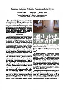
2.1. Preoperative Intervention Planning First the tracheobronchial tree, with special attention to the extraction of lower generations, is segmented using the segmentation component. The result is transferred into a graph-based description.8 At the same time a region of interest (e.g. carcinoma) can be segmented by the use of interactive tools. With the help of 2Dand 3D-visualizations the lung specialist defines interactively the desired target bronchus inside the preoperative planning component. Thereupon the path from the trachea to the target is calculated using the graph-based description of the bronchial tree (s. fig. 1) and directly displayed. Afterwards the intervention can be simulated showing a virtual bronchoscopy. The irradiation planning for brachytherapy can be done in an independent application afterwards using the already extracted information e.g. segmentation of the carcinoma. At last three or more characteristic positions, for example the three first bifurcations, inside the virtual bronchial tree have to be chosen. With these positions, the initial registration of the virtuality to reality is done during the intervention.
2.2. Realtime Navigation Shortly before the intervention, three sensors from a commercial electromagnetic tracking system (e.g. Aurora, NDI) are fastened to the chest of the patient to build up points of reference inside the electromagnetic field. Another sensor is inserted into the tip of the bronchoscope, or irradiation catheter, to measure its position. Then the characteristic positions, chosen inside the planning component, have to be encountered inside of the bronchial tree to initially correlate the virtual with real images. From there on the navigation component gradually enhances the accuracy through storing the already covered path of the sensor to a graph description. The description then is mapped to the preoperatively generated graph based bronchial tree description. This prevents the calculated position of the tip from leaving the inside of the bronchial tree and also weakens it to jump into a different branch of the tree, especially if it has a different shape. So the longer the covered distance, the better is the accuracy of the navigation compound. This corrected position is then sent to the intervention component, which manages differnet widgets to satisfy the needs of the lung specialist.
Figure 1. Visualization of the graph based representation of a bronchial tree (yellow) and a carcinoma (purpel) within the preoperative planning component. The physician interactively selects the bronchi he wants to be led to. The path from that bronchi to the trachea is shown thereafter (green).
2.3. Merging Virtual Reality with Video Usually only a minimal change of a good working system permits to improve it. Since, for the examination of the inner lung, the use of a video bronchoscope is highly established, a slight change would be the merging of the video image with additional information, not covering any important structures (augmented reality). Several arrows or a line, representing the path to a target, would represent such additional information. The images taken from such a bronchoscope are heavily distorted to gather more information of the surrounding area. Thus the additional information, here the virtual line or a transparent surface representing a carcinoma inside the surrounding tissue, has to be distorted the same way, to correlate virtual with real data. Furthermore the scene has to be cropped to only show the area, that is visible on the video. For example the virtual path has to be cut exactly where a bronchus turns. Finally this all has to be done in real-time to obtain the quality of the system. To realize this, we expanded MITK in the following manner: To access the current video frame of the bronchoscope and to maintain cross-platform ability, different classes wrapping application program interfaces (API) for multimedia (e.g. vfw: video for windows) were implemented. So during application runtime a video can be grabbed and displayed in the background of a virtual scene. To be able to distort everything inside the scene but the video (path, surfaces etc.), the visualization pipeline had to be adapted. MITK uses VTK (Visualization Toolkit, Kitware Inc.9 ) which uses, apart others, OpenGL10 to render a virtual scene. OpenGL permits the use of alpha testing, which is used in this context to separate the virtual data from the video. This is realized by first copying the drawn virtual scene to texture memory and building up the scene for augmented reality. For this the OpenGL scene is set to orthographic projection, and two planes are drawn one behind the other. The first plane is textured with the copied image of the virtual scene and the second is textured with the current frame of the video. Through alpha testing, the first plane is hidden where no virtual object was drawn, so the plane behind it is visible. This way we could realize to always show the virtual objects in front of the video.
Figure 2. For merging virtual path with the distorted video of the bronchoscope, the path has to be distorted as well. The left side shows an undistorted image, right side shows the same image distorted by the implemented distortion mechanism.
To distort the texture on the plane, the plane itself had to be divided into several adjacent tiles, each textured with a part of the copied virtual scene (s. fig. 2). The corners of the tiles are repositioned according to a calculation described by Mei.11 Afterwards the tiles in the middle of the compound are bigger in size than the tiles on the edge. Since the texture gets stretched on an expanded tile and compressed on a shrunken tile, the sum of all textured tiles shows the desired distorted image. Now to crop the virtual objects to only show the area, that is visible on the video (e.g. turning of a curve of a bronchus), the surface of the segmented bronchial tree is used. The order of the rendering of the virtual data is changed so that the surface of the bronchial tree is rendered first. Through disabling the OpenGL color buffer, the surface only gets rendered into the depth buffer and is not visible inside the scene. After enabling color buffer again, depth testing leads to the desired results. The virtual objects are cropped by the invisible surface of the bronchial tree, so that the path, shown in figure 4 on the lower left, is cut to correlate to the real image on the video.
2.4. Test Environment To test the whole environment, a flexible lung model, consisting of plastic tubes that are welded together according to the structure of a human bronchial tree, was constructed (s. fig. 3). The tubes are transparent so the tip of the catheter or bronchoscope can be seen to verify the position of real and virtual data. The transparent tubes lower the quality of the video taken by the bronchoscope, because they don’t reflect the emitted light. To also test the system with a good video refresh rate, one branch was obscured by insulating tape. This still allows a positioning control not supported by other lung models. The branches were hung up with elastic rubber band onto a movable panel, which can be used to simulate the respiration cycle of a tracheobronchial tree. Because of using non-metal materials, a CT dataset of the model could be acquired. Within the segmentation component, the inside of the tube was extracted and converted into the graph based description. Furthermore the surface could be extracted (s. fig. 3 upper widgets showing the surface in 3D with transparency to also see the path inside). For testing the navigation system, the extracted data can now be loaded into the planning and the guidance component. For this test, three sensors are used as markers and fastened onto the model. As described, a forth sensor is located in the inside of a bronchoscope. Now all four tracked positions can be read from the tracking system into the navigation component. The three positions from the markers build up points of reference and the forth sensor is gradually correlated to the virtual
Figure 3. Self-made flexible lung model with a movable panel to simulate the respiration cycle. One navigation sensor for point of reference is visible on the left side. The two others are fastened behind the movable panel.
data. The adapted coordinate is then send to the guidance component, where it then is visualized relatively to the virtual data of the model in real-time.
3. RESULTS First experiments on the self constructed transformable lung model showed, that the gradually registering navigation component overcomes the usual registration problem of a moving soft tissue and improves the accuracy highly needed in the periphery of the lung. Without this improvement, the virtual data can not be correlated precisely with the real data, so that a led intervention by means of augmented reality is not possible. The tests were made with the MicroBird system from Ascension, with the conventional Aurora system from NDI, and its new flat panel 6D update. Tests with 5D sensors instead of 6D showed the necessity of all 6 dimensions. With the missing information of rotation of the bronchoscope, either the bronchoscope must not be rotated (unacceptable) or the virtual position has to be adapted once more using image registration (no real-time). Using a 6D sensor the rotation of the bronchoscope as well is detected and thus the orientation of the virtual scene is properly adapted. Further tests are about to come. The navigation and registration mechanism has been patented under the number PCT/EP2005/002244 and its implementation is under continuous development. The presented system combines all components necessary for a navigated brachytherapy (apart irradiation planning) and is adaptable to the viewing habits of the lung specialist. Therefore several widgets (2D, 3D, video, virtual bronchoscopy also in stereo) are available (s. fig. 4 and 5). Our initial tests also showed, that the use of an image guided navigation system, which allows a fusion of a video-bronchoscope with a target-path, represents a major improvement for a navigation inside the lung. Since the information is merged with the video, the lung specialist doesn’t have to look on additional monitors, and thus can concentrate on the intervention. Furthermore the target path, as additional information, improves the complex and time-consuming location of a region of interest and so reduce the load of the patient by shorter anaesthesia.
Figure 4. Example of the different views in the intraoperative guidance component. Loaded is a CT-data taken from the self constructed transformable lung model. Top left and right: 3D views showing a transparent surface of the bronchial tree, the path to the preoperatively defined bronchi, and a cone representing the tip of the catheter; bottom right: virtual bronchoscopy with the target-path; bottom left: video of bronchoscope merged with target-path.
Through distorting the virtual scene, the correlation between the virtual data and the video of the bronchoscope is made possible. Future test will also analyse the accuracy of the correlation. The implemented distortion algorithm is processed by the graphical processor unit (GPU). A standard graphic card can easily manage the additional computation. Since exact values are not only dependant on the hardware used, but also on the dedicated refresh rate of the tracking system, and the video grabbing card, further values for performance (frames per second etc.) are marginal. The navigation system is develpoed to be independant on any bronchoscope or tracking system manufacturer. But tests showed, that the quality of the hardware, for example the material of the tip of the bronchoscope and the accuracy of the tracking system near metal parts, influences the system. A good setup was found with the 6D tracking system Aurora from NDI and a fiber optic bronchoscope with the tip made out of stainless steel.
4. CONCLUSION Because of the direct guidance, operation-time can be shortened, and due to the ability to be movementindependent, the anesthesia and high frequency jet respiration can be reduced. The lung specialist won’t have to adapt to a new technique because the path is merged with the video of the bronchoscope he already is familiar with. Also the immersion into the augmented reality is, as required, small, since only a thin virtual path is drawn into the video of the bronchoscope. Even if the bronchoscope can not penetrate all the way to the target bronchus due to its size, the improved registration process allows pushing forward the smaller irradiation catheter with the inserted tracking-sensor by means of virtual bronchoscopy. To be able to follow the specialist’s habits, several widgets (2D, 3D, AR-video, VB, also red-green stereo) are available. Furthermore the acquirement of
Figure 5. Three orthogonal 2D views of the intraoperative guidance component showing the CT-slices taken from the self-made lung model.
CT-data, for checking the right position of the catheter, can be reduced. Also the same position can be located over and over again, for example for an after-treatment.
5. DISCUSSION The navigation component gradually registers the already covered path of the tip with the preoperatively extracted, graph based description of the bronchial tree. This rises the accuracy of the navigation system but also assumes that the tip of the bronchoscope is still inside the bronchus. If this is not the case, for example because of a too strong pushing of the bonchoscope, the lung specialist has to stop the procedure and to continue the conventional way. Leading manufacturers concentrate on enhancing the resolution of the images, taken by bronchoscopes. To achieve this, the development of flexible fiber optic bronchoscopes seems to be replaced with the development of flexible digital bronchoscopes with a high resolution. Maintaining the image quality and the size of the working channel, but still reducing the diameter of a fiber optic bronchoscope seems to be challenging. At present manufacturers offer fiber optic bronchoscopes with a diameter around 5mm. Digital high resolution bronchoscopes still are of diameter around 6mm. Furthermore the electronic circuit (CCD-sensors etc.) inside the tip of an digital high resolution bronchoscope is more likely to interfere with the tracking system. Thus the problem of reaching peripheral lesions with a bronchoscope will not be solved by a smaller diameter in the near future. This leads to the importance of a image guided navigation system, which offers to also navigate in a virtual environment. As long as the examined structure is wide enough to allow the insertion of the bronchoscope, the lung specialist will favor the video. But if the bronchus gets too thin, a catheter with an inserted navigation sensor, can be pushed further. Here virtual bronchoscopy will be a way to orientate in a similar manner.
Regarding the merge of the virtual scene with the video, we first used OpenGL stencil buffer to prohibit the drawing of the video where virtual objects have been drawn. This also led to the required results. But to realize a distorted virtual scene with the unchanged video in the background, alpha-testing was essential because of the use of textured tiles. The adaptation of the visualization pipeline didn’t cause a perceivable change in rendering performance. To realize the distortion of the virtual scene, the camera parameters of the bronchoscope have to be calculated. In most cases, manufacturers of bronchoscopes hardly can feature the parameters needed. A calibration procedure, which is described by Mei,11 provides the acquisition of several screenshots of the bronchoscopic video to gather all necessary information of the optical characteristic. Then a free Matlab plugin is used to correlate the screenshots. The procedure results in the camera parameters of this brand and type of bronchoscope. These parameters are then used to calculate the new coordinates of the corners of the tiles. But since this procedure is well documented and only done once, the amount of work is justifiable. Plans for the future contain, amongst others, precise tests of the navigation component with the use of animal data inside the operation room and an enhancement of the semiautomatic segmentation process to be able to segment even more fine structures of the bronchial tree.
REFERENCES 1. K. Stanley, “Prognostic factors for survival in patients with inoperable lung cancer.,” J Natl Cancer Inst (65), pp. 25–32, 1980. 2. W. Harms, H. Becker, R. Krempien, and M. Wannenmacher, “Contemporary role of modern brachytherapy techniques in the management of malignant thoracic tumors.,” Semin Surg Oncol (20), pp. 57–65, 2001. 3. Superdimension. http://www.superdimension.com/, 2005. Herzliy, Israel. 4. Q. Schwarz, A. Mehta, A. Ernst, F. Herth, A. Engel, D. Besser, and H. Becker, “Elektromagnetic navigation during flexible bronchoskopy.,” Respiration (70), pp. 516–522, 2003. 5. T. Gildea, P. Mazzone, and A. Mehta, “Adapting gps-like technology to bronchoscopy: Elektromagnetic navigation,” Respiratory Exchange Fall 2005, The Cleveland Clinic Foundation. 6. M. Vetter, I. Wolf, P. Hassenpflug, M. Hastenteufel, R. Ludwig, L. Grenacher, G. Richter, W. Uhl, M. Bchler, and H. Meinzer, “Navigationaids and real-time deformation modeling for open liver surgery.,” SPIE Medical Imaging (5029), pp. 58–68, 2003. 7. I. Wolf, M. Vetter, I. Wegner, M. Nolden, T. Bttger, M. Hastenteufel, M. Schobinger, T. Kunert, and H. Meinzer, “The medical imaging interaction toolkit (mitk).,” SPIE (5367), pp. 16–27, 2004. 8. M. Schoebinger, M. Thorn, M. Vetter, C. Cardenas, P. Hassenpflug, I. Wolf, and H. Meinzer, “Robuste analyse von gefstrukturen auf basis einer 3d-skelettierung.,” Bildverarbeitung fr die Medizin (BVM) , pp. 76– 80, 2003. 9. W. Schroeder, K. Martin, and B. Lorensen, The Visualization Toolkit An Object-Oriented Approach To 3D Graphics, 3rd Edition, Kitware Inc., 2005. 10. M.Woo, J.Neider, and T.Davis, OpenGL Programming Guide Second Edition (RedBook), Addison Wesley, 1996. 11. C. Mei, “Camera calibration toolbox for matlab.” 2005, INRIA Sophia-Antropolis. http://www.vision.caltech.edu/bouguetj/calib doc/index.html.