Sep 3, 2015 - 1The Pharmaceutical College, Barpali, Bargarh, Odisha, 768029. 2School of Pharmaceutical Sciences, Siksha 'O' Anusandhan University, ...
Available online on www.ijppr.com International Journal of Pharmacognosy and Phytochemical Research 2015; 7(5); 934-942 ISSN: 0975-4873 Research Article
Development of Quality Control Parameters for Standardization of Triumfetta rhomboidea Chandan Das1, Prateekshya Mishra1, Debajyoti Das2, Laxmidhar Maharana2, Goutam Ghosh2* 1
The Pharmaceutical College, Barpali, Bargarh, Odisha, 768029 School of Pharmaceutical Sciences, Siksha ‘O’ Anusandhan University, Kalinga Nagar, Ghatikia, Bhubaneswar, Odisha, India 751003
2
Available Online:3rd September, 2015 ABSTRACT Triumfetta rhomboidea is a herbaceous perennial plant found abundantly throughout India. The plant is used in the treatment of dysentery, diarrhoe, ulcer, leprosy and gonorrhoea. The present study comprises taxonomic details, macro and microscopical characters of parts used, and physico-chemical details. This study will be helpful in setting some pharmacopoeial standards and preparation of monograph of this plant. Key words: Triumfetta rhomboidea, Microscopical character, Powder microscopy, Physico-chemical study. INTRODUCTION The herbal drugs can be used safely if their standard parameters such as authentification, safety, efficacy and quality are maintained as per specifications. In view of the growing demand and use of herbal drugs, it is crucial to ascertain standard samples of herbal drugs for future reference1. In recent years, the rapid development of herbal drugs provides a systematic approach to evaluate these drugs in modern pharmacognosy by qualitative and quantitative means2. Nowadays, analytical instruments are playing an important role in the production and evaluation of new herbal for the consumers and the environment. The use of sophisticated instruments is a fascinating measure of chemical analysis. Though, several modern instrumental techniques are required to solve analytical problems still the value of classical methods can’t be over looked. Therefore, it is necessary to perform some physical and chemical operations on the drug sample prior to the actual analysis. Triumfetta rhomboidea (family-Tiliaceae) is a herbaceous perennial plant. It is distributed throughout tropical and subtropical India3. It is 0.6 to 1.5m in height; branches are slender, more or less pubescent with simple hairs. Leaves are variable, stipulate, 3-lobed, irregularly serrate, clothed with simple and stellate hairs on both surfaces. Flower is yellow in colour, dense terminal and leaf opposed cymes. Buds are oblong apiculate, peduncles and pedicels are very short; bracts are subulate. Sepals are oblong, hooded and apiculate at the apex. Petals are shorter than the sepals, obovate-oblong and ciliate at the base. Fruits are 4mm in diameter, pubescent and spines are glabrous4. The leaf, bark, root, flower and fruit of the plant are traditionally used for treatment of various diseases. In Ayurveda the root is bitter and acrid; aphrodisiac, tonic,
*Author for Correspondence
cooling; useful in dysentery. The leaves and stem are used as a poultice on tumor5. Powdered leaf infusions of Triumfetta rhomboidea are drunk represents for the treatment of anemia in different regions of East Africa 6. Triumfetta(Tiliaceae) species are used in the folk medicine forthe treatment of various diseases, such as diabetes, leprosy, diarrohea, demulcent, etc. Triumfetta rhomboidea is locally used as antidiabetic, aphrodisiac, tonic, galactogenic, roots asdiuretic, barks in diarrheoa, leaves and flowers as astringent7. Pounded roots are given in the treatment of intestinal ulcer. Leaves, flowers and fruit are mucilaginous demulcent, astringent, and also used in gonorrhoea and against leprosy8. MATERIALS AND METHODS Collection and preparation of specimen The plant specimens for the proposed study were collected from Bargarh, Odisha. The required sample of different organs were cut and remove from the plant and fixed on FAA (Formalin-5ml + Acetic acid-5ml +70% Ethyl alcohol-90 ml) after 24 hrs of fixing, the specimen were dehydrated with graded series of tertiary-butyl alcohol as per the scheduled given by Sass,1940. Infiltration of the specimen was carried by gradual addition of paraffin wax (melting pointing 580 to 600C) until solution attained super saturation. Then the specimen was cast into paraffin wax blocks. Sectioning and staining The paraffin embedded specimen was sectioned with help of Rotary Micrometer. The thickness of the specimen was 10-12µm9. The section was stained with safranin ana fast green. The dye render deep red color to lignin and bright green color to cellulose10. For studying the stomatal
Das et al. / Development of Quality…
Table 1: Dimention of various leaf constants of Triumfetta rhomboidea Parameter Maximum Minimum Stomatal index 50 30 Length of stomata 120 µm 97.5 µm Width of stomata 75 µm 52.5 µm Length of stomata pore 75 µm 52.5 µm Width of stomata pore 22.5 µm 15 µm Palisade cell 11.5 8.25 Vein-islet 40 30 Vein termination 110 70 Length of trichome 900 µm 225 µm Diameter of starch grain of stem 45 µm 22.5 µm Diameter of starch grain of root 75 µm 22.5 µm Length of phloem fibre in stem 525 µm 105 µm Length of phloem fibre in root 525 µm 120 µm
1(a)
1(b)
Average 38.28 109.5 µm 68.25 µm 63 µm 15.75µm 9.58 35 95 555.75 µm 29.62 µm 38.25 µm 310.8 µm 327 µm
1(c)
1(d) 1(e) 1(f) Figure 1(a): Transverse section of leaf at magnification 5x, 10x; Figure 1(b): Transverse section of leaf at magnification 10x, 10x., Figure (c, d, e &f): Transverse section of leaf at magnification 5x, 45x. Abbreviations: U.Epi-Upper epidermis, Par-Parenchyma, Phl-Phloem Scl-Sclerenchyma, C.Tr-Covering trichome, GtGround tissues, Pal- Palisade cells, Sp. par- Spongy parenchyma, P.xy-Proto xylem, M.xylem-Meta xylem morphology, venation pattern and trichome distribution, paradermal section as well as clearing of leaf with 5% sodium hydroxide or epidermal peeling by maceration Jeffrey’s maceration fluid were prepared11. Photomicrographs Photographs of different magnifications were taken with Nikon Labphoto 2 microscopic unit. For normal observation bright field was used. For the study of crystals, starch grains and lignified cells, polarized light was employed. Since these structure have birefringent property under polarized light. They appear bright against dark
background. Descriptive terms of the anatomical features are as given in the standard anatomy books12. Quantitative determination was carried out for measurement of diameter of starch grain and length of phloem fibre13, physico-chemical studies12, behavior of powder drug towards different chemical reagent14, fluorescence analysis15. Preliminary phytochemical screening Preliminary phytochemical screening was carried out using the standard methods16. RESULTS
IJPPR, Volume 7, Issue 5, October 2015- November 2015
Page 935
Das et al. / Development of Quality…
Quantitative microscopy
2(a) 2(b) Figure 2(a): Transeverse section of lamina at magnification 5x,10x Figure 2(b): Transeverse section of lamina at magnification 5x,45x Abbreviation: U.Epi-Upper epidermis, L.Epi-Lower epidermis. Sp par-Spongy parenchyma. Pal-Palisade cell. Epidermis is wavy walled and also straight walled polygonal. It shows the presence of anomocytic and paracytic stomata. Epidermal surface shows presence of both glandular and covering trichonmes. Covering trichomes are uniseriate, unicellular, thick wall with acute apex. It shows the presence of stellate trichomes. Vein-islet and vein termination are prominent. Veins are lignified. Macroscopical characters Leaves are green in colour with characteristic taste and slight odour. The colour of stem is brown and taste is bitter with slight odour.The roots are found light brown in colour with characteristic taste. Microscopical study of leaf Upper epidermis is single layered, polygonal parenchymatous cells. Covering trichomes are present above the epidermal cell. Epidermal layer is followed by 9-10 layers of parenchymatous cell. Vascular bundle is arc shaped and is more towards dorsal side. It is bicollateral (xylem is surrounded by phloem). Xylem is lignified and phloem is non-lignified. Vascular bundle is surrounded by pericycle fibre. Pericycle consists of sclerenchymatous cells which are highly lignified. Vascular bundle is surrounded by ground tissue. It consists of thin walled parenchymatous cell (round or polygonal). Some are small in size and some are bigger in size. Ground tissue covers the dorsal and ventral side.
Transeverse section of lamina Lamina is dorsiventral in nature. Upper epidermis is single layer and consists of polygonal cell. Covering trichomes are present on the epidermis. Mesophyll region contain palisade cells and spongy parenchymatous cells. Palisade cells are single layered, compactly arranged, radially elongated cells containing brownish matter. Palisade cells are followed by spongy parenchyma. It consists of 3-4 layer of loosely arranged parenchymatous cells. Lower epidermis is similar to upper epidermis. Transeverse section of petiol Epidermis is singly layered, tangentially elongated, not covered by cuticle. These are polygonal cells contain covering trichomes. Epidermal layer is followed by cortex zone. Below the epidermis about two layer of thick walled collenchymatous cells are present. Collenchymatous cell is followed by parenchymatous cell. Smaller and bigger cells are present towards inner side. Few cells contain clusture of crystals. Five number of vascular bundle are present, out of which one vascular bundle is bigger than others. Three vascular bundle are fully developed and two are less developed. Xylem is lignified and phloem is non lignified. Xylem is endarch (protoxylem lies towards centre and meta xylem lies towards periphery). Growth of xylem is centrifugal. Arrangement of vascular bundle is bicollateral. Pericycle fibre consists of sclerenchymatous cell, which are lignified and are arranged as crown above phloem region of vascular bundle. At the centre pith is present. It consists of large thin walled parenchymatous cells. Transeverse section of bark Cork cell is about 2-3 layers, consists of radish brown matter. The cells are oval to rectangular shapes. It is followed by stratified cortex. These are thin walled parenchymatous cells of rectangular shapes. It consists of many layers of parenchymatous cell , extend to the next region. It contains squarish / prismatic calcium oxalate crystals. Group of pericycle fibres are appeared in the cortex region. Each group contains approximately 9-16 of sclerenchymatous cells. These are highly lignified when treated phloroglucinol and concentrated hydrochloric acid. Group of pericycle fibres become more diverge towards the centre which gives the appearance of divergent rays. Transeverse section of stem Cork consists of few layer of irregular shape and contain reddish brown matter. Below the cork region stratified
3(a) 3(b) 3(c) 3(d) Figure 3(a): Transverse section of petiol at magnification 10x,10x; Figure 3(b, c & d): Transverse section of petiol at magnification 5x, 45x. Abbreviations: Par fib-Pericycle fibre, Phl-Phloem, Xy-Xylem, Cr-Crystal, Scl-Sclerenchyma, Gt- Ground tissue, TrTrichomes, Par-Parenchyma, Xy-Xylem, V.B-Vascualr bundle, Col-Collenchyma.
IJPPR, Volume 7, Issue 5, October 2015- November 2015
Page 936
Das et al. / Development of Quality…
4(a) 4(b) 4(c) 4(d) Figure 4(a): Transverse section of stem at magnification 5x, 10x; Figure 4(b, c & d): Transverse section of stem at magnification 5x, 45x. Abbreviations: Epi-Epidermis, Co-Cork, Cor-Cortex, Cr-Crystal, Par-Parenchyma, Scl-Sclerenchyma, Per fib-Pericycle fibre.
5(a) 5(b) 5(c) 5(d) Figure 5(a): Transverse section of stem at magnification 5x, 10x; Figure 5(b,c & d): Transverse section of stem at magnification 5x,450x. Abbreviation: Co-Cork, Cor-Cortex,, Cam-Cambium, Xy par-Xylem parenchyma, , Par-Parenchyma, SclSclerenchymatous, Phl-Phloem, Xy-Xylem, Per Fib-Pericycle fibre, Cr-Crystal, X.V-Xylem vessel.
6(a) 6(b) 6(c) 6(d) Figure 6(a): Transverse section of root at magnification 5x, 10x; Figure 6(b): Transverse section of root at magnification 10x, 10x; Fig. 6(c& d): Transverse section of root at magnification 5x, 45x Abbreviation: Co-Cork, Cor-Cortex, Phl-Phloem, Md.ray-Medullary rays, X.V-Xylem vessel, Xy.par-Xylem parenchyma.
7(a)
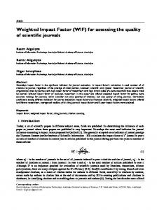
7(b) 7(c) 7(d) Figure 7: Powder Microscopy of Leaf, Figure 7(a &b): Trichomes Figure 7(c): Stomata; Figure 7(d): Crystal, Figure 7(e): Starch granule
cortex is present. These are large thin walled parenchymatous cells. The cells contains clusture of calcium oxalate crystals. Cambium is present in between
7(e)
cortex and vascular bundle. It is about 2-3 layers and consists of rectangular cells. Vascular bundle consists of xylem and phloem. It open, due to presence of cambium.
IJPPR, Volume 7, Issue 5, October 2015- November 2015
Page 937
Das et al. / Development of Quality…
8(a)
8(b)
8(c)
8(d) 8(e) 8(f) Figure 8: Powder Microscopy of Stem, (a): Fibre, (b): Cortex, (c,d,e,f): Tracheal element, (g,h,i): Starch granules.
9(a)
9(b)
9(c)
9(d) 9(e) 9(f) Figure 9: Powder characteristic of root, (a,b,c): Cork cell, (d,e,f): Cortex, (g,h): Fibre, (I,j,k,l): Xylem vessels, (m,n,o): Crystals, (p,q): Starch granules
IJPPR, Volume 7, Issue 5, October 2015- November 2015
Page 938
Das et al. / Development of Quality…
Table 2: Determination of physico-chemical parameters Parts used Parameter %w/w Ash value Total ash 10.33 Leaf Water soluble 4 ash Acid in soluble 2.2 ash Sulphated ash 12.2 Total ash 7 Stem Water soluble 3 ash Acid in soluble 1.5 ash Sulphated ash 11.5 Total ash 7 Root Water soluble 4.5 ash Acid in soluble 4 ash Sulphated ash 11 Extractive value Chloroform 3.2 Leaf Acetone 5.6 Methanol 19.2 Water 32 Chloroform 3.2 Stem Acetone 0.4 Methanol 1.4 Water 5 Chloroform 1 Root Acetone 0.66 Methanol 2.6 Water 6 Loss on drying Leaf 9.5 Stem 10 Root 7.5 Arrangement of vascular bundle if collateral. Xylem is lignified. Xylem vessels are large in size. Xylem is surrounded by thin walled non lignified xylem parenchyma. Phloem is present as group above the xylem parenchyma. Phloem is nonlignified. Patches of pericycle fibre are present above the vascular bundle. Each group contains about 9-16 no of sclerenchymatous cell. Medullary rays is absent. Pith occupies large portion at the centre. It consists of thin walled polygonal parenchymatous cells. Cells are closely arranged without having intercellular spaces. Transeverse section of root Cork consists of 2-3 layers of tabular cells, irregular shape. Cork region followed by cortex. It consists of 3-4 layers of slightly flattened parenchymatous cells. Cells contain reddish brown matter. Vascular bundle is present below cortex. Arrangement of vascular bundle is collateral. Xylem is lignified and phloem is non lignified. Xylem vessels may be spiral, annular, scleriform, reticulat or pitted. Most of the vessels are bigger in size. Most of the
xylem vessels are singly and large while few xylem vessels are compound. Vessels are surrounded by xylem parenchyma. Phloem is present above the xylem and is 78 layer closely arranged. It consists of thin walled parenchymatous cells of irregular shape. Medullary rays are multiserriate. Medullary rays narrows towards centre and becomes diverge towards periphery. It consists of squarish and rectangular shape of parenchymatous cell. Pith is absent. Powder characteristic of leaf Trichomes are simple and stellate (branched) type. Uniseriate, unicellular and multicellular covering trichomes with pointed ends are found. The trichomes are thick walled and lignified. Paracytic type of stomata is found. Prismatic calcium oxalate crystals and are present frequently. Starch grains are not found frequently. These occur in few groups and are spherical shape. The diameter of starch grains varies from 15µ-45µ. Powder characteristic of stem Fragment of cork consists of rectangular or squarish parenchymatous cell. Cortex is made up of polygonal parenchymatous cells. Few parenchymatous cells contain starch grains. Both spiral and bordered pitted vessels are found.Both lignified and non lignified fibres are found. These are large number of thick walled elongated fibres. The length of phloem fibres varies from 105µ-525µ. Starch granules are abundant, spherical and compound. The diameter is from 22.5µ-45µ. Crystals are large in size. These are appeared in squarish or prism. Powder characteristic of root The cork contains thin walled rectangular parenchymatous cell. Fragment of cortex shows several layer of thin walled squarish rectangular parenchymatous cell. These are brown to dark brown in colour. Wood element consists of border pitted xylem vessels. They are well developed. Prismatic crystals are found. Starch granules are not frequently found. These are spherical, few starch grains are bi-head. The diameter varies from 22.5µ-75µ. Determination of physico-chemical parameters Ash value Ash value is a measure of the quality and purity of the drug. The total ash, water soluble ash, acid insoluble ash and sulphated ash of Triumfetta rhomboidea of leaf were found to be 10.33%w/w, 4%w/w, 2.2%w/w and 12.2%w/w. Total ash of Triumfetta rhomboidea leaf were found to be more than water soluble ash and acid insoluble ash. Acid insoluble ash was found to be very less than total ash, water soluble ash and sulphated ash. Sulphated ash was found to be more than total ash, water soluble ash acid insoluble. The total ash, water soluble ash, acid insoluble ash and sulphated ash of Triumfetta rhomboidea of stem were found to be 7%w/w, 3%w/w, 1.5%w/w and 11.5%. The total ash and water soluble ash value of Triumfetta rhomboidea stem powder were found to be more. Sulphated ash was found to be more than total ash and water soluble ash. Acid insoluble ash was very less than total ash, water soluble ash and sulphated ash. The total ash, water soluble ash, acid insoluble ash and sulphated ash of Triumfetta rhomboidea root were found to be 7%w/w, 4.5%w/w, 4%w/w and 11%w/w. Toatal ash was more than
IJPPR, Volume 7, Issue 5, October 2015- November 2015
Page 939
Das et al. / Development of Quality…
Table 3: Behavior of powdered leaf, stem and root of Triumfetta rhomboidea with chemical reagent Acid/Reagent Observation Leaf Stem Root Powder as such Green Light brown Pale yellow Powder + Picric acid Yellow Yellow Yellow Powder + Con.Nitric acid Light orange Yellowish orange Yellowish orange Powder + Con.HCL Green Green Yellowish brown Powder + Con.H2SO4 Black Black Deep brown Powder + Glacial acetic acid Yellowish green Light green Light brown Powder + 5% FeCl3 Light green Green Light green Powder + NaOH (5N) Green Yellowish green Light brown Powder +KOH(5%) Yellowish green Yellowish green Light brown Powder + Iodine/20 Reddish brown Reddish brown Reddish brown Table 4: Fluorescence analysis of powder of Triumfetta rhomboidea Reagent Leaf Stem Day light Short wave Day light Powder as Pale green Green Dull green such Powder + 1N Light green Light green Light green NaOH in methanol Powder + 1N Light green Green Light green NaOH Powder + Light green Light green Yellowish Ethanol brown Powder Light green Green Yellowish +HNO3 + NH3 brown solution Powder + 50% Yellowish Green Yellowish HNO3 green brown Powder + 1N Green Green Light yellow HCL Powder + Light green Green Yellowish HCL brown Powder + Black Dark green Brown H2SO4 Powder + 50% Light green Light green Light green H2SO4 Powder + Light green Greenish Yellowish Glacial acetic black brown acid Powder + Green Light green Yellow HNO3 ater soluble and acid insoluble ash. Sulphatated ash was more than total ash water soluble ash and acid insoluble ash. Acid insoluble ash was very less than other ash values. Total extractive values The extractive values were determined to find out the amount of soluble compounds. The chloroform, acetone, methanol and water extractive values of leaf of Triumfetta rhomboidea were 3.2%w/w, 5.6%w/w, 19.2%w/w and 32%w/w. The leaf shows more amount of water soluble compound than chloroform, acetone and methanol extract. The chloroform, acetone, methanol and water extractive values of stem of Triumfetta rhomboidea were 3.2%w/w,0.4%w/w, 1.4%w/w and 5%w/w. The stem showed more amount of water and chloroform soluble
Short wave Green
Root Day light Pale yellow
Short wave Green
Light green
Yellowish brown
Light green
Green
Yellowish brown Yellowish brown Yellowish brown
Light green
Yellowish brown Yellow
Light green
Yellowish brown Deep brown
Light green
Yellowish brown Yellowish brown
Light green
Yellowish brown
Light green
Light green Green
Green Light green Light green Black Green Green
Green
Light green Deep green
Light green
Black
Light green
component than acetone and methanol extract. The chloroform, acetone, methanol and water extractive values of root of Triumfetta rhomboidea were 1%w/w, 0.66%w/w, 2.6%w/w and 6%w/w. The root showed more amount of water and methanol soluble components than chloroform and acetone extract. Loss on drying The moisture content of leaf, stem and root were found to be 9.5%w/w, 10%w/w and 7.5%w/w. Stem has more moisture content than leaf and root. Behavior of powdered materials towards chemical reagent The behavior of the powdered leaf, stem and root were treated with picric acid, con. Sulphuric acid, con. Hydrochloric acid, con. Nitric acid, glacial acetic acid, 5%
IJPPR, Volume 7, Issue 5, October 2015- November 2015
Page 940
Das et al. / Development of Quality…
ferric chloride, sodium hydroxide (5N),potassium hydroxide (5N), iodine/20 solution were observed. Fluorescence analysis of powder of Triumfetta rhomboidea Fluorescence analysis of entire leaf, stem and root has been carried out in day light and under UV light. The powders were treated with differing organic solvents and solutions and observed in normal day light and under UV light. DISCUSSION In view of the commercialization of formulations of traditional plants and for the development of new chemotherapeutic agents, quality control of medicinal plants used in traditional medicine is becoming more important. Adulterated and substituted medicinal plants may produce severe health related problems when taken by the patients and may cause legal problems in the pharmaceutical industries17. The botanical identification and physico-chemical characters may be helpful for pharmacognostical study and standardization of herbal drugs. It may be considered as the diagnostic tool for the researchers who are involved in the evaluation of herbal drugs from indigenous source. The microscopical study of medicinal plants is a major tool for the authentification of drugs especially for identification of powdered drugs, because in these cases most of the morphological diagnostic features are lost18. The moisture content of leaf, stem and root were found to be 9.5%w/w, 10%w/w and 7.5%w/w. The less value of moisture content of drugs could prevent content bacterial, fungal or yeast growth through storage19. The total ash, water soluble ash, acid insoluble ash and sulphated ash of Triumfetta rhomboidea of leaf were found to be 10.33%w/w, 4%w/w, 2.2%w/w and 12.2%w/w respectivey. Ash values used to find out quality, authenticity and purity of unsophisticated drug and also these values are important quantitative standards20. The extractive values of the drug are valuable to estimate the chemical constituents present in the drug and also help to evaluate certain phytoconstituents soluble in a particular solvent21. Phytochemical screening gives the information of nature of chemical constituents of the drug. Preliminary phytochemical screening revealed the presence of carbohydrate glycosides, phytosterol, steroids, flavonoids, tannin & phenolic compounds and triterpenoids in this plant. The crude drugs do not produce fluorescence in daylight, but they can produce fluorescence, when observed in ultra violet light. The powder crude drug does not produce fluorescence on their own, but they may be converted into fluorescent derivatives in presence of different solvents and reagents. The fluorescent method is helpful in determination of the drug sample over a satisfactory concentration range without several time consuming dilution steps prior to the analysis. So, this fluorescence study could be one of the parameter for evaluation of crude drugs19.
CONCLUSION This research paper discusses not only pharmacognostical and phytochemical characteris but also microscopic and fluorescence characters of the different parts of the plant. These characteristics can be used further as identification and authentication parameters of the plant. The data could be useful to differentiate closely related plant species having similar phytoconstituents and pharmacological activities. REFERENCES 1. Ansari MY, Wadud A, Ehteshamudddin, Bano H. Pharmacognostical evaluation of root of Gumma (Leucas cephalotes Spreng). Indian Journal of Natural Products and Resources 2013; 4: 88-95. 2. Shinde V, Dhalwal K. General Review, Pharmacognosy: The changing Scenario. Pharmacog Review 2007; 1: 1-5. 3. Joshi SG. Medicinal plants. Oxford & IBH publishing Co Pvt. Ltd. New Delhi 2000; 392. 4. Kirtikar KR, Basu BD.Indian Medicinal Plants. International Book distributors. Dehradun 1975; 1: 395. 5. Kirtikar KR, Basu BD.Indian Medicinal Plants. International Book distributors. Dehradun 1998; 4: 2738-2739. 6. Chabbra SC, Mahunnah RLA, Mshiu EN. Plants used in traditional medicinein Eastern Tanzania. VI. Angiosperms (Sapotaceae to Zingiberaceae. Journal of Ethanopharmacology 1993; 39: 83-103. 7. Khare CP. Indian Medicinal Plants, Springer Publications, USA 2007; 677. 8. Chattergee A, Chandra Prakash S. The Treatise of Indian Medicinal Plants. National Institute of Science and Communication. CSIR, New Delhi 1992; 170-171. 9. Sass JE. Elements of Botanical Microtechnique. Mc Graw Hill Book Co, New York 1940; 222. 10. Johansen DA. Plant Microtechnique. Mc Graw Hill Book Co, New York 1940; 523. 11. Rastogi RP, Mehrotra BN, Editors. Compendium of Indian Medicinal Plants. Central Drug Research Institute, Lucknow, CSIR, New Delhi 1991. 12. Khandelwal KR. Practical Pharmacognosy Techniques and Experiments. 12thed. Nirali prakashan, Pune 1996; 15-163. 13. Esau K. Plant Anatomy. John Wiley and Sons, New York 1965; 767. 14. Rayner RW. A Mycological Colour Chart. Common Wealth Mycological Institute. Kew, Surrey and British Mycological Society. England 1970. 15. Pratt RJ, Chase CR. Fluorescence of powder vegetable drugs with particular reference to development of identification. Journal of American Pharmaceutical Association 1949; 38:324-333. 16. Kokate CK. Practical Pharmacognosy. 1st ed. Vallabh Prakashan, New Delhi 1994; 107. 17. Kumar S, Kumar V, Prakash O. Microscopic evaluation and physiochemical analysis of Dillenia
IJPPR, Volume 7, Issue 5, October 2015- November 2015
Page 941
Das et al. / Development of Quality…
indica leaf. Asian Pacific Journal of Tropical Biomedicine 2011; 1:337-340. 18. Periyanayagam K, Gopalakrishnan S, Karthikeyan V. Pharmacognostical, phytochemical studies on the leaves of psidium guajava linn– anakapalli varieity. Innovare Journal of Health Science 2013; 1:10-13. 19. Chanda S. Importance of pharmacognostic study of medicinal plants: An overview, Journal of Pharmacognosy and Phytochemistry 2014; 2: 69-73.
20. Swamy P, Mulla SK. Preliminary Pharmacognostical and Phytochemical Evaluation of Portulaca quadrifida Linn”, International Journal of PharmTech Research 2010; 2: 1699-1702. 21. Sanmugarajah V, Thabrew I, Sivapalan SR. Phyto Physicochemical Standardization of Medicinal Plant Enicostemma Littorale, Blume, IOSR Journal of Pharmacy 2013; 3: 52-58.
IJPPR, Volume 7, Issue 5, October 2015- November 2015
Page 942