JOURNAL OF CLINICAL MICROBIOLOGY, Sept. 1998, p. 2778–2781 0095-1137/98/$04.0010 Copyright © 1998, American Society for Microbiology. All Rights Reserved.
Vol. 36, No. 9
Development of Specific Nested Oligonucleotide PCR Primers for the Streptococcus iniae 16S-23S Ribosomal DNA Intergenic Spacer BRIAN R. BERRIDGE,1* JEFFREY D. FULLER,2 JOYCE DE AZAVEDO,2 DONALD E. LOW,2 HERVE BERCOVIER,3 AND PAUL F. FRELIER1 Department of Veterinary Pathobiology, College of Veterinary Medicine, Texas A&M University, College Station, Texas1; Department of Microbiology, University of Toronto, and Mount Sinai and Princess Margaret Hospitals, Toronto, Ontario, Canada2; and Department of Clinical Microbiology, The Hebrew UniversityHadassah Medical School, Jerusalem, Israel3 Received 17 February 1998/Returned for modification 22 April 1998/Accepted 22 June 1998
Streptococcus iniae is a cause of septicemia, meningoencephalitis, and death in farmed fish and of cellulitis in human beings. A set of nested oligonucleotide PCR primers that specifically amplified a 373-bp subunit from a variety of clinical isolates from farmed fish and human patients were constructed from a 524-bp consensus sequence of the S. iniae 16S-23S ribosomal DNA intergenic spacer. Streptococcus iniae is an encapsulated, non-Lancefield group, beta-hemolytic, gram-positive coccus first isolated and described from subcutaneous abscesses in a captive Amazon freshwater dolphin (Inia geoffrensis) (14). More recently, it has been described as a cause of septicemia and meningoencephalitis in cultured hybrid tilapia (Oreochromis nilotica X Oreochromis aurea), hybrid striped bass (Morone saxatilis X Morone chrysops), rainbow trout (Oncorhynchus mykiss), and yellowtail (Seriola quinqueradiata) (6, 13, 16). S. iniae has also emerged as a zoonotic agent, with a recent report of its isolation from nine human patients in Canada with localized cellulitis (20). Eight of the patients related a recent history of injury sustained while handling or cleaning fresh fish purchased from a local market. Six of the patients confirmed the identity of the fish as tilapia. A retrospective study revealed two previous isolations of S. iniae from human patients in Canada and Texas. S. iniae is currently an economically significant cause of death of tilapia and striped bass in the United States and rainbow trout and tilapia in Israel. Although S. iniae is phenotypically well characterized, laboratory detection and identification is complicated by slow growth (up to 48 h for visible growth on solid medium when incubated at room temperature—i.e., fish cultures), lack of Lancefield group-specific antiserum reactivity, and morphologic and biochemical similarities to other, more common human pathogens. Early in vitro growth exhibits small colonies with weak beta-hemolysis that can be easily confused with alpha-hemolytic viridans Streptococcus spp. Biochemically, S. iniae may be mistakenly identified as Streptococcus uberis with commercially available assay systems (20). Additionally, small numbers of the organism on the skin of farmed fish are often difficult to detect with routine bacterial culture because of rapid overgrowth by other contaminating bacteria and fungi. Nucleic acid-based diagnostic assays are increasingly being used in clinical bacteriology. The rapidity, specificity, and sen-
sitivity of these techniques often surpass those offered by conventional in vitro culture and biochemical characterization. The prokaryotic ribosomal DNA (rDNA) operon is a particularly useful target for the development of nucleic acid hybridization- and PCR-based assays, and it has been well characterized in a significant number of important pathogens of animals and humans (3, 10, 19). Although the 16S rRNA gene has been most widely used, the 16S-23S rDNA intergenic spacer has received increased attention as a target in molecular detection and identification schemes (7, 8, 17). This approach is facilitated by previously described oligonucleotide primers complementary to sequence subunits at the 39 end of the 16S rRNA gene and the 59 end of the 23S rRNA gene that allow nonspecific PCR amplification of the 16S-23S spacer from a wide range of eubacteria (2). The 16S-23S rDNA intergenic spacer varies more significantly in size and sequence among closely related bacterial species than the more evolutionarily constrained 16S and 23S rRNA genes. This variability may extend to individual copies of the often multicopied spacer, with species or strain-specific amplicon polymorphisms exhibited when this region is PCR amplified with nonspecific primers. These polymorphisms are due, in part, to the variable presence of tRNA genes within the intergenic spacer regions (2, 9). PCR amplicon heterogeneity among 16S-23S spacers has been successfully used to differentiate bacterial species within the Streptococcus milleri group (21), to identify individual strains of methicillin-resistant Staphylococcus aureus (11), and to differentiate clinically significant species of the genus Enterococcus (17). The purpose of this investigation was to sequence the 16S23S rDNA intergenic spacer of S. iniae, to compare the sequence to that reported for phylogenetically related prokaryotes, and to develop a specific set of nested oligonucleotide primers that could be used in a PCR assay to identify suspect in vitro isolates or detect the organism in clinical samples from fish and human beings. The S. iniae type strain, ATCC 29178, was used for nucleic acid sequencing of the 16S-23S rDNA intergenic spacer for specific primer design. Eleven aquatic isolates of S. iniae obtained from a variety of fish species from various geographic locations (Table 1) and 11 clinical isolates of S. iniae from the
* Corresponding author. Mailing address: Department of Veterinary Pathobiology, College of Veterinary Medicine, Texas A&M University, College Station, TX 77843. Phone: (409) 845-5066. Fax: (409) 862-6682. E-mail:
[email protected]. 2778
VOL. 36, 1998
NOTES TABLE 1. S. iniae aquatic isolates
Isolate
2779
TABLE 3. PCR primers
Source
Primer
ATCC 29178..........................Type strain; Amazon freshwater dolphin 4B10M60 ................................Tilapia, brain; Texas M34.........................................Tilapia, brain; Texas M32B ......................................Tilapia, brain; Texas Idaho ......................................Tilapia, kidney; Idaho M43B ......................................Tilapia, brain; Texas Dan 12....................................Rainbow trout, brain; Israel M45 Red ................................Red tilapia, brain; Texas M132B ....................................Hybrid striped bass; Massachusetts ND5C .....................................Tilapia, brain; Israel Dan 1......................................Rainbow trout, brain; Israel M52.........................................Tilapia, brain; Texas
Canadian and Texas patients (Table 2) were used to demonstrate the broad applicability of the specific primers. A wide variety of ATCC and clinical isolates of related Streptococcus spp. and commonly encountered aquatic bacterial pathogens were used to evaluate the specificity of the S. iniae-specific primers (Table 2). A variety of methods were used for preparation of bacterial genomic DNA for PCR. The methods ranged from modifications of a protocol previously described (22) for recovery of relatively pure DNA to a rapid protocol for preparation of crude bacterial lysate for direct use as a template in a PCR. Briefly, individual isolates were cultivated aerobically overnight in 1.5 ml of brain heart infusion broth (Difco Laboratories, Detroit, Mich.) at 24 or 37°C. Bacterial cells were lysed with either a 4-h incubation at 24°C with 6 ml of ampicillin (50 mg/ml) followed by pelleting and resuspension in digestion buffer (50 mM Tris, 100 mM NaCl, 20 mM EDTA, 0.05% sodium dodecyl sulfate [pH 8.5]) with 4 ml of proteinase K (100 mg/ml) for an additional 24 h at 37°C or pelleting with resus-
TABLE 2. Bacterial isolates examined for primer set specificity Species
No. examined
Source
S. iniae Streptococcus porcinus S. uberis Streptococcus parauberis Streptococcus difficile S. dysgalactiae S. agalactiae S. pyogenes Streptococcus sanguis Streptococcus mitis S. salivarius S. pneumoniae S. bovis Streptococcus anginosus Aerococcus sp. Vagococcus sp. Enterococcus sp. Enterococcus seriolicida Enterococcus faecium Edwardsiella tarda Edwardsiella ictaluri Aeromonas hydrophila Aeromonas salmonicida Vibrio parahaemolyticus Vibrio alginolyticus E. coli Listonella anguillarum
11 1 2 1 1 1 3 10 18 16 8 7 4 3 3 4 4 1 1 1 1 1 1 1 1 1 1
Human clinical ATCC ATCC; human clinical (1) ATCC ATCC ATCC ATCC; bovine (2) Human clinical Human clinical Human clinical Human clinical Human clinical Human clinical Human clinical Human clinical Human clinical Human clinical ATCC ATCC ATCC ATCC ATCC Fish clinical ATCC ATCC ATCC ATCC
Sequence
A1
59AGTCGTAACAAGGTAAGCCG39
B1
59C T/C A/G T/C TGCCAAGCATCCA CT39 59CAGCTATCACCATGATTACG39 59GTTTTCCCAGTCACGACGT39 59GGAAAGAGACGCAGTGTCAAAA CAC39 59CTTACCTTAGCCCCAGTCTAAGG AC39
M13 reverse M13 forward 59144 39516
Use
Nonspecific 16S-23S PCR Nonspecific 16S-23S PCR Sequencing Sequencing S. iniae-specific PCR S. iniae-specific PCR
pension in 0.5 ml of lysis mixture (50 mM glucose, 25 mM Tris [pH 8.0], 10 mM EDTA [pH 8.0], 150 mM NaCl, 100 mg of RNase, 50 mg of mutanolysin, 500 mg of lysozyme) for 1.5 h at 37°C. Cells resuspended in lysis mixture were additionally treated with 30 ml of 10% sodium dodecyl sulfate and proteinase K (50 mg) and incubated for an additional 1.5 h at 45°C. Bacterial polysaccharides were precipitated with 100 ml of 5 M NaCl and 80 ml of hexadecyltrimethylammonium bromide (CTAB)-NaCl (10% CTAB in 0.7 M NaCl) at 65°C for 10 min. The resultant lysates were extracted with phenol-chloroform (25:24) and either precipitated with ice-cold 90% ethanol or purified in a Chroma Spin TE-1000 column (Clontech Laboratories, Inc., Palo Alto, Calif.). Alternatively, crude lysates were prepared by suspending 5 to 10 isolated colonies of overnight growth on Columbia nutrient agar supplemented with 5% sheep’s blood (Becton Dickinson Microbiology Systems, Cockeysville, Md.) in 250 ml of Tris-EDTA buffer (pH 8.0) followed by pelleting with centrifugation. The bacterial pellets were resuspended in 100 ml of lysis solution (100 mM NaCl, 10 mM Tris-HCl [pH 8.3], 1 mM EDTA [pH 8.0], 1% Triton X-100), boiled for 10 min, and then cooled to room temperature. The lysates were diluted to 1.0 ml with sterile distilled water. PCR was used to characterize the 16S-23S rDNA spacer from each of the S. iniae isolates examined (Tables 1 and 2) by using the previously reported nonspecific oligonucleotide primers (Table 3) (2) to produce an amplicon template for subsequent cloning and nucleotide sequencing and to assess the specificity of the oligonucleotide primers designed in this study. The contents of the PCR reagent mixtures used with both the nonspecific and S. iniae-specific primer pairs were identical. Fiftymicroliter reaction mixtures contained 13 reaction buffer (100 mM Tris-HCl [pH 9.0], 500 mM KCl, 1% Triton X-100) (Promega Corporation, Madison, Wis.); 2.5 mM MgCl2; 0.8 mM (each) dATP, dCTP, dGTP, and dTTP; 0.2 mM each oligonucleotide primer; 0.5 ml of bovine serum albumin (10 mg/ml); 1.25 U of Taq polymerase (Promega Corporation); 50 to 100 ng of template DNA, and sterile distilled water (to 50 ml total volume). The reaction mixtures containing the nonspecific 16S-23S primers were cycled 35 times at 94°C for 1 min, 50°C for 1 min, and 72°C for 1 min, with a final extension for 5 min at 72°C. The annealing temperature was adjusted from 50°C to 60°C for the S. iniae-specific primers. PCR amplification products were examined by electrophoresis on a 1.5% agarose gel containing ethidium bromide. An approximately 550-bp nonspecific 16S-23S spacer amplicon from the S. iniae type strain was gel purified and ligated into the pCR II plasmid vector, and competent INVaF9 One Shot cells were transformed with an Invitrogen (San Diego, Calif.) Original TA cloning kit. The transformants were screened by a complementation and restriction analysis (15). Briefly, the
2780
NOTES
transformants were plated and incubated overnight at 37°C on Luria-Bertani agar plates (Becton Dickinson Microbiology Systems) containing 50 mg of ampicillin/ml and impregnated with 40 ml of X-Gal (5-bromo-4-chloro-3-indolyl-b-D-galactopyranoside) (40 mg/ml) (Boehringer Mannheim, Indianapolis, Ind.). Individual white colonies were selected and cultivated overnight at 37°C in 3 ml of Luria-Bertani agar broth containing 50 mg of ampicillin/ml. The resultant bacterial growth was pelleted by centrifugation, and plasmids were purified with a Wizard Plus Minipreps DNA purification system (Promega). An aliquot of purified plasmid (20 ml) was digested with EcoRI endonuclease (New England Biolabs, Beverly, Mass.) and screened by agar gel electrophoresis for the presence of an approximately 550-bp insert. The remaining purified plasmid was sequenced with an Applied Biosystems model 377 automated sequencer. Both strands of the double-stranded insert were sequenced twice with universal M13 forward and reverse sequencing primers (Table 3) (Invitrogen Corporation). The resultant nucleotide sequences were aligned with the MacVector sequence analysis software version 6.0 (Oxford Molecular Group, Inc., Campbell, Calif.) alignment application, and a consensus sequence was determined. The consensus sequence determined for the S. iniae 16S-23S rDNA spacer was examined for sequence homology with those of other prokaryotes by using the National Center for Biotechnology Information (NCBI) GenBank BLAST function (1). The resultant sequence alignments were examined visually for nucleotide segments with species-specific sequence variability. The primer design function of MacVector was applied to the S. iniae intergenic nucleotide sequence to generate a selection of oligonucleotide primers that satisfied predetermined criteria (18 to 25 bp in length, 55 to 80°C melting temperature, 45 to 55% G1C content, and a 100- to 400-bp amplicon). Primers that were complementary to regions in which species-specific variability was identified were selected for specificity determination. The selected primers were obtained from a commercial source (Genosys Biotechnologies, Inc., The Woodlands, Tex.). The specificity of the selected primer pair was tested against purified genomic DNA from a variety of bacterial species, including the ATCC type strain of S. iniae (29178), a selection of S. iniae isolates from a variety of cultured fish species (Table 1), S. iniae isolates from the Canadian and Texas patients, a variety of human clinical and ATCC type strain isolates of related Streptococcus spp., and ATCC type strains of a selection of commonly encountered aquatic bacterial pathogens (Table 2). Genomic DNA was harvested from each of the isolates as described above, and an aliquot (50 to 100 ng) was used as the template in a PCR as described above. PCR products were examined by electrophoresis on a 1.5% agarose gel containing ethidium bromide. S. iniae isolates from varied sources were also examined for nonspecific 16S-23S amplicon polymorphisms in an attempt to identify strain differences. Purified genomic DNA from each was used as the template in a PCR with the nonspecific 16S23S intergenic primers as described above. PCR products were examined by electrophoresis on a 1.5% agarose gel containing ethidium bromide. Consistent PCR amplification of the 16S-23S intergenic rDNA was accomplished from template bacterial genomic DNA prepared by each of the methods described above. Agar gel electrophoresis of PCR products from the type strain of S. iniae with the nonspecific 16S-23S intergenic primers consistently yielded a single amplicon of approximately 550 bp. In contrast, two amplicons—a major product of approximately 550 bp and a minor product of approximately 390 bp in length—were consistently exhibited by a variety of S. iniae
J. CLIN. MICROBIOL.
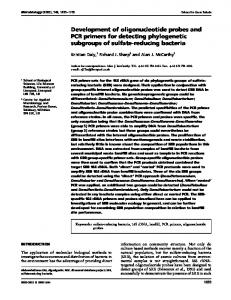
clinical isolates obtained from the United States and Israel and from the Canadian and Texas human patients. The 550-bp type strain product common to each of the isolates examined was cloned and sequenced. A 524-bp consensus sequence was derived. This nucleotide sequence was compared to available sequences in the NCBI GenBank database by using the BLAST function. Subunits of the S. iniae intergenic spacer sequence demonstrated consistent identity and significant homology with 16S-23S intergenic sequences listed for other Streptococcus spp. deposited in GenBank (S. uberis [GenBank accession no., U39765], S. dysgalactiae [U39767], S. agalactiae [L31412 and U39765], S. bovis [U39766], S. pneumoniae [M60763 and L31413], S. salivarius [X83760], and S. thermophilus [U32965]) and for Enterococcus faecalis [L16515]. Furthermore, the distribution of those regions of homology were generally in agreement with that reported by Forsman et al. when they examined sequence data for streptococcal agents of bovine mastitis (i.e., S. agalactiae, S. dysgalactiae, and S. uberis) (7). In brief, the regions of greatest conservation were noted near the 59 termini of the aligned sequences and within the tRNAAla gene present in all of the Streptococcus spp. intergenic spacer sequences thus far reported. More variability was observed at the 39 termini of the reported sequences. Of note was the presence of an approximately 160-bp insertion within the S. iniae spacer, between the homologous 59 terminus and the tRNAAla gene, that is not present in any of the other Streptococcus spp. These regions of variability were the subsequent focus for the development of nested S. iniaespecific primers. The S. iniae 16S-23S rDNA spacer consensus sequence was next searched with the MacVector primer design function to identify nested primers that would define an easily recognizable amplicon (i.e., 200 to 400 bp) and meet the design criteria described above. A set of oligonucleotide primers (59144 and 39516 [Table 3]) was selected that defined a 373-bp subunit of the S. iniae intergenic spacer and annealed to regions outside those identified in the BLAST search as areas of consistent sequence homology among those bacterial genome sequences available in the NCBI database. This primer set consistently produced a single 373-bp PCR amplicon from all of the S. iniae isolates examined (ATCC, fish, and human). No amplicon was produced from any of the other bacterial species examined, with the exception of Streptococcus pyogenes, from which a 250bp amplicon was produced from all 10 of the human clinical isolates tested. Discussion. The advent of PCR and the increasing availability of bacterial genome sequence data has facilitated the development of molecular identification schemes and diagnostics. Accordingly, genome segments with species-specific sequence variability are targeted for sequence analysis and complementary oligonucleotide probe and primer design. The 16S-23S rDNA intergenic spacer has received increased attention as a suitable target for molecular bacterial identification and detection techniques. Two unique oligonucleotide subsequences were identified within the 16S-23S rDNA spacer of S. iniae by aligning the nucleotide sequence of the spacer with sequence data for the homologous region in other Streptococcus spp. deposited in the NCBI GenBank. Oligonucleotide primers complementary to opposing strands of these subsequences allowed PCR amplification of a product unique to S. iniae isolated from a variety of fish species from several geographic locations as well as human patients. Figure 1 diagrammatically illustrates the design scheme. 16S-23S spacer amplicons of unequal length, like those detected in S. iniae, have been observed in other bacteria and
VOL. 36, 1998
NOTES
2781
This work was supported by a United States-Israel Binational Agriculture and Development Fund Grant (IS-2307-93). REFERENCES
FIG. 1. S. iniae-specific PCR primer design scheme. The 16S-23S rDNA intergenic spacer was nonspecifically PCR amplified from the S. iniae rRNA operon. S. iniae-specific oligonucleotide primers 59144 and 39516 define a 373-bp subunit.
have been shown to represent heterogeneity among the spacers within the various copies of the rRNA operon present within the bacterial genome (2, 4, 9, 17, 21). This heterogeneity has made this region useful as a means of differentiating closely related bacterial species. The larger amplicon from S. iniae was chosen for sequencing and analysis, since it consistently appeared both in clinical isolates and in the type strain. The presence of a single tRNAAla gene within the 16S-23S spacer of S. iniae is consistent with what has been observed in other Streptococcus spp. for which sequence data is available. This is in contrast to the presence of tandem tRNAIle and tRNAAla genes observed in some of the rRNA operons of Bacillus subtilis (18) and Escherichia coli (12). Although a nonspecific 16S-23S amplicon polymorphism was observed that distinguished the type strain of S. iniae isolated from an Amazon freshwater dolphin from our clinical isolates from fish and human patients, no polymorphisms were observed among our clinical isolates. Some differentiation of individual S. iniae strains has been successful with pulsed-field gel electrophoresis (20) and ribotyping (5). These S. iniae-specific oligonucleotide primers should be useful in PCR-based methods for rapid and specific identification of suspect bacterial isolates as well as for specific detection of S. iniae in clinical samples from infected fish and human patients. Identification of S. iniae isolated in vitro from human patients is complicated by its phenotypic similarities to other streptococcal pathogens (20). Rapid detection and identification of bacterial cultures collected from diseased fish are hampered by slow growth, which may require 48 h for adequate evaluation of colony morphology and in vitro biochemical characterization. Skin swab cultures collected from local market fish were used in epidemiologic studies associated with the outbreak of S. iniae cellulitis in human patients in Toronto (20). Detection of S. iniae in skin swabs, in particular, can be complicated by overgrowth of other bacterial and fungal flora. The rapidity and sensitivity of PCR coupled with the specificity of the primers designed in this study could circumvent many of these difficulties. Specific detection of S. iniae would be important in efforts to prevent introduction of infected fish into a native fish production unit or the human food supply. Nucleotide sequence accession number. The consensus sequence described in this paper has been deposited with GenBank under accession no. AF048773.
1. Altschul, S. F., W. Gish, W. Miller, E. W. Myers, and D. J. Lipman. 1990. Basic local alignment search tool. J. Mol. Biol. 215:403–410. 2. Barry, T., G. Colleran, M. Glennon, L. K. Dunican, and F. Gannon. 1991. The 16S/23S ribosomal spacer region as a target for DNA probes to identify eubacteria. PCR Methods Appl. 1:51–56. 3. Bentley, R. W., J. A. Leigh, and M. D. Collins. 1993. Development and use of species-specific oligonucleotide probes for differentiation of Streptococcus uberis and Streptococcus parauberis. J. Clin. Microbiol. 31:57–60. 4. Dolzani, L., E. Tonin, C. Lagatolla, and C. Monti-Bragadin. 1994. Typing of Staphylococcus aureus by amplification of the 16S-23S rRNA intergenic spacer sequences. FEMS Microbiol. Lett. 119:167–174. 5. Eldar, A., S. Lawhon, P. F. Frelier, L. Assenta, B. R. Simpson, P. W. Varner, and H. Bercovier. 1997. Restriction fragment length polymorphisms of 16S rDNA and of whole rRNA genes (ribotyping) of Streptococcus iniae strains from the United States and Israel. FEMS Microbiol. Lett. 151:155–162. 6. Eldar, A., Y. Bejerano, and H. Bercovier. 1994. Streptococcus shiloi and Streptococcus difficile: two new streptococcal species causing a meningoencephalitis in fish. Curr. Microbiol. 28:139–143. 7. Forsman, P., A. Tilsala-Timisjarvi, and T. Alatossava. 1997. Identification of staphylococcal and streptococcal causes of bovine mastitis using 16S-23S rRNA spacer regions. Microbiology 143:3491–3500. 8. Gurtler, V., and V. A. Stanisich. 1996. New approaches to typing and identification of bacteria using the 16S-23S rDNA spacer region. Microbiology 142:3–16. 9. Jensen, M. A., J. A. Webster, and N. Strauss. 1993. Rapid identification of bacteria on the basis of polymerase chain reaction-amplified ribosomal DNA spacer polymorphisms. Appl. Environ. Microbiol. 59:945–952. 10. Jurtshuk, R. J., M. Blick, J. Bresser, G. E. Fox, and P. Jurtshuk, Jr. 1992. Rapid in situ hybridization technique using 16S rRNA segments for detecting and differentiating the closely related gram-positive organisms Bacillus polymyxa and Bacillus macerans. Appl. Environ. Microbiol. 58:2571–2578. 11. Kumari, D. N. P., V. Keer, P. M. Hawkey, P. Parnell, N. Joseph, J. F. Richardson, and B. Cookson. 1997. Comparison and application of ribosome spacer DNA amplicon polymorphisms and pulsed-field gel electrophoresis for differentiation of methicillin-resistant Staphylococcus aureus strains. J. Clin. Microbiol. 35:881–885. 12. Lund, E., J. E. Dahlberg, L. Lindahl, S. R. Jaskunuas, P. P. Dennis, and M. Nomura. 1976. Transfer RNA genes between 16S and 23S rRNA genes in rRNA transcription units of E. coli. Cell 7:165–177. 13. Perera, R. P., S. K. Johnson, M. D. Collins, and D. H. Lewis. 1994. Streptococcus iniae associated with mortality of Tilapia nilotica X T. aurea hybrids. J. Aquat. Anim. Health 6:335–340. 14. Pier, G. B., and S. H. Madin. 1976. Streptococcus iniae sp. nov., a betahemolytic streptococcus isolated from an Amazon freshwater dolphin, Inia geoffrensis. Int. J. Syst. Bacteriol. 26:545–553. 15. Sambrook, J., E. F. Fritsch, and T. Maniatis. 1989. Identification of bacterial colonies that contain recombinant plasmids, p. 1.85–1.87. In J. Sambrook, E. F. Fritsch, and T. Maniatis (ed.), Molecular cloning: a laboratory manual, 2nd ed. Cold Spring Harbor Laboratory Press, Cold Spring Harbor, N.Y. 16. Stoffregen, D. A., S. C. Backman, R. E. Perham, P. R. Bowser, and J. G. Babish. 1996. Initial disease report of Streptococcus iniae infection in hybrid striped (sunshine) bass and successful therapeutic intervention with the fluoroquinolone antibacterial enrofloxacin. J. World Aquacult. Soc. 27:420– 434. 17. Tyrrell, G. J., R. H. Bethune, B. Willey, and D. E. Low. 1997. Species identification of enterococci via intergenic ribosomal PCR. J. Clin. Microbiol. 35:1054–1060. 18. Vold, B. S., C. J. Green, N. Narasimhan, M. Strem, and J. N. Hansen. 1988. Transcriptional analysis of Bacillus subtilis rRNA-tRNA operons. J. Biol. Chem. 263:14485–14490. 19. Walker, J., and G. Dougen. 1989. DNA probes: a new role in diagnostic microbiology. J. Appl. Bacteriol. 67:229–238. 20. Weinstein, M. R., M. Litt, D. A. Kertesz, P. Wyper, D. Rose, M. Coulter, A. McGeer, R. Facklam, C. Ostach, B. M. Willey, A. Borczyk, and D. E. Low. 1997. Invasive infections due to a fish pathogen, Streptococcus iniae. N. Engl. J. Med. 337:589–594. 21. Whiley, R. A., B. Duke, J. M. Hardie, and L. M. C. Hall. 1995. Heterogeneity among 16S-23S rRNA intergenic spacers of species within the ‘Streptococcus milleri group’. Microbiology 141:1461–1467. 22. Wilson, K. 1994. Preparation of genomic DNA from bacteria, p. 2.4.1–2.4.5. In F. M. Ausubel, R. Brent, R. E. Kingston, D. D. Moore, J. G. Seidman, J. A. Smith, and K. Struhl (ed.), Current protocols in molecular biology. Greene Publishing Associates and Wiley Interscience, New York, N.Y.