0013-7227/07/$15.00/0 Printed in U.S.A.
Endocrinology 148(7):3532–3540 Copyright © 2007 by The Endocrine Society doi: 10.1210/en.2007-0339
Developmental Programming: Follicular Persistence in Prenatal Testosterone-Treated Sheep Is Not Programmed by Androgenic Actions of Testosterone Teresa Steckler, Mohan Manikkam, E. Keith Inskeep, and Vasantha Padmanabhan Departments of Pediatrics (T.S., M.M., V.P.) and Molecular and Integrative Physiology (V.P.) and the Reproductive Sciences Program (T.S., M.M., V.P.), University of Michigan, Ann Arbor, Michigan 48109-0404; and Division of Animal and Veterinary Sciences (E.K.I.), West Virginia University, Morgantown, West Virginia 26506 Testosterone (T) treatment during early-midgestation (30 –90 d; term is 147 d) leads to reproductive cycle defects. Daily ultrasonography in prenatal T-treated female sheep during the first two breeding seasons revealed an increase in the number of large follicles and follicular persistence. The objective of this study was to determine whether follicular persistence in prenatal T-treated females was programmed by the androgenic actions of T. Pregnant Suffolk ewes were injected with 100 mg (im; twice weekly) of T propionate or dihydrotestosterone (DHT, a nonaromatizable androgen) in cottonseed oil from d 30 to d 90 of gestation. Prior to daily transrectal ovarian ultrasonography, estrus was synchronized with two injections of 20 mg of prostaglandin F2␣ (PGF2␣) given 11 d apart in two consecutive years. In yr 1 ultrasonography began 14 d after PGF2␣, during the presumptive luteal phase, and
I
NAPPROPRIATE STEROID EXPOSURE, in the context of timing, duration, and level of exposure can have detrimental effects on the developing fetus. Many studies documented deleterious effects of exposure to excess prenatal testosterone (T) during a critical period of development (d 30 –90 of gestation) on postnatal neuroendocrine, ovarian, and metabolic function in female sheep (reviewed in Ref. 1), the cumulative effect being early reproductive senescence (2– 4). At the neuroendocrine level, all three feedback systems are affected by prenatal T exposure; decreased responsiveness to estradiol-positive (5–7) and -negative feedbacks (5, 8) and progesterone (P4)-negative feedback (9). A key feature resulting from prenatal exposure to excess T in sheep is the development of enlarged, multifollicular ovaries (10). The multifollicular phenotype may result from failure of follicles to regress (follicular persistence) or increased follicular recruitment. Morphometric analysis of fetal ovaries near term revealed a decrease in percentage of primordial follicles with a reciprocal increase in more advanced follicles, suggestive of enhanced follicular recruitment (11). Our recent studies, through long-term daily transrectal ultrasonography, clearly demonstrated that development of the multi-
First Published Online April 19, 2007 Abbreviations: C, Control; CL, corpus luteum; DHT, dihydrotestosterone; P4, progesterone; PCOS, polycystic ovary syndrome; PGF2␣, prostaglandin F2␣; T, testosterone. Endocrinology is published monthly by The Endocrine Society (http:// www.endo-society.org), the foremost professional society serving the endocrine community.
continued until subsequent ovulation and corpora lutea were detected (10 –13 d). In yr 2, ultrasonography began 2 d before the last PGF2␣ injection and concluded 25 d after the last PGF2␣ injection. Daily changes in appearance and disappearance of ovarian follicles and follicular sizes were assessed. Prenatal DHT, but not prenatal T, treatment increased the total number of follicles by increasing the number of small follicles. Prenatal T, but not DHT, treatment increased (P < 0.05) the number of large follicles with the majority of prenatal T-treated females manifesting follicular persistence. The data indicate that occurrence of large-sized follicles and follicular persistence in prenatal T-treated females are not programmed by androgenic actions but likely are programmed by estrogenic actions stemming from aromatization of T to estradiol. (Endocrinology 148: 3532–3540, 2007)
follicular phenotype is, in part, due to follicular persistence (2). The neuroendocrine, ovarian, and metabolic perturbations resulting from fetal exposure to excess T may be programed by T acting as either an androgen or estrogen because T can be aromatized to estradiol. Machinery is in place for conversion of T to estradiol as well as androgenic and estrogenic programing at the fetal level. Aromatase expression begins early in gestation (d 30) in the placenta (12) and developing gonad (d 32–35) (13, 14). The developing gonad also expresses androgen and estrogen receptors beginning at discrete time points during fetal ontogeny (15). In the context of steroidal programming of reproductive neuroendocrine function, disruption of the positive feedback actions of estradiol (5–7) appears to be programmed by conversion of T to estradiol because such disruptions did not follow prenatal treatment with dihydrotestosterone (DHT, a nonaromatizable androgen) (16, 17). On the contrary, disruption of estradiol-negative feedback appears to be programmed by androgenic actions of T because prenatal treatment with either T or DHT disrupted this feedback loop (5, 8, 18, 19). In contrast to the wealth of knowledge at the neuroendocrine level, it is unclear whether the programming of ovarian follicular recruitment (11) and persistence (2) contributing to multifollicular development (10) is programmed by androgenic or estrogenic actions of T. Studies of West et al. (10) demonstrated that the multifollicular phenotype programmed by prenatal T excess did not occur in prenatal DHT-treated females. It is therefore conceivable that the increased number of large folli-
3532
Steckler et al. • Programming of Ovarian Follicular Persistence
cles and follicular persistence observed in prenatal T-treated females (2) are not programmed by androgenic actions of T. In this study, using a subtractive approach of comparing follicular dynamics in prenatal T- and DHT-treated females, we tested the hypothesis that follicular persistence and increased presence of larger-sized follicles found in prenatal T females are not programmed by its androgenic action. Dissimilar phenotypes between prenatal T- and DHT-treated females would be suggestive of estrogenic programming because DHT cannot be aromatized. Materials and Methods Breeding and prenatal treatment All procedures were approved by the University Animal Care and Use Committee at the University of Michigan. Adult Suffolk (breeding) ewes of proven fertility were purchased locally and moved to a U.S. Department of Agriculture-inspected and University of Michigan Department of Laboratory Animal Medicine-approved farm for breeding in the fall of 2003. Starting 2–3 wk before and continuing until the time of breeding, ewes were group fed daily with 0.5 kg shelled corn and 1.0 –1.5 kg alfalfa hay/ewe to increase energy balance. Breeding ewes were mated to fertility-proven, raddled Suffolk rams. Day of mating was determined by visual confirmation of paint markings left on the rumps of breeding ewes by the raddled rams or by copulation. After breeding, mated ewes were maintained on pasture and supplemented with 1.25 kg alfalfa/brome mix hay/ewe. Starting 6 wk before lambing and continuing until lambing, pregnant ewes were group fed 0.5 kg shelled corn, 2 kg alfalfa hay, and 250 mg aureomycin crumbles (chlortetracycline) per ewe per day. The aureomycin crumbles are given to reduce the incidence of abortion caused by Campylobacter fetus infection. All lambs were born early in 2004 (February 25 to March 18). Just before and for the first 3 d after lambing, ewes and lambs were penned separately from the herd after which all ewes and lambs were group housed. Lactating ewes were provided a ration of 1 kg shelled corn and 2–2.5 kg alfalfa hay/ewe/d. Lambs had ad libitum access to commercial feed pellets (Shur-Gain, Elma, NY) containing 18% crude protein and alfalfa hay. All lambs and ewes were provided with water and minerals ad libitum and were treated regularly with antihelminthics to minimize parasitic infection. To minimize influence of maternal body condition on the developing fetus, ewes were distributed within control (C), T, or DHT treatment groups based on body weight and body condition score. More ewes were assigned to the T and DHT treatment groups (C: T/DHT ratio ⫽ 1:1.2) to maximize the number of treated females born. Initiation of treatment was based on day of mating. Pregnant ewes received twice-weekly injections (100 mg, im) of T propionate (Sigma-Aldrich Corp., St. Louis, MO) or DHT (Steraloids, Inc., Newport, RI) suspended in cottonseed oil (Sigma-Aldrich) from d 30 to 90 of gestation (term 147 d). Administration of this dose of T has been shown to achieve levels of T in female fetuses that are comparable with that of male fetuses (20). Control ewes did not receive vehicle because no differences have been observed in biweekly P4 patterns between females born at the research facility receiving vehicle and those that did not. Female offspring from three C, seven T, and five DHT dams were available for this study (mother is the experimental unit). Additional age-matched control lambs with their mothers (n ⫽ 4) were purchased at approximately 5 wk of age from the source that provided the breeder ewes to be raised in parallel with in-house-born C lambs. No significant differences were observed in the growth trajectory of those born at the facility and the purchased lambs, nor were there differences in timing of onset of puberty.
Maintenance of experimental females All lambs were weaned at 8 wk of age and maintained outdoors at the Sheep Research Facility (Ann Arbor, MI; 42° 18⬘N). The female lambs were fed commercial feed pellets ad libitum until they attained 40 kg body weight and then switched to a diet providing 15% crude protein until 6 months of age. After 6 months of age and during spring/summer months between the first and second breeding seasons, the ewes were maintained on pasture under natural photoperiod. During the winter
Endocrinology, July 2007, 148(7):3532–3540
3533
months, the ewes were fed hay and corn in covered areas. The ewes were provided with water and minerals ad libitum and were treated regularly to minimize parasitic infection.
Progestogenic cycles and ultrasonography Twice-weekly P4 measures in yr 1 began on July 8, 2004, and concluded March 29, 2005. Sample collection in yr 2 began on July 18, 2005, and ceased after November 24, 2005, when the ewes were moved to a terminal study. P4 measures from these twice-weekly samples were used to determine onset of progestogenic cycles (puberty in yr 1 and resumption of cyclicity in yr 2), duration, and end of the breeding season (yr 1 only) and percentage of ewes with repetitive progestogenic cycles of normal duration (16 –18 d; yr 1 and 2). At approximately 36 wk of age, early November of yr 1, estrus was synchronized in all ewes with two 20-mg injections of prostaglandin F2␣ (PGF2␣, 5 mg/ml Lutalyse; Pfizer Animal Health, Kalamazoo, MI) administered 11 d apart. Daily transrectal ultrasonography commenced 14 d after the last PGF2␣ injection and concluded when an ovulatorysized follicle disappeared and subsequent detection of a new corpus luteum (CL) or after 13 d. The scanning period included the late luteal phase of the synchronized estrus and the following follicular phase. In yr 2, as in yr 1, estrus was synchronized with two injections of PGF2␣, and scanning was performed daily throughout the month of November. Daily transrectal ultrasonography began 2 d before the second PGF2␣ injection and concluded when either a new CL was detected after the second follicular phase or 25 d after the last PGF2␣ injection. The scanning period encompassed the synchronized follicular period, the following luteal phase, and a second follicular phase. During ultrasonographic examination, sheep were restrained in a crate in the standing position while both ovaries were examined using a rigid-mounted 7.5 MHz linear-array transducer connected to an Aloka SSD-900V ultrasound machine (Aloka Co. Ltd., Wallington, CT). A DCR-TRV33 (Sony Corp. of America, New York, NY) was used to record the digital video output for each ovary to document follicular and luteal changes. To minimize subjectivity of measures, two investigators were present during all ultrasonographic examinations. The diameter and relative positions of all antral follicles 2 mm or larger in diameter and CL on both ovaries were sketched daily and used to assess changes in follicular dynamics. Follicles were tracked across successive days using landmark structures, e.g. CL in cycling females and the largest follicles in anovulatory prenatal T-treated females. Transrectal ultrasonography is a wellaccepted approach (21–24) that provides a better indication of follicular/CL changes than limited time sampling by laparotomy or laparoscopy. The approach has been used extensively to document growth and regression of follicles (21–24). Daily blood samples were collected during both scanning periods for P4 measurements to confirm CL presence and functionality.
RIAs Plasma concentrations of P4 were measured using a commercial RIA kit (Coat-A-Count P4; Diagnostic Products Corp., Los Angeles, CA). Validation of this assay for sheep plasma has been described elsewhere (25). Sensitivity of this assay was 0.083 ⫾ 0.02 ng/ml (mean ⫾ sem). The intraassay coefficients of variation based on two quality control pools measuring 1.45 ⫾ 0.05 and 12.99 ⫾ 0.31 ng/ml were 4.3 and 3.0%, respectively. The interassay coefficients of variation for the same quality control pools were 10.8 and 8.2%, respectively.
Statistical analysis For analyses of cycle data, a progestogenic cycle was defined as P4 concentrations 0.5 ng/ml or greater for a minimum of two consecutive twice-weekly samples. The duration of each uninterrupted progestogenic cycle (some cycles were interrupted with PGF2␣ for inclusion in neuroendocrine studies) was calculated from the day of P4 rise greater than 0.5 ng/ml to the day when P4 concentration fell below this value. Only progestogenic cycles of 16 –18 d duration were considered normal. End of the breeding season was defined as the first of five consecutive samples with P4 concentrations less than 0.5 ng/ml. Duration of the breeding season was defined as the difference between age at onset and end of the breeding season. The number of normal duration (16 –18 d)
3534
Endocrinology, July 2007, 148(7):3532–3540
cycles was determined for each ewe taking into account only the uninterrupted progestogenic cycles. Ewes with at least 75% of uninterrupted cycles of normal duration (16 –18 d) was considered to be cycling normally. Onset, end, and duration of the first breeding season were analyzed by ANOVA. At the onset of the first breeding season, seven C, seven prenatal T-, and five prenatal DHT-treated females were available to study. One prenatal T-treated sheep did not achieve puberty during the first year and was excluded from first breeding season calculations of onset, end, and duration. Two prenatal DHT ewes were euthanized before the end of the first breeding season due to a systemic infection they developed. Because prenatal T-treated females were studied extensively in detail earlier (2), to conserve resources, two random C and prenatal T-treated females were euthanized for tissue procurement before the end of the first breeding season. Therefore, for the first breeding season, onset is based on seven C, seven prenatal T-, and five prenatal DHT-treated females, whereas end and duration is based on five C, four prenatal T-, and three prenatal DHT-treated females. Second breeding season dynamics are based on five C, five prenatal T-, and three prenatal DHT-treated females. During the first breeding season, the proportion of ewes with greater than 75% cycles of normal duration and those without was compared by Fisher’s exact test. For both years, only follicles that grew to 3 mm or greater in diameter and present on the ovary for 2 d or more were used to examine follicular dynamics as reported by Ginther et al. (26) and validated by Schrick et al. (27). Follicles 2 mm or greater and less than 3 mm were included only in calculation of the total number of follicles. Follicles were classified into following size classes: 3– 4 mm or less, greater than 4 to 8 mm or less, and greater than 8 mm in diameter. These size classes, 3– 4 mm or less, greater than 4 to 8 mm or less, and greater than 8 mm, correspond to gonadotropin-dependent recruited follicles, those selected to become ovulatory-sized follicles, and follicles larger than ovulatory size follicles, respectively (28 –30). Follicle duration, calculated from 3 mm through growth to maximum diameter and back to 3 mm, was determined for all follicles 3 mm or greater. Not all follicles were observed from 3 mm through growth and back to 3 mm, so duration from 3 mm to 3 mm was estimated for all follicles 4 mm or greater on the first and last scanning day. The estimated follicle duration was based on the assumption that follicles grow and regress at a constant rate of approximately 1 mm/d (26, 27). The number, diameter, and duration of follicles were calculated for both years. Follicles were considered persistent if they were present on the ovary for 12 d or longer (31). The proportion of ewes that ovulated,
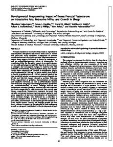
FIG. 1. Patterns of twice-weekly plasma P4 from three each of C, prenatal T-, and DHT-treated sheep during the first two breeding seasons (yr 1 and 2) are shown in the top panels. Arrows indicate PGF2␣ administration whereas the shaded area indicates an artificially induced progestogenic cycle (part of neuroendocrine studies). Onset, duration, and end of the breeding season and percentage of ewes with regular cycles during the first breeding season are presented in the bottom histograms (C: white columns; prenatal T: black columns; prenatal DHT: striped columns). Start and end of the breeding season for those ewes that had elevated concentrations of P4 (⬎0.5 ng/ml) at the initiation and/or conclusion of twice-weekly sampling was assigned first and last day of sampling as start and end of breeding season, a conservative estimate. Asterisks, Significant differences from C (*, P ⬍ 0.05; **, P ⬍ 0.01).
Steckler et al. • Programming of Ovarian Follicular Persistence
the diameter and duration of ovulatory follicles, and number of CL were also calculated for both years. Duration of ovulatory follicles was calculated from the time they were 3 mm until ovulation, which was confirmed by presence of a new CL with a corresponding increase in plasma concentrations of P4. Follicular counts of individual sizes and within size classes during the 13-d (yr 1) and 25-d (yr 2) scanning periods were modeled as a function of treatment, using a Poisson regression (32), which used the log of the number of days scanned as an offset to take into account the differing number of days scanned for each ewe. The percentage of missed ovarian scans in yr 1 and 2 were 3.4 and 3.2%, respectively. However, one of the prenatal T-treated females was excluded from all analyses that relied on ovarian observation due to repeated scanning difficulties; upon autopsy, this ewe was found to have many adhesions and a rather large, fluidfilled uterus. Thus, transrectal ultrasonography was performed in yr 1 on seven C, six prenatal T-, and five prenatal DHT-treated females and five C, four prenatal T-, and three prenatal DHT-treated females in yr 2. A one-way ANOVA was carried out to compare the mean number and duration of follicles for C, prenatal T-, and DHT-treated animals. Post hoc tests were carried out using the Dunnett adjustment for multiple comparisons (C vs. prenatal T- and prenatal DHT-treated females). Because some of the follicles began growing before the scanning period, and others continued to persist after the end of the scanning period, a survival analysis was used, which allowed left-truncated and right-truncated observations, using Proc Lifereg in SAS (33). The proportion of ewes that ovulated was compared by Fisher’s exact test for both years. Diameter and duration of ovulatory follicles and number of CL from ewes that ovulated were analyzed by ANOVA. Ovulatory and CL data were collected from one ovulatory period in yr 1 but two ovulatory periods during the 25-d scanning period in yr 2. All analyses were performed using SAS for Windows (release 9.1.3; SAS Institute Inc., Cary, NC).
Results Progestogenic cycles in yr 1 and 2
Figure 1 (top panel) shows progestogenic cycles of C, prenatal T-, and DHT-treated females during the first and second breeding season. During both years more than 75% of uninterrupted cycles examined in C ewes were of normal duration. In contrast, most prenatal T-treated females
Steckler et al. • Programming of Ovarian Follicular Persistence
Follicular dynamics in C, prenatal T-, and DHT-treated sheep
Follicular dynamics (follicles ⱖ 3 mm and present for ⱖ 2 d) from both ovaries of two representative ewes of each treatment group (C, prenatal T-, and DHT-treated ewes) during the 13-d scanning period (yr 1) are depicted in Fig. 2. Follicular dynamics from left and right ovaries of two representative C, prenatal T-, and DHT-treated females for the 25-d scanning period (yr 2) are shown in Fig. 3. P4 patterns are shown in the background. Follicular diameter and duration appear to be clearly increased in the prenatal T-treated females when compared with C. Within the prenatal T-treated group, follicular diameter was greater in the females with constant low P4 (e.g. ewe 415) than those with normal P4 (e.g. ewe 437). The total number of 3-mm follicles from both ovaries ranged from 15 to 20 in C females vs. 25 to 30 in the prenatal DHT-treated females (histogram).
3535
Control
8
Ewe 445
Ewe 442
4 2
Follicle diameter (mm)
showed considerable cycle disruptions, which included absent or cycles of longer duration. In yr 1, even though all five prenatal DHT-treated females had P4 increases, three of the five prenatal DHT-treated females had high P4 at the initiation of sample collection (July 8) and had persistently elevated P4 during part of the first breeding season. This acyclic pattern of P4 was again evident in yr 2. All three remaining prenatal DHT-treated females showed P4 increases in yr 2, but only one of these three exhibited repetitive cycles of normal duration. The remaining two prenatal DHT-treated ewes, as in yr 1, manifested persistently elevated concentrations of P4. Summary statistics for onset, duration, and end of the first breeding season are shown in Fig. 1 (bottom panels). Date (mean ⫾ se) of onset of the first breeding season (first P4 increase) was October 8, 2004 ⫾ 3.3 d, October 13, 2004 ⫾ 18.8 d, and August 15, 2004 ⫾ 19.9 d for C, prenatal T-, and prenatal DHT-treated females, respectively. The elevated P4 at start of sampling in three of the five prenatal DHT-treated females tended to advance (P ⫽ 0.06) the timing of P4 increase relative to C females. Duration of the first breeding season, on the basis of elevated P4, was increased for the prenatal DHT- but not the prenatal T-treated females (C vs. DHT: P ⫽ 0.02). As seen with onset and duration, the end of the first breeding season was similar for C (February 20, 2005 ⫾ 7.8 d) and prenatal T-treated (February 7, 2005 ⫾ 24.9 d) females but significantly delayed in the prenatal DHT-treated group (C vs. DHT: P ⬍ 0.01); all three surviving prenatal DHT-treated females had high P4 on the last sample day (March 28, 2005). Onset of the second breeding season did not differ between C (September 12, 2005 ⫾ 9.5 d) and prenatal T-treated (August 23, 2005 ⫾ 11.3 d) females. In yr 2, two of the three prenatal DHT-treated sheep had elevated P4 in the first sample taken on July 17, 2005. Whereas 100% of C exhibited regular cycles, only 40% prenatal DHT-treated (C vs. DHT: P ⬍ 0.05) and 57% of prenatal T-treated females (C vs. T; NS) exhibited regular cycles (Fig. 1, bottom panel). As reported previously (2), there was a numerical increase from yr 1 to 2 in the percentage (43 and 60%, respectively) of prenatal T-treated females failing to exhibit regular cycles, but this did not achieve statistical significance.
Endocrinology, July 2007, 148(7):3532–3540
Prenatal T
8
Ewe 437
Ewe 413
4 2
Prenatal DHT
8
Ewe 410
Ewe 402
4 2
1
6
12
1
6
12
Days scanned
FIG. 2. Ovarian follicular dynamics of both ovaries from two each of C, prenatal T-, and DHT-treated sheep in yr 1. Each line represents only one follicle and both ovaries are shown within a panel. Only follicles that were 3 mm or greater and present for a minimum of 2 d are shown. Arrows indicate follicles that ovulated. Ovulatory follicles were not detected in one C (ewe 445: P4 fell at the expected time but an ovulation was not detected), one prenatal T- (ewe 437: persistently low levels), and both DHT-treated sheep (persistently elevated levels of P4 during the scanning period).
Number of follicles
The number of follicles in different diameter classes for all treatment groups in yr 1 and 2 is summarized in Fig. 4. During the first breeding season, more total number of follicles (ⱖ3 mm) were observed on the ovaries of prenatal DHT-treated females than on the ovaries of C (P ⬍ 0.05). This increase appears to be due to increased number of follicles in the 3- to 4-mm range (P ⬍ 0.07). During the second breeding season, the trend for increased number of follicles (P ⫽ 0.08) in the prenatal DHT-treated group continued, even though only three sheep were available for ultrasonography. As in yr 1, the increase in total number of follicles in yr 2 appears to be due to a numerical increase of 3- to 4-mm follicles (C: 33.9 ⫾ 4.2 vs. DHT: 50.7 ⫾ 7.0). There was a greater number (P ⬍ 0.05) of 7-mm follicles in the prenatal T-treated females, compared with C in yr 1 (see inset in Fig. 4). Follicles greater than 8 mm in size were present predominantly in the prenatal T-treated females during yr 2 of life. None of the controls had a follicle greater than 8 mm during yr 2 of life. One of the three surviving prenatal DHT-treated females had a single 9-mm-size follicle. Follicular persistence
The number, diameter, and duration of persistent (duration ⱖ 12 d) follicles are shown in Fig. 5. During the first breeding season, 33% of prenatal T-treated females had persistent follicles as opposed to 14% of C females. No prenatal DHT-treated sheep had persistent follicles. In yr 2 no C, one
3536
Endocrinology, July 2007, 148(7):3532–3540
Steckler et al. • Programming of Ovarian Follicular Persistence
FIG. 3. Ovarian follicular dynamics determined by ultrasonography for 25 d in both ovaries of two each of C, prenatal T-, and DHT-treated (C: white columns; prenatal T: black columns; prenatal DHT: striped columns) sheep in yr 2 overlaid on daily P4 (shaded areas). Each line represents growth and disappearance of one follicle, and follicles from the left and right ovaries are shown in separate panels. Only follicles that reached a diameter of 3 mm and persisted for at least 2 d are shown. One prenatal T-treated ewe (ewe 415) did not ovulate, but a persistent follicle luteinized (double-lined arrow). One prenatal DHT-treated sheep (ewe 410) failed to ovulate and had persistently elevated levels of P4 during the entire scanning period. The inlaid histogram shows the total number of 3-mm follicles present for less than 2 d from both ovaries for the given ewe during the entire scanning period. Prenatal T-treated females had larger-sized follicles that lasted on the ovary longer as compared with C sheep. The vertical dashed line represents the second of two PGF2␣ injections, administered 11 d apart.
Steckler et al. • Programming of Ovarian Follicular Persistence
Endocrinology, July 2007, 148(7):3532–3540
3537
FIG. 5. Number, diameter, and duration of persistent (lasting longer than 12 d) follicles in ovaries of C (white columns), prenatal T- (black columns), and DHT-treated (striped columns) sheep in yr 1 and 2. Only one C sheep in yr 1 and one prenatal DHT-treated sheep in yr 2 had a single persistent follicle. Follicles in prenatal T-treated females persisted on the average for 15 and 19 d in yr 1 and 2, respectively. The single persistent follicle in one C in yr 1 and one prenatal DHT-treated female in yr 2 persisted for 13 d. FIG. 4. Total number of follicles and follicles in size classes of 3– 4 mm (gonadotropin-dependent recruited follicles), greater than 4 – 8 mm (follicles selected to become ovulatory sized), and greater than 8 mm (larger than ovulatory sized follicles) detected by ultrasonography in ovaries of C, prenatal T-, and DHT-treated sheep in yr 1 (13 d scan; C: white columns; prenatal T: black columns; prenatal DHT: striped columns) and yr 2 (23–25 d scan). The inlaid histogram shows the total number of 7-mm-size follicles. Asterisks, Significant differences from C (P ⬍ 0.05). Only one C in yr 1 and one prenatal DHT-treated female in yr 2 had follicles greater than 8 mm.
prenatal DHT-treated, and 75% of prenatal T-treated females had persistent follicles. Unlike C and prenatal DHT-treated females, prenatal T-treated females had persistent follicles during both scanning periods. There was an increase in number of persistent follicles in the prenatal T-treated group from yr 1 to yr 2 (P ⫽ 0.01). The average diameter of the persistent follicle present on the ovaries of prenatal T-treated females was greater (P ⬍ 0.05) in yr 2 (9.4 ⫾ 0.9 mm) than yr 1 (7.6 ⫾ 0.1 mm). Duration of these persistent follicles in yr 1 and 2 was 15.5 ⫾ 1.5 and 18.7 ⫾ 2.6 d, respectively, and did not differ significantly. There was one persistent follicle of 7.6 mm size in one prenatal DHT sheep in yr 2, which persisted for 13 d. Ovulatory follicle and CL dynamics
Scanning during the first breeding season began during the late luteal phase (14 d after the second PGF2␣ injection)
of the synchronized estrous cycle and continued through the subsequent follicular phase. At the start of the scanning period, 100% of C, 80% of prenatal T-, and 67% of prenatal DHT-treated sheep had CL present. More CL were present on the ovaries of both prenatal T- and DHT-treated females relative to C (P ⬍ 0.05, P ⬍ 0.01, respectively; Table 1) when scanning began. During the scanning period, 71% of C, 67% of prenatal T-, and 40% of prenatal DHT-treated females ovulated. Of the two prenatal T-treated females that failed to ovulate, one had persistently low P4, indicative of anovulatory condition. Three prenatal DHT-treated sheep that did not ovulate had either persistently elevated (n ⫽ 2) or low (n ⫽ 1) P4 during the entire scanning period. Diameter and duration of ovulatory follicles and number of CL formed in those that ovulated in yr 1 were similar between C, prenatal T-, and prenatal DHT-treated sheep. Scanning during yr 2 (second breeding season) encompassed two potential ovulatory periods, the first one immediately after PGF2␣ synchronization and a second one after the ensuing luteal phase. Concentrations of P4 were high on the day of the second PGF2␣ in all C and prenatal DHTtreated but in only three of five prenatal T-treated sheep. After PGF2␣, P4 declined in all C, two prenatal T-, and two prenatal DHT-treated sheep (Table 1). Overall, 100, 50, and 67% of C, prenatal T-, and prenatal DHT-treated sheep, re-
3538
Endocrinology, July 2007, 148(7):3532–3540
Steckler et al. • Programming of Ovarian Follicular Persistence
TABLE 1. Ovulatory follicle and CL characteristics Control
Prenatal T
Prenatal DHT
Year 1 n No. CL per ewe at start No. of ewes that ovulated Persistent high or low P4 Diameter of ovulatory follicled Duration of ovulatory follicled No. CL per ewe after ovulationd
7 1.0 ⫾ 0 5 0 5.7 ⫾ 0.1 5.6 ⫾ 1.3 1.0 ⫾ 0
6a 1.75 ⫾ 0.25b 4 2 6.0 ⫾ 0.6 5.5 ⫾ 1.1 1.5 ⫾ 0.3
5 2.25 ⫾ 0.25c 2 3 5.8 6.0 1.5
Year 2 n No. CL per ewe at start Synchronization achieved with PGF2␣ Persistent high or low P4 No. of ewes ovulating Diameter of ovulatory folliclea Duration of ovulatory folliclea No. CL per ewe after ovulationa
5 1.4 ⫾ 0.2 5 0 5 5.6 ⫾ 0.2 5.8 ⫾ 0.3 1.0 ⫾ 0
4 1.5 ⫾ 0.5 2 2 2 6.3 6.5 2.3
3 2.0 ⫾ 0.0 2 1 2 6.5 6.0 2.0
a
Excludes one ewe that did not achieve puberty. P ⬍ 0.05, significant difference from C. c P ⬍ 0.01, significant difference from C. d Derived from those that ovulated. b
spectively, ovulated. The diameter and duration of the ovulatory follicles and average number of CL per sheep in those that ovulated were similar across treatment groups. Discussion
Findings from this study demonstrate the divergence in programing effects of prenatal T and DHT treatment on ovarian follicular dynamics and cyclicity with prenatal T treatment programing follicular persistence and prenatal DHT programming increased number of small follicles. Differential effects of prenatal T and DHT programming in the context of androgenic or estrogenic programing of follicular growth and development and its implications are discussed below. Consistent with our earlier findings (2), follicles persisted longer than 12 d in 33% of prenatal T-treated animals in yr 1 and 75% of prenatal T animals in yr 2. The average diameter of follicles that persisted in prenatal T-treated females was 7.6 ⫾ 0.1 mm in yr 1. Furthermore, as reported earlier (2), follicles greater than 8 mm in size were clearly evident by yr 2 of life in prenatal T-treated females. Follicles in prenatal T-treated sheep reached sizes up to 12 mm that persisted on an average for approximately 15 d in yr 1 and approximately 19 d in yr 2. Prenatal T-treated animals in this study differed from those in previous study (2) in that follicles greater than 8 mm in size were not evident in yr 1 of life. However, a greater number of follicles in the 7-mm range was present in the prenatal T-treated females (see inset in Fig. 4). A reduction in number of 3- to 4-mm follicles in yr 2 was also not seen in the present study as evidenced in prenatal T-treated females of the previous study (2). Such differences between the current study and our previous study (2) may relate to: 1) differences in severity of disruptions and/or the percentage of animals exhibiting disruptions in the two studies, 2) geneenvironment interaction, and/or 3) maternal body condition; the breeder ewes in this study were of better body condition than the previous study. Irrespective of such differences, findings from this study confirm our earlier findings (2) that
prenatal T excess programs follicular persistence and accumulation of large-sized follicles. In contrast, follicular persistence was not evident in any of the prenatal DHT-treated females (n ⫽ 5) in yr 1 of life and two of the three surviving prenatal DHT-treated females in yr 2. One prenatal DHT-treated female in yr 2 of life had a single follicle of 7.6 mm size that persisted for 13 d, 1 d longer than the cut-off (12 d) used for estimating follicular persistence. Existence of an occasional large persistent follicle such as those seen in one control (yr 1) and one prenatal DHT (yr 2) sheep may be similar to what has been reported in normoandrogenic women (34, 35). The subtractive method implemented in this study comparing follicular dynamics of prenatal T- and prenatal DHTtreated females across 2 yr provides evidence that programming of follicular persistence (evidenced both years) in prenatal T-treated females but not prenatal DHT-treated females (except one DHT female in yr 2) is likely not programmed by androgenic actions of T but rather by conversion of T to estradiol. Clearly the requisite machinery is in place at both the placental and fetal ovarian levels to facilitate conversion of T to estradiol and initiate estrogenic signaling. The fetal villi of the ovine cotyledon express aromatase as early as d 30 – 40 of gestation (12). In addition, aromatase mRNA is expressed in the fetal ovary as early as d 32–35 of gestation (13, 14). To orchestrate estrogen signaling, estrogen receptor- mRNA and protein and estrogen receptor-␣ protein are expressed by the fetal ovary as early as d 30 of fetal life (15) when T treatment is initiated. Our earlier morphometric studies that documented multifollicular phenotype in prenatal T-treated but not DHT-treated females (10) are consistent with estradiol programming of increased follicular size and persistence. Development of paraovarian cysts after prenatal exposure to estrogens in rodents (36, 37) is also supportive of this premise. Prenatal DHT-treated females, although not characterized by follicular persistence, had a larger number of follicles in
Steckler et al. • Programming of Ovarian Follicular Persistence
the 3- to 4-mm range in yr 1. In yr 2, despite fewer prenatal DHT-treated females available to study, a numerical increase in the number of 3- to 4-mm follicles was still evident. However, the small number of prenatal DHT-treated females available for study in the second breeding season did preclude assessing the severity of follicular defects with advancing age. Irrespective of this, the increased presence of 3to 4-mm-size antral follicles in both years (with the same trend in yr 2) that do not persist in prenatal DHT-treated females is consistent with androgenic programming of early follicular differentiation (38, 39). Postnatally androgens are implicated in early follicular differentiation with both T and DHT treatment significantly increasing the number of small antral follicles (38, 39). It remains to be determined whether androgens also play a modulatory role in developmentally programming such a phenotype. Earlier studies have implicated low P4 milieu in the development of persistent follicles (40 – 42). Comparison of follicular dynamics in prenatal T- and DHT-treated females indicates that the size of follicular growth during estrous cycles in these two groups may reflect their P4 environment. Consistent with this premise, prenatal T-treated females often had subluteal or low P4 and larger than ovulatory-sized follicles that failed to ovulate. Within the prenatal T-treated group, the diameter of the largest follicle was greater when P4 was low (Fig. 3, ewe 415), compared with the diameter of the largest follicle when P4 was normal (Fig. 3, ewe 437). Development of these large follicles in prenatal T-treated females may be a consequence of hypergonadotropism (5, 8) that these ewes manifest because LH administration has been shown to enhance development of large follicles (43). In contrast, most prenatal DHT-treated females studied had persistently elevated P4 but increased numbers of smaller (3– 4 mm) follicles. Because subluteal levels of progesterone promote development of persistent follicles while reducing the number of small follicles (40 – 42) and administration of supraphysiological levels of progesterone increases the number of small follicles in hypophysectomized hamsters (44), it is conceivable that sustained elevated levels of progesterone in the prenatal DHT-treated ewes may have contributed to the increased number of small follicles and preventing development of persistent follicles. To what extent the changes in follicular dynamics of prenatal T- and DHT-treated females influence maintenance of cyclic function is unclear. As reported previously (2), prenatal T treatment resulted in oligo- or anovulation, a feature prenatal T-treated females share with women with polycystic ovary syndrome (PCOS) (1, 45– 47), and luteal defects. Luteal defects were also evident in prenatal DHT-treated females, even in the absence of follicular persistence. The early increase in P4 during pubertal maturation and start of second breeding season and persistently elevated P4 during the breeding season in the prenatal DHT-treated females may be reflective of P4 release from a luteinized follicle rather than CL. These findings in concert with the increase in number of small follicles are reminiscent of premature follicle luteinization, such as that seen in small follicles of PCOS women (48, 49). Alternatively, the luteolytic mechanism was aberrant sustaining the secretion of P4. This is supported by the failure of some prenatal DHT-treated sheep to reduce P4
Endocrinology, July 2007, 148(7):3532–3540
3539
after administration of PGF2␣. CL, not luteinized follicles, were also observed to remain on the ovary of prenatal DHTtreated females during the entire ultrasonographic scanning period in yr 2. Another possibility is that the persistent P4 may be of adrenal origin. Whereas this possibility was not tested in this study, adrenals of prenatal T-treated females at fetal d 140 (11) and 2 yr (50) were larger, suggestive of altered adrenal function (11). The relevance of programming of follicular persistence in prenatal T-treated females as they relate to PCOS has been discussed in detail in an earlier paper (2). Importantly, follicular persistence such as that seen in the prenatal T-treated sheep is likely to influence the quality of oocytes and impair fertility. Recently using the fetal 60- to 90-d T treatment paradigm, we found that prenatal T excess compromised fertility (51). The mechanism by which prenatal T treatment programs adult follicular persistence is paradoxical. A possibility to consider is that exposure to consistently high concentrations of aromatized estradiol beyond when the decline in estradiol normally occurs during fetal development (d 55) (13, 14) may aberrantly program expression of key genes in the differentiating follicles, predisposing them for enhanced growth and persistence later in life. Recent studies suggest that environmental estrogens can induce epigenetic alterations in the developing fetus culminating in atypical adult responses (52–54). In summary, findings from this study provide evidence that androgens and estrogens program different aspects of follicular development and that the follicular persistence in prenatal Ttreated females is not programmed by androgenic actions of T but likely facilitated via aromatization of T to estradiol. Acknowledgments We are grateful to Mr. Douglas Doop for providing quality care and maintenance of animals used in this study. We thank Mrs. Carol Herkimer, Ms. Eila Roberts, Ms. Erica LaVire, Mr. Michael Zakalik, Ms. Allie Spencer, and Mr. Jim Lee for assistance with twice-weekly blood sampling and Mrs. Pam Olton, Mrs. Carol Herkimer, and Ms. Meha Muralidharan for performing progesterone assays. Received March 12, 2007. Accepted April 11, 2007. Address all correspondence and requests for reprints to: Vasantha Padmanabhan, Departments of Pediatrics, Obstetrics, and Gynecology, and Molecular and Integrative Physiology and the Reproductive Sciences Program, University of Michigan, 300 North Ingalls Building, Room 1109, Ann Arbor, Michigan 48109-0404. E-mail:
[email protected]. This work was supported by U.S. Public Health Service Grant P01HD44232 (to V.P.). Disclosure Statement: The authors have nothing to disclose.
References 1. Padmanabhan V, Manikkam M, Recabarren S, Foster D 2006 Prenatal testosterone excess programs reproductive and metabolic dysfunction in the female. Mol Cell Endocrinol 246:165–174 2. Manikkam M, Steckler TL, Welch KB, Inskeep EK, Padmanabhan V 2006 Fetal programming: prenatal testosterone treatment leads to follicular persistence/luteal defects: partial restoration of ovarian function by cyclic progesterone treatment. Endocrinology 147:1997–2007 3. Birch RA, Padmanabhan V, Foster DL, Robinson JE 2003 Prenatal programming of reproductive neuroendocrine function: fetal androgen exposure produces progressive disruption of reproductive cycles in sheep. Endocrinology 144:1426 –1434 4. Clarke IJ, Scaramuzzi RJ, Short RV 1977 Ovulation in prenatal androgenized ewes. J Endocrinol 73:385–389 5. Wood RI, Foster DL 1998 Sexual differentiation of reproductive neuroendocrine function. Rev Reprod 3:130 –140
3540
Endocrinology, July 2007, 148(7):3532–3540
6. Sharma TP, Herkimer C, West C, Ye W, Birch R, Robinson JE, Foster DL, Padmanabhan V 2002 Fetal programming: prenatal androgen disrupts positive feedback actions of estradiol but does not affect timing of puberty in female sheep. Biol Reprod 66:924 –933 7. Unsworth WP, Taylor JA, Robinson JE 2005 Prenatal programming of reproductive neuroendocrine function: the effect of prenatal androgens on the development of estrogen positive feedback and ovarian cycles in the ewe. Biol Reprod 72:619 – 627 8. Sarma HN, Manikkam M, Herkimer C, Dell’Orco J, Welch KB, Foster DL, Padmanabhan V 2005 Fetal programming: excess prenatal testosterone reduces postnatal LH, but not FSH responsiveness to estradiol negative feedback in the female. Endocrinology 146:4281– 4291 9. Robinson JE, Forsdike RA, Taylor JA 1999 In utero exposure of female lambs to testosterone reduces the sensitivity of the GnRH neuronal network to inhibition by progesterone. Endocrinology 140:5797–5805 10. West C, Foster DL, Evans NP, Robinson J, Padmanabhan V 2001 Intrafollicular activin availability is altered in prenatally androgenized lambs. Mol Cell Endocrinol 185:51–59 11. Steckler T, Wang J, Bartol FF, Roy SK, Padmanabhan V 2005 Fetal programming: prenatal testosterone treatment causes intrauterine growth retardation, reduces ovarian reserve and increases ovarian follicular recruitment. Endocrinology 146:3185–3193 12. Leung ST Reynolds TS, Wathes DC 1998 Regulation of oxytocin receptor in the placentome capsule throughout the pregnancy in the ewe: the possible role of estrogen receptor, progesterone receptor and aromatase. J Endocrinol 158:173–181 13. Juengel LJ, Sawyer HR, Smith PR, Quirkle LD, Heath DA, Lun S, Wakfield St. J, McNatty KP 2002 Origins of follicular cells and ontogeny of steroidogenesis in ovine fetal ovaries. Mol Cell Endocrinol 191:1–10 14. Quirke LD, Juengel JL, Tisdall DJ, Lun S, Heath DA, McNatty KP 2001 Ontogeny of steroidogenesis in the fetal sheep gonad. Biol Rep 65:216 –228 15. Juengel JL, Heath DA, Quirke LD, McNatty KP 2006 Oestrogen receptor ␣ and , androgen receptor and progesterone receptor mRNA and protein localization within the developing ovary and in small growing follicles of sheep. Reproduction 131:81–92 16. Masek KS, Wood RI, Foster DL 1999 Prenatal dihydrotestosterone differentially masculinizes tonic and surge modes of luteinizing hormone secretion in sheep. Endocrinology 140:3459 –3466 17. Astapova O, Herkimer CH, Olton PO, Lee JS, Flak J Padmanabhan V 2006 Developmental Programming: defects in preovulatory estradiol rise and gonadotropin surge dynamics in prenatal testosterone-treated ewes are not programmed by androgenic action of testosterone. Biol Reprod Special Issue p 77 (Abstract 28) 18. Foster DL, Jackson LJ, Padmanabhan V 2006 Programming of GnRH feedback controls timing puberty and adult reproductive activity. Mol Cell Endocrinol 254 –255:109 –119 19. Lee JS, Aizenberg E, Djoumbi D, Foster DL, Padmanabhan V 2005 Fetal programming of the postnatal responsiveness of LH to estradiol negative feedback in sheep: time and duration of exposure and quality of prenatal steroid. Biol Reprod Special Issue p 227 (Abstract 660) 20. Wood RI, Ebling FJ, I’Anson H, Bucholtz DC, Yellon SM, Foster DL 1991 Prenatal androgens time neuroendocrine sexual maturation. Endocrinology 128:2457–2468 21. Ginther OJ, Kot K 1994 Follicular dynamics during the ovulatory season in goats. Theriogenology 42:987–1001 22. Ginther OJ, Beg MA, Bergfelt DR, Donadeu FX, Kot K 2001 Follicle selection in monovular species. Biol Reprod 65:638 – 647 23. Lopez-Sebastian A, Gonzalez de Bulnes A, Santiago Moreno J, GomezBrunet A, Townsend EC, Inskeep EK 1997 Patterns of follicular development during the estrous cycle in monovular Merino del Paris ewes. Anim Reprod Sci 48:279 –291 24. Duggavathi R, Bartlewski PM, Barrett DM, Gratton C, Bagu ET, Rawlings NC 2004 Patterns of antral follicular wave dynamics and accompanying endocrine changes in cyclic and seasonally anestrous ewes treated with exogenous ovine follicle-stimulating hormone during the inter-wave interval. Biol Reprod 70:821– 827 25. Padmanabhan V, Evans NP, Dahl GE, McFadden KL, Mauger DT, Karsch FJ 1995 Evidence for short or ultrashort loop negative feedback of GnRH secretion. Neuroendocrinology 62:248 –258 26. Ginther OJ, Kot K, Wiltbank MC 1995 Association between emergence of follicular waves and fluctuations in FSH concentrations during the estrous cycle in ewes. Theriogenology 43:689 –703 27. Schrick FN, Surface RA, Pritchard JY, Dailey RA, Townsend EC, Inskeep EK 1993 Ovarian structures during the estrous cycle and early pregnancy in ewes. Biol Reprod 49:1133–1140 28. Campbell BK, Scaramuzzi RJ, Webb R 1995 Control of antral follicle development and selection in sheep and cattle. J Reprod Fertil 49(Suppl):335–350 29. Driancourt MA 2001 Regulation of ovarian follicular dynamics in farm animals. Implications for manipulation of reproduction. Theriogenology 55:1211–1239
Steckler et al. • Programming of Ovarian Follicular Persistence
30. Hunter MG, Robinson RS, Mann GE, Webb R 2004 Endocrine and paracrine control of follicular development and ovulation rate in farm species. Anim Reprod Sci 82– 83:461– 477 31. Flynn JD, Duffy P, Boland MP, Evans AC 2000 Progestagen synchronisation in the absence of a corpus luteum results in the ovulation of a persistent follicle in cyclic ewe lambs. Anim Reprod Sci 62:285–296 32. Allison PD 1999 Logistic regression using the SAS system: theory and application. Cary, NC: SAS Institute; 304 33. Allison PD 1995 Survival analysis using the SAS system: a practical guide. Cary, NC: SAS Institute; 292 34. Polson DW, Adams J, Wadsworth J, Franks S 1988 Polycystic ovaries—a common finding in normal women. Lancet 1:870 – 872 35. Phy J, Foong S, Session D, Thornhill A, Tummon I, Dumesic D 2004 Transvaginal ultrasound detection of multifollicular ovaries in non-hirsute ovulatory women. Ultrasound Obstet Gynecol 23:183–187 36. Tchernitchin AN, Tchernitchin N 1992 Imprinting of paths of heterodifferentiation by prenatal or neonatal exposure to hormone, pharmaceuticals, pollutants and other agents or conditions. Med Sci Res 20:391–397 37. Tchernitchin AN, Tchernitchin NN, Mena MA, Unda C, Soto J 1999 Imprinting: perinatal exposures cause the development of diseases during the adult age. Acta Biol Hung 50:425– 440 38. Vendola KA, Zhou J, Adesanya OO, Weil SJ, Bondy CA 1998 Androgens stimulate early stages of follicular growth in the primate ovary. J Clin Invest 101:2622–2629 39. Louvet JP, Harman SM, Schrieber JR, Ross GT 1975 Evidence of a role of adrogens in follicular maturation. Endocrinology 97:366 –372 40. Johnson SK, Dailey RA, Inskeep EK, Lewis PE 1996 Effect of peripheral concentrations of progesterone on follicular growth and fertility in ewes. Domest Anim Endocrinol 13:69 –79 41. Vinoles C, Meikle A, Forsberg M, Rubianes E 1999 The effect of subluteal levels of exogenous progesterone on follicular dynamics and endocrine patterns during early luteal phase of the ewe. Theriogenology 51:1351–1361 42. Turnbull KE, Mattner PE, George JM, Scaramuzzi RJ 1978 The relation between patterns of ovarian follicle growth and ovulation rate in sheep. Aust J Biol Sci 31:649 – 655 43. Filicori M, Cognigni GE, Samara A, Melappioni S, Perri T, Cantelli B, Parmegiani L, Pelusi G, DeAloysio D 2002 The use of LH activity to drive folliculogenesis: exploring uncharted territories in ovulation induction. Hum Reprod Update 8:543–557 44. Kim I, Greenwald GS 1989 Effects of progesterone or luteinizing hormone on folliculogenesis in intact or hypophysectomized immature hamsters. Biol Reprod 41:74 –78 45. Dunaif A, Thomas A 2001 Current concepts in the polycystic ovary syndrome. Ann Rev Med 52:401– 419 46. Chang WY, Knochenhauer ES, Bartolucci AA, Azziz R 2005 Phenotypic spectrum of polycystic ovary syndrome: clinical and biochemical characterization of the three major clinical subgroups. Fertil Steril 83:1717–1723 47. Abbott DH, Dumesic DA, Levine JE, Dunaif A, Padmanabhan V 2007 Animal models and fetal programming of PCOS. In: Azziz R, Nestler JE, Dewailly D, eds. Contemporary endocrinology: androgen excess disorders in women: polycystic ovary syndrome and other disorders. 2nd ed. Totowa, NJ: Humana Press Inc.; 259 –272 48. Lidor AL, Goldenberg M, Cohen SB, Seidman DS, Mashiach S, Rabinovici J 2000 Management of women with polycystic ovary syndrome who experienced premature luteinization during clomiphene citrate treatment. Fertil Steril 74:749 –752 49. Foong SC, Abbott DH, Zschunke MA, Lesnick TG, Phy JL, Dumesic DA 2006 Follicle luteinization in hyperandrogenic follicles of polycystic ovary syndrome patients undergoing gonadotropin therapy for in vitro fertilization. J Clin Endocrinol Metab 91:2327–2333 50. Padmanabhan V, Lee JS, Anthony RV, Mohankumar S, Mohankumar PS, Fetal programming: prenatal testosterone excess leads to compromised cardiac development. Program of the 86th Annual Meeting of The Endocrine Society, New Orleans, LA, 2004 (Abstract P12-68) p 322 51. Steckler TL, Roberts EK, Doop DD, Lee TM, Padmanabhan V 2007 Developmental programming in sheep: administration of testosterone during 60 to 90 days of pregnancy reduces breeding success and pregnancy outcome. Theriogenology 67:459 – 467 52. Crews D, McLachlan JA 2006 Epigenetics, endocrine disruption, heath, and disease. Endocrinology 147:S4 –S10 53. Contractor RG, Foran CM, Shuanfang L, Willett L 2004 Evidence of genderand tissue-specific promoter methylation and the potential for ethynylestradiol-induced changes in Japanese Medaks (Ooryzias laypes) estrogen receptor and aromatase genes. J Toxicology Environ Health 67:1–22 54. Carlos Guerrero-Bosagna C, Sabat P, Valladares L 2005 Environmental signaling and evolutionary change: can exposure of pregnant mammals to environmental estrogens lead to epigenetically induced evolutionary changes in embryos? Evol Dev 7:341–350
Endocrinology is published monthly by The Endocrine Society (http://www.endo-society.org), the foremost professional society serving the endocrine community.