Appl Phys A (2013) 113:327–332 DOI 10.1007/s00339-013-7970-2
R A P I D C O M M U N I C AT I O N S
Diatom-templated TiO2 with enhanced photocatalytic activity: biomimetics of photonic crystals Jiao He · Daomei Chen · Yongli Li · Junlong Shao · Jiao Xie · Yuejuan Sun · Zhiying Yan · Jiaqiang Wang
Received: 27 June 2013 / Accepted: 4 September 2013 / Published online: 18 September 2013 © Springer-Verlag Berlin Heidelberg 2013
Abstract The siliceous frustules with sophisticated optical structure endow diatoms with superior solar light-harvesting abilities for effective photosynthesis. The preserved frustules of diatom (Cocconeis placentula) cells, as biophotonic crystals, were thus employed as both hard templates and silicon resources to synthesize TiO2 photocatalyst. Characterizations by a combination of physicochemical techniques proved that the bio-inspired sample is TiO2 -coated SiO2 with biogenic C self-doped in. It was found that the synthesized composites exhibited similar morphologies to the original diatom templates. In comparison with commercial Degussa P25 TiO2 , the C-doped TiO2 /SiO2 catalyst exhibited more light absorption in the visible region and higher photocatalytic efficiency for photodegradation of rhodamine B under visible light due to the biomorphic hierarchical structures, TiO2 coating and C-doping.
1 Introduction Photocatalytic processes under solar light illumination have been shown to be potentially advantageous and useful when we have to tackle serious energy and environmental problems [1, 2]. For practical applications, TiO2 has been conJ. He · D. Chen · Y. Li · J. Shao · J. Xie · Y. Sun · Z. Yan (B) · J. Wang The Universities’ Center for Photocatalytic Treatment of Pollutants in Yunnan Province, Key Laboratory of Medicinal Chemistry for Natural Resources, Ministry of Education, School of Chemical Sciences & Technology, Yunnan University, Kunming 650091, P. R. China e-mail:
[email protected] Fax: +86-871-65031567 J. Wang (B) e-mail:
[email protected]
sidered as one of the most effective photocatalysts and enormous efforts have been devoted to improve its photocatalytic activity under visible-light irradiation [3]. However, efforts to improve TiO2 photocatalytic efficiency by improving light-harvesting were quite limited. In recent years, notable progress has been made in the shape-controlled synthesis of photocatalytic materials and investigations of the relationship between the morphological or structural characteristics and the photocatalytic properties. Particular emphasis has been placed on complex or hierarchical heteronanostructures for facilitating the separation of photogenerated electron–hole pairs in order to further improve the photocatalytic activity [4]. For example, the synthesis of hollow titania spheres with tunable interior structure and urchin-like morphology enhanced photocatalytic activity of TiO2 [5]. But, some hierarchical hetero-nanostructures are barely attainable by traditional means. Biotemplating might come in handy here as an effective strategy to obtain morphologycontrollable materials with structural specialty, complexity and related unique properties [6]. Diatoms are microscopic, unicellular algae which possess intricate exoskeletons made of amorphous silica (called frustules) and are natural hierarchical porous silica structures. It is possible to design and produce specific frustule morphologies that have potential applications [7, 8]. Diatom-templated macroporous carbon [9], metal–nanoparticle assemblies [10], titanates [11], anatase assemblies [12] and so forth have been successfully synthesized but the photocatalytic properties of these materials have been seldom explored. Moreover, the silica exoskeleton of a diatom can be regarded as a photonic crystal slab waveguide with moderate refractive-index contrast [13], which has spatially ordered and periodic nanostructures and can control the propagation of light, only allowing certain wavelengths to pass through the crystal. Photosynthetic receptors of di-
328
atoms are located in chloroplasts close to the silica wall, so the light channeling and focusing properties of their silica structure could help the transmission and collection of more light into the photoreceptors to improve their photosynthetic efficiencies [14]. Design and synthesis of photocatalysts that mimic the architectures of diatoms with their light-harvesting properties and are able to operate effectively under visible-light irradiation are highly appealing. Herein we propose living diatom cells as biotemplates to replicate them on functional materials. The frustules were preserved and TiO2 -coated SiO2 catalyst was thereby obtained. Diatoms played the roles both biotemplates and resources of silica in the synthetic process. Meanwhile, carbon contained in diatom cells (not only silica frustules) could be partially preserved and doped into the product. The combination of the diatom structure and the existence of the carbon in the as-prepared TiO2 /SiO2 composite render it an improved photocatalytic performance under visible-light irradiation.
2 Experimental The diatoms (identified as Cocconeis placentula) were firstly washed with distilled water and immersed in 4 % glutaraldehyde solution for 12 h. After rinsing by distilled water, they were treated with 2 M HCl for another 12 h. Then, the templates were dehydrated gradually by ethanol and transferred into isopropanol via a gradual solvent exchange process. A titania coating process was carried out by immersing the as-treated templates into a closed vessel containing 5 % titanium tetraisopropoxide (TTIP) in isopropanol for 24 h. After that, the samples were filtered, held in air for 48 h and calcined at 773 K in the air for 6 h to remove biotemplates. The grey-white product is denoted as D-TiO2 /SiO2 . Scanning electron microscopy (SEM) images of the samples were taken on a FEI Quanta 200FEG microscope. X-ray fluorescence (XRF) spectra were estimated on a Rigaku ZSX100E spectrometer. A wide-angle X-ray powder diffraction (XRD) experiment was conducted on a D/max-3B spectrometer with Cu Kα radiation. Fourier transform infrared (FT-IR) measurement was performed on a Thermo Nicolet AVATAR FT-IR 360 instrument. N2 adsorption/desorption measurements were carried out using a NOVA 2000e gas sorption analyzer. UV-Vis diffuse reflectance spectra (DRS) were measured on a Shimadzu UV2401PC photometer. Photocatalytic degradation of rhodamine B (RhB, C28 H31 ClN2 O3 ) was carried out on an XPA-7 photochemical reactor (Xujiang Electromechanical Plant, China) equipped with an 800W Xe illuminator and a cut-off filter to completely remove wavelengths less than 420 nm. A suspended solution containing 50 mL 10 ppm RhB solution and 20 mg
J. He et al.
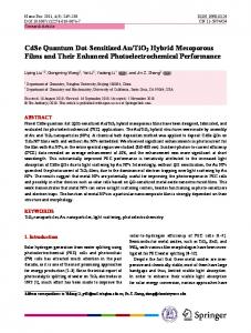
Fig. 1 XRD pattern of D-TiO2 /SiO2
photocatalyst was stirred in the dark for 12 h. After the system reached adsorption equilibrium, it was exposed for 6 h under visible-light irradiation. The first-order rate constant of degradation is reported as ln(C0 /C). C is the absorption of RhB at each irradiated time interval of the maximum peak of the absorption spectrum at wavelength 554 nm. C0 is the absorption of the starting concentration when adsorption/desorption equilibrium was achieved.
3 Results and discussion XRF measurement revealed that the percentages of O, Ti, Si and C in D-TiO2 /SiO2 are 47.7 %, 36.1 %, 4.3 % and 2.6 %, respectively. The rest of the weight is taken up by other trace elements. This confirms that SiO2 in templates was preserved and the C element from diatom cells has been self-doped in D-TiO2 /SiO2 . The calculated molar ratio of Ti, Si and C is approximately 8:1:4, which shows an extremely higher Ti/Si molar ratio [15]. The XRD pattern of the sample prepared by using diatoms as template is shown in Fig. 1. The peaks at 25.3°, 37.7°, 48.0°, 53.8°, 55.0°, 62.6°, 68.6°, 70.2°, 74.9° and 82.5° correspond to the diffractions of the (101), (004), (200), (105), (211), (204), (116), (220), (215) and (224) planes of anatase (JCPDS No. 71-1167), respectively. It is seen that D-TiO2 /SiO2 has only anatase phase even after calcination at 773 K for 6 h. This could be owing to enhancement of the thermal stability for phase transformation from anatase to rutile caused by SiO2 incorporation [16–19]. The crystalline size calculated by using the Scherrer equation according to (101) diffraction is about 16 nm. In addition, crystalline SiO2 and carbonaceous species have not been detected, indicating the amorphous states of SiO2 . According to the FT-IR spectra of D-TiO2 /SiO2 between 400 and 4000 cm−1 (Fig. 2), the broad band from 500 to 590 cm−1 is a typical Ti–O–Ti stretching vibration and is attributed to the anatase phase. The peaks near 3430 and
Diatom-templated TiO2 with enhanced photocatalytic activity: biomimetics of photonic crystals
Fig. 2 FT-IR spectrum of D-TiO2 /SiO2
Fig. 3 SEM images of (a) diatom (Cocconeis placentula) cells, (b, c) D-TiO2 /SiO2 , (d) HR-TEM image of D-TiO2 /SiO2
1630 cm−1 correspond to the stretching vibrations and bending vibrations of the O–H linkage on the surface of the sample. The peak at 1120 cm−1 reveals the antisymmetric Si– O–Si vibration caused by silica frustules of diatoms. However, it is worthwhile to note that the band at 910–960 cm−1 , which is widely accepted as the characteristic vibration due to the formation of Ti–O–Si bonds, was not observed. This confirms that D-TiO2 /SiO2 consists of matrix-isolated TiO2 layers on the surface of SiO2 frustules. Cocconeis placentula is an epiphytic diatom abundant on leaves of submerged aquatic plants [20]. Figure 3a shows the SEM image of the templates showing morphology of diatom (Cocconeis placentula) cells. The epitheca and hypotheca constitute an oval cell and the edges of each valve gather to form a girdle band. The cells are about 10 to 20 µm in length
329
Fig. 4 N2 adsorption/desorption isotherm and the BJH pore-size distribution (inset) of D-TiO2 /SiO2
and the width is about half of the corresponding length. Morphology of D-TiO2 /SiO2 is shown in Figs. 3b and c; the entire shapes of the oval cells remain intact in D-TiO2 /SiO2 . In the cell wall of valves and girdles, very regular arrays of chambers (areolae) and pores (cribra) form periodic patterns. The as-synthesized samples exhibit similar morphology to that of the original diatom templates, revealing that the morphologies of diatom templates were inherited to the samples through a biotemplating process. The only difference is whether the regular patterns of valve faces could be seen clearly. The surfaces of diatom cells are relatively rough because of organic substances covering them, which constitute organic cell architectures such as cell membranes and chromatoplasts. After the sol–gel and calcination processes, the organics could be removed. The frustules were exposed and coated by TiO2 ; the fine structures of the frustules therefore turned up clearly in D-TiO2 /SiO2 . It is clearly seen that the raphes on the valves have a simple slit with straight and parallel sides. The high-resolution TEM (HRTEM) image (Fig. 3d) reveals the structure of single TiO2 nanoparticles coated on frustules. The size of a single TiO2 particle is about 10 nm in diameter, which is in agreement with TEM measurement. The lattice spacing of 0.35 nm corresponds to the interplanar distance between adjacent (101) crystallographic planes of anatase TiO2 . It can be inferred that the diatom templates entailed their hierarchical structures to the resultant D-TiO2 /SiO2 . The Barrett–Joyner–Halenda (BJH) pore-size distribution of D-TiO2 /SiO2 (Fig. 4, inset) shows primary pore-size distributions in the region between 3 and 25 nm, indicating that the sample has mesoporous channels. As shown in Fig. 4, the N2 adsorption/desorption isotherm of DTiO2 /SiO2 can be classified as IUPAC type IV, suggesting the presence of mesopores [21]. The shape of the hysteresis loops is of type H3, associated with mesopores present in aggregates composed of primary particles, giving rise to pile-up pores [22]. The Brunauer–Emmett–Teller (BET) surface area and average pore volume of D-TiO2 /SiO2 were
330
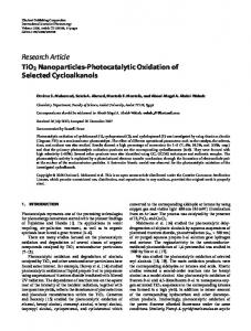
Fig. 5 The UV-Vis diffuse reflectance spectra of D-TiO2 /SiO2 , calcined diatom templates and Degussa P25 TiO2
41.7 m2 /g and 0.22 cm3 /g, respectively. It has been reported that a significant increase for a specific surface area could be achieved by introducing SiO2 into pure TiO2 [15, 23]. The intricate three-dimensional microstructure of diatoms could also result in a higher surface area formed by myriad accessible pores and channels [24, 25]. Regarding the prepared D-TiO2 /SiO2 , however, the specific surface area was slightly less than pure TiO2 catalyst Degussa P25(50 ± 5m2 /g). It is proposed that the TiO2 coating decreased the surface area of frustules by probable blocking of the pores and channels, unlike replicas of frustules [25], which transform silica into other compounds. The results from the above-mentioned structural and compositional analysis are also in accordance with the titania coating process. According to the sol–gel coating, titania precursors in the solution could interact with the surfaces of diatom cells and be adsorbed. After removing excessive solution, the precursors were hydrolyzed by moisture in the air and aggregated to form a titania layer. Due to the titania coating, the frustules and biogenic C could not be completely removed by calcination. As a result, the TiO2 -coated SiO2 structures templated by diatoms are formed, which are characterized by a matrix-isolated TiO2 layer coating and an inner self-doped C. Although TiO2 -coated substances, especially TiO2 -coated SiO2 , had been studied for a variety of uses mainly because of their photocatalytic and photovoltaic properties [26–29], TiO2 -coated SiO2 derived from a biotemplate with C-doping has not been reported yet. The algal templates sacrificed themselves as both templates and resources of doped elements. The UV-Vis diffuse reflectance spectra (DRS) of DTiO2 /SiO2 , calcined diatoms and Degussa P25 TiO2 are shown in Fig. 5. In comparison with P25 (a pure UV TiO2 photocatalyst), the diatom-templating approach significantly increased the light absorption of TiO2 in the visible range (from 400 to 800 nm) and the absorption edge of D-TiO2 /SiO2 showed a clear red shift with decreasing
J. He et al.
Fig. 6 Pseudo-first-order plots of photocatalytic degradation of RhB in the presence of D-TiO2 /SiO2 and P25 TiO2 under visible-light irradiation (λ > 420 nm)
band gap. The broad absorption of calcined diatom templates from 200 to 800 nm proved that the increase in visible range should be mainly attributed to diatom templating. Doped carbon species could narrow the band gap of pristine TiO2 to contribute to the red shift [30–33] and the ability to absorb visible light could also be significantly improved [32–34]. Biogenic C may substitute for some of the lattice oxygen atoms on the interface of SiO2 frustules and TiO2 coatings; therefore, the red shift of the absorption edge occurred in D-TiO2 /SiO2 . Meanwhile, it has been reported that introducing SiO2 into the TiO2 matrix could cause a decrease in visible-light absorption and a blue shift in the absorption band [16, 35, 36], mainly due to dispersion and refinement of TiO2 particles in SiO2 . By contrast, a decrease in visible-light absorption and a blue shift in the absorption band are not observed in D-TiO2 /SiO2 . This again strongly suggests that D-TiO2 /SiO2 is very different from conventional titania–silica obtained by introducing SiO2 into the TiO2 matrix because the hierarchical structures derived from diatom templates capture light more effectively as diatom cells. The photocatalytic performances of D-TiO2 /SiO2 and P25 TiO2 were evaluated by the degradation of RhB. As shown in Fig. 6, a fairly good correlation to the pseudo-firstorder reaction kinetics could be found. The determined reaction rate constant k for RhB degradation is 0.12 and 0.01 h−1 for TiO2 prepared by using diatoms as template and P25 TiO2 , respectively. It is interesting to evoke some reasons why D-TiO2 /SiO2 exhibited the reasonable visible-light activity. The first explanation is that it shifted the absorption edge to the visiblelight range, narrowing the band gap, and had definite absorptions in the visible region. Furthermore, the unique morphology and structure of D-TiO2 /SiO2 is important for it to possess considerable photocatalytic activities [37, 38]. Based on the character of the regular arrays of chambers (areolae) and pores (cribra) structure (Fig. 4), it is proposed that the inner
Diatom-templated TiO2 with enhanced photocatalytic activity: biomimetics of photonic crystals
chambers can trap more dye molecules and yield a multiplereflection effect on visible light that cooperatively enhances the photocatalytic activity of D-TiO2 /SiO2 [39]. On one hand, the dye molecules can pass through the porous walls of hollow chambers and are trapped in the chambers. Compared with the relatively free dye molecules outside the hollow chambers, the target molecules in the chambers have a higher probability of making contact with the D-TiO2 /SiO2 wall and being degraded. On the other hand, the visible light will be more effectively utilized through a multiplereflection effect of the chamber structure [5, 40, 41]. Even though incorporation of silica did not provide a high surface area for adsorption and reaction sites, the DTiO2 /SiO2 inherited its morphology from diatoms due to preserved silica frustules which are exactly the key of superiority in light-harvesting of diatoms [13, 14]. The frustules supported the hierarchical structures of the D-TiO2 /SiO2 after a titania coating process. Meanwhile, like diatom cells, the frustules, can control the propagation of light, so the light channeling and focusing properties of their silica structure could help the transmission and collection of more light into the light-sensitive TiO2 coatings to enhance the lightharvesting ability; the photocatalytic activity was therefore improved [42].
4 Conclusions In summary, with the facile sol–gel process we developed, diatom cells were successfully used as biotemplates to prepare C-self-doped TiO2 /SiO2 catalyst. The frustules were conserved to form the skeletons of the hierarchical structures derived from diatoms. The templated morphologies in conjunction with C self-doping contributed to light absorption in a wide wavelength range. Thus, a considerable visible-light photocatalytic activity of the diatom-templated TiO2 /SiO2 was obtained. The strategy presented here promoted a new way to prepare photocatalysts utilizing smart biological structures and, more importantly, to harvest solar light effectively. Acknowledgements The authors thank the National Natural Science Foundation of China (Project NSFC-YN U1033603, 21063016) and the Program for Innovative Research Teams (in Science and Technology) in the University of Yunnan Province (IRTSTYN) for financial support.
References 1. M. Matsuoka, M. Kitano, M. Takeuchi, K. Tsujimaru, M. Anpo, J.M. Thomas, Catal. Today 122, 51 (2007) 2. M.R. Hoffmann, S.T. Martin, W. Choi, D.W. Bahnemann, Chem. Rev. 95, 69 (1995) 3. L. Jing, Y. Qu, B. Wang, S. Li, B. Jiang, L. Yang, W. Fu, H. Fu, J. Sun, Sol. Energy Mater. Sol. Cells 90, 1773 (2006)
331
4. H. Tong, S. Ouyang, Y. Bi, N. Umezawa, M. Oshikiri, J. Ye, Adv. Mater. 24, 229 (2012) 5. H. Li, Z. Bian, J. Zhu, D. Zhang, G. Li, Y. Huo, H. Li, Y. Lu, J. Am. Chem. Soc. 129, 8406 (2007) 6. T. Fan, S.K. Chow, D. Zhang, Prog. Mater. Sci. 54, 542 (2009) 7. J. Parkinson, R. Gordon, Trends Biotechnol. 17, 190 (1999) 8. A.R. Parker, H.E. Townley, Nat. Nanotechnol. 2, 347 (2007) 9. M. Pérez-Cabero, V. Puchol, D. Beltrán, P. Amorós, Carbon 46, 297 (2008) 10. Z. Bao, E.M. Ernst, S. Yoo, K.H. Sandhage, Adv. Mater. 21, 474 (2009) 11. S. Dudley, T. Kalem, M. Akinc, J. Am. Ceram. Soc. 89, 2434 (2006) 12. R.R. Unocic, F.M. Zalar, P.M. Sarosi, Y. Cai, K.H. Sandhage, Chem. Commun. 7, 796 (2004) 13. T. Fuhrmann, S. Landwehr, M. El Rharbi-Kucki, M. Sumper, Appl. Phys. B, Lasers Opt. 78, 257 (2004) 14. R. Gordon, D. Losic, M.A. Tiffany, S.S. Nagy, F.A. Sterrenburg, Trends Biotechnol. 27, 116 (2009) 15. Z. Zhai, Y. Miao, Q. Sun, H. Tao, W. Wang, J. Wang, Catal. Lett. 131, 538 (2009) 16. Y. Hou, X. Wang, L. Wu, X. Chen, Z. Ding, X. Wang, X. Fu, Chemosphere 72, 414 (2008) 17. X. Chen, X. Wang, X. Fu, Energy Environ. Sci. 2, 872 (2009) 18. K. Balachandran, R. Venckatesh, R. Sivaraj, Int. J. Eng. Sci. Technol. 2, 3695 (2010) 19. C. Anderson, A.J. Bard, J. Phys. Chem. B 101, 2611 (1997) 20. D. Gillan, G. Cadée, J. Sea Res. 43, 83 (2000) 21. G. Leofanti, M. Padovan, G. Tozzola, B. Venturelli, Catal. Today 41, 207 (1998) 22. X. Yu, J. Yu, B. Cheng, M. Jaroniec, J. Phys. Chem. C 113, 17527 (2009) 23. J. Wang, S. Uma, K.J. Klabunde, Appl. Catal. B, Environ. 48, 151 (2004) 24. H.E. Townley, A.R. Parker, H. White-Cooper, Adv. Funct. Mater. 18, 369 (2008) 25. Z. Bao, M.R. Weatherspoon, S. Shian, Y. Cai, P.D. Graham, S.M. Allan, G. Ahmad, M.B. Dickerson, B.C. Church, Z. Kang, H.W. Abernathy 3rd, C.J. Summers, M. Liu, K.H. Sandhage, Nature 446, 172 (2007) 26. L.W. Miller, M.I. Tejedor-Tejedor, M.A. Anderson, Environ. Sci. Technol. 33, 2070 (1999) 27. M. Holgado, A. Cintas, M. Ibisate, C.J. Serna, C. Lopez, F. Meseguer, J. Colloid Interface Sci. 229, 6 (2000) 28. X. Fu, S. Qutubuddin, Colloids Surf. A, Physicochem. Eng. Asp. 179, 65 (2001) 29. K.D. Kim, H.J. Bae, H.T. Kim, Colloids Surf. A 221, 163 (2003) 30. Y. Choi, T. Umebayashi, M. Yoshikawa, J. Mater. Sci. 39, 1837 (2004) 31. W. Ren, Z. Ai, F. Jia, L. Zhang, X. Fan, Z. Zou, Appl. Catal. B, Environ. 69, 138 (2007) 32. J.-W. Shi, X. Zong, X. Wu, H.-J. Cui, B. Xu, L. Wang, M.-L. Fu, ChemCatChem 4, 488 (2012) 33. F. Dong, H. Wang, Z. Wu, J. Phys. Chem. C 113, 16717 (2009) 34. Q. Wang, Z. Jiang, Y. Wang, D. Chen, D. Yang, J. Nanopart. Res. 11, 375 (2008) 35. J. Bahadur, D. Sen, S. Mazumder, P.U. Sastry, B. Paul, H. Bhatt, S.G. Singh, Langmuir 28, 11343 (2012) 36. H.S. Kibombo, D. Zhao, A. Gonshorowski, S. Budhi, M.D. Koppang, R.T. Koodali, J. Phys. Chem. C 115, 6126 (2011) 37. N. Shi, X. Li, T. Fan, H. Zhou, J. Ding, D. Zhang, H. Zhu, Energy Environ. Sci. 4, 172 (2011)
332 38. X. Sun, C. Zheng, M. Qiao, J. Yan, X. Wang, N. Guan, Chem. Commun. 31, 4750 (2009) 39. T. Zhao, Z. Liu, K. Nakata, S. Nishimoto, T. Murakami, Y. Zhao, L. Jiang, A. Fujishima, J. Mater. Chem. 20, 5095 (2010) 40. S. Liu, J. Yu, J. Solid State Chem. 181, 1048 (2008)
J. He et al. 41. X. Wang, J.C. Yu, C. Ho, Y. Hou, X. Fu, Langmuir 21, 2552 (2005) 42. C.B. Field, M.J. Behrenfeld, J.T. Randerson, P. Falkowski, Science 281, 237 (1998)