Department of Medicine and the Sammy Davis, Jr. National Liver Institute, University of Medicine and. Dentistry of New Jersey-New Jersey Medical School, ...
Differing effects of cholesterol and taurocholate on steady state hepatic HMG-CoA reductase and cholesterol 7~hydroxylaseactivities and mRNA levels in the rat Sarah Shefer,l Lien B. Nguyen, Gerald Salen, Gene C. Ness, Indu R. Chowdhary, Susan Lerner, Ashok K. Batta, and G. Stephen Tint Department of Medicine and the Sammy Davis, Jr. National Liver Institute, University of Medicine and Dentistry of New Jersey-New Jersey Medical School, Newark, NJ 07103; Veterans Administration Medical Center, East Orange, NJ 07019; and Department of Biochemistry and Molecular Biology, College of Medicine, University of South Florida, Tampa, FL 33612
Supplementary key words HMG-CoA reductase activity HMGCoA reductase mRNA cholesterol 7a-hydroxylase activity cholesterol 7a-hydroxylase mRNA cholestyramine bile acids
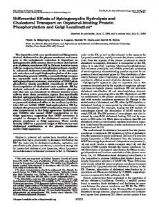
Cholesterol is the obligate precursor of bile acids in all mammalian species (1). During bile acid synthesis, the nonpolar cholesterol molecule is transformed into a water-soluble bile acid. The key reaction in this pathway (Fig. 1) is the 7a-hydroxylation of cholesterol that is catalyzed by the enzyme cholesterol 7a-hydroxylase (EC 1.14.13.17). According to current information, this reaction is rate-limiting (2, 3). The preferred substrate pool for cholesterol 7a-hydroxylase is newly synthesized cholesterol (4), and therefore, bile acid synthesis is controlled, not only by the activity of cholesterol 7a-hydroxylase, but also by the supply of the substrate, cholesterol. Thus, bile acid synthesis is coordinately regulated with cholesterol synthesis, which is controlled by HMG-CoA reductase (EC 1.1.1.23). Both HMG-CoA reductase and cholesterol 7a-hydroxylase activities vary diurnally (5-7) and are believed to be under the feedback control of the hepatic cholesterol pool and the flux of bile acids through the liver (8-14). Recently, HMG-CoA reductase and cholesterol 7a-hydroxylase have been purified and their cDNA sequences have been elucidated so that probes are available to quantitate mRNA levels for each enzyme under various experimental conditions (6, 11-18). In this study, we have determined HMG-CoA reductase and cholesterol 7a-hydroxylase activities and relative levels of mRNA in
Abbreviations: HMG, 3-hydroxy-3-methylglutaryl;TLC, thin-layer chromatography. 'To whom correspondence should be addressed at: Department of Medicine, MSB H-534, UMDNJ-NJ Medical School, 185 South Orange Avenue, Newark, NJ 07103.
Journal of Lipid Research Volume 33, 1992
1193
Downloaded from www.jlr.org by guest, on July 21, 2011
Abstract We investigated the effects of cholesterol, cholestyramine, and taurocholate feeding on steady state specific activities and mRNA levels of hepatic 3-hydroxy-3-methylglutaryl (HMG)-CoA reductase and cholesterol 7a-hydroxylase in the rat. Interruption of the enterohepatic circulation of bile acids (cholestyramine feeding) increased total HMG-CoA reductase activity 5-fold. Cholesterol and taurocholate administration suppressed total microsomal HMG-CoA reductase activities 87 % and 65%, respectively. HMG-CoA reductase mRNA levels increased 3-fold with cholestyramine, did not decrease significantly with cholesterol feeding, but were markedly decreased after taurocholate treatment. Cholesterol 7a-hydroxylase activity increased 4-fold with cholestyramine and 29% during cholesterol feeding, but decreased 64% with taurocholate. Cholesterol 7ahydroxylase mRNA levels rose 150% and 50% with cholestyramine and cholesterol feeding, respectively, but decreased 73% with taurocholate. The administration of cholesterol together with taurocholate prevented the decline in cholesterol 7ahydroxylase mRNA levels, but inhibition of enzyme activity persisted ( - 76%). Hepatic microsomal cholesterol concentrations increased 2-fold with cholesterol feeding but did not change with taurocholate or cholestyramine treatment. These results demonstrate that mRNA levels of HMG-CoA reductase are controlled by the hepatic taurocholate flux, whereas mRNA levels of cholesterol 7a-hydroxylase are controlled by the cholesterol substrate supply. These end products, cholesterol and bile acids, exert post-transcriptional regulation on HMG-CoA reductase and cholesterol 7a-hydroxylase, respectively.-- Shefer, S., L. B. Nguyen, G . Salen, G . C. Ness, I. R. Chowdhary, S. Lerner, A. K. Batta, and G. S. Tint. Differing effects of cholesterol and taurocholate on steady state hepatic HMG-CoA reductase and cholesterol 7ar-hydroxylase activities and mRNA levels in the rat. J Lipid Res. 1992. 33: 1193-1200.
ACETATE
i
,HMGS
HMG-COA
lMcR
MEVALONIC ACID
tions. To minimize diurnal variations in cholesterol and bile acid synthesis (19, 20), the animals were killed at about 10 AM. The research protocol was approved by the Institutional Animal Care and Use Committee of the University of Medicine and Dentistry of New Jersey-New Jersey Medical School, Newark, NJ. Materials
MATERIALS AND METHODS
Assays for total HMG-CoA reductase and cholesterol 7a-hydroxylase activities
i CHOLESTEROL
I-
7cL-HYD ROX YCHO L ESTEROL
i
BILE A C I D S Fig. 1. Key reactions in the cholesterol and bile acid biosynthetic pathways. HMGS, hydroxymethylglutaryl-CoAsynthase; HMGR, hydroxymethylglutaryl-CoA reductase; CH7aH, cholesterol 'la-hydroxylase.
Animals and experimental design Male Sprague-Dawley rats (Taconic Farms, Inc., Germantown, NY) that weighed 150-180 g were fed rat chow (Purina Mills Inc., Saint Louis, MO) and water ad lib. After 1 week the rats were divided into five groups of six to ten rats each and fed rat chow diets that were supplemented with: 2 % cholesterol, 2.5% cholestyramine, 1% taurocholate, or the combination of 1% taurocholate and 2 % cholesterol. A control group received only rat chow. All treatment groups ate the same amounts of food and gained similar weight during the feeding period. When food intake or weight gain of an experimental animal differed by more than 10% from the average of the control group, the animal was excluded from the study. The diets were continued for 7 days after which five rats from each group were anesthetized with sodium pentobarbital (Fort Dodge Laboratories, Inc., Fort Dodge, IA), common bile ducts were cannulated, and bile was collected for a period of 30 min. All rats were then killed by decapitation and their livers were excised, weighed, and stored at -70°C until used for sterol, enzymatic, and mRNA determina-
1194
Journal of Lipid Research Volume 33, 1992
Hepatic microsomes were prepared by differential ultracentrifugation (23), and the protein was determined according to Lowry et al. (24). The assay for HMG-CoA reductase activity was carried out as described previously (25). Cholesterol 7a-hydroxylase activity was measured by the isotope incorporation method of Shefer, Salen, and Batta (23). In cholesterol- and taurocholate-fed rats where hepatic cholesterol concentrations increased, cholesterol 7a-hydroxylase activity was assayed in a reconstituted system after removal of endogenous cholesterol by acetone treatment (26, 27) and compared to similarly assayed control microsomes. This method eliminates the confounding effect of endogenous cholesterol, measures cholesterol 7a-hydroxylase activity with the enzyme fully saturated with substrate (zero-order kinetics), and gives results comparable to intact microsomes (26, 27). Isolation and quantitation of mRNA Total RNA was isolated from 1-g pieces of rat liver by a low temperature modification of the guanidinium thiocyanate extraction procedure (28) with the addition of a lithium chloride extraction step to remove glycogen (29).
Downloaded from www.jlr.org by guest, on July 21, 2011
livers from rats fed diets supplemented with cholesterol, cholestyramine, taurocholate, or the combination of cholesterol and taurocholate. Our objective was to elucidate the role of cholesterol and bile acids as regulators of these rate-controlling enzymes.
Cholesterol (Sigma Chemical Co., St. Louis, MO) and taurocholate (Calbiochem Co., La Jolla, CA) were >98% pure. Cholestyramine was a gift from Merck Sharp and Dohme Research Laboratories, Rahway, NJ. [3-'4C]HMGCoA (Amersham, Arlington Heights, IL) was diluted with unlabeled HMG-CoA to a specific activity of 50 dpm/pmol. [4-1*C]Cholesterol(Du Pont Co., New England Nuclear Research Products, Boston MA) was diluted to a specific activity of 2 x 10⁴ dpm/nmol and purified by chromatography on a silicic acid column (21). The purified cholesterol contained less than 0.06% 7 a hydroxycholesterol. A mixture of 7a- and 7P-hydroxycholesterol, used as reference standards for TLC, was obtained by the reduction of 7-ketocholesterol (Schwarz/ Mann, Orangeburg, NY) with sodium borohydride (22). The cDNA probes for hamster HMG-CoA reductase (pRED 227) and human catalase (pCAT 10) were purchased from American Type Culture Collection (Rockville, MD). The probe for rat liver cholesterol 7 a hydroxylase (7a6) was obtained from Dr. John Y. L. Chiang (Northeastern Ohio Universities College of Medicine, Rootstown, OH).
4
*
Determination of hepatic total and microsomal cholesterol For the determination of free and esterified cholesterol in the liver, aliquots of whole liver homogenates (50-200 mg in 0.5 ml) were extracted with 20 volumes of chloroform-methanol 2:l (vol/vol) after the addition of [3H]cholesterol and [3H]cholesteryloleate (1.1 x lo6 dpm each) as recovery standards. The extract was separated by TLC on silica gel G plates with hexane-ethyl ether-acetic acid 85:15:0.5 (vol/vol/vol). The bands corresponding to free and esterified fractions (Rf= 0.09 and 0.87, respectively) were scraped and eluted with ethyl acetate-methanol 85:15 (vol/vol). The esterified cholesterol fractions were hydrolyzed at 6OoC for 1 h in 10% ethanolic KOH and extracted with n-hexane. Both free and esterified cholesterol fractions were counted and analyzed by capillary gas-liquid chromatography as the trimethylsilyl ether derivatives using 5a-cholestane as internal standard (34). For the determination of total cholesterol concentrations in the microsomes, aliquots of microsomal suspensions (2-5 mg) were saponified in 10% ethanolic KOH at 6OoC for 2 h with 30,000 dpm [3H]cholesterol as internal recovery standard. Cholesterol was extracted with nhexane and quantitated by capillary gas-liquid chromatography as described above. Determination of biliary bile acids Bile acid composition was determined in bile specimens obtained at the end of each treatment period, using tauro-
Shejier et al.
ursodeoxycholic acid as internal recovery standard (9, 35). The bile acids were quantitated as the methyl ester trimethylsilyl ether derivatives by capillary gas-liquid chromatography on a wall-coated open tubular fused silica column (0.22 mm x 25 m), coated with a 0.12-pm film of C P Sil 5 CB (Chrompak, Inc. Bridgewater, NJ). The chromatograph was operated at a column temperature of 265OC and a helium flow of 1.3 ml/min. The retention times of the bile acid methyl ester trimethylsilyl ethers relative to 5a-cholestane (13.1 min) were: deoxycholic acid, 1.49; chenodeoxycholic acid, 1.54; a-muricholic acid, 1.56; cholic acid, 1.57; ursodeoxycholic acid, 1.63; and 6-muricholic acid, 1.83. Statistical analysis Data were analyzed statistically by comparing the 95 % and 99% confidence intervals for the means of the various treatment groups according to Altman and Gardner (36).
RESULTS In Fig. 2A are presented measurements of total HMGCoA reductase activities during the various treatments. After cholesterol and taurocholate administration, total HMG-CoA reductase activities declined 87% and 65%) respectively. When cholesterol was combined with taurocholate, HMG-CoA reductase activity was reduced 93 %. In contrast, cholestyramine treatment stimulated HMGCoA reductase activity 5-fold. In Fig. 2B are presented measurements of cholesterol 7a-hydroxylase activities. Cholestyramine treatment increased cholesterol 7a-hydroxylase activity 4-fold as compared to a 29% increase when cholesterol was administered. In contrast, taurocholate inhibited cholesterol 7a-hydroxylase activity 64%, while the combination of taurocholate plus cholesterol resulted in a 76% decline. Thus, the hepatic bile acid flux appears to be a potent down-regulator of HMG-CoA reductase and cholesterol 7a-hydroxylase activities, while cholesterol inhibits the activity of HMG-CoA reductase but stimulates slightly the activity of cholesterol 7a-hydroxylase. Estimates of relative steady state mRNA levels for HMG-CoA reductase, determined by densitometric scanning and corrected for catalase mRNA recovery (25, 37), are presented in Table 1. Taurocholate virtually abolished HMG-CoA reductase mRNA as compared with an insignificant decrease observed with cholesterol feeding (37). When taurocholate was combined with cholesterol, HMG-CoA reductase mRNA was also barely detected. In contrast, cholestyramine treatment increased HMG-CoA reductase mRNA levels over 2-fold. Thus, hepatic bile acid depletion stimulates HMG-CoA reductase mRNA levels while taurocholate virtually eliminates the message for enzyme protein synthesis. Interestingly, cholesterol
Feedback control of cholesterol and bile acid synthesis
1195
Downloaded from www.jlr.org by guest, on July 21, 2011
Poly A' RNA was isolated by oligo (dT) cellulose chromatography (30). Ten-pg aliquots of poly A' RNA were denatured and electrophoresed in 1% agarose gels containing 0.02 M borate, pH 8.3, 0.2 mM ethylene diamine tetraacetic acid, and 3% formaldehyde (31). The separated RNAs were transferred to Gene Screen Plus membranes by capillary blotting and the RNA was fixed by baking under vacuum for 2 h at 8OOC. The cDNA probes were labeled with 32P to specific activities ranging from 2 x 10⁷ to 4 x 10⁹ cpm/pg by nick translation. The hybridizations were carried out as previously described (32), except that a hybridization incubator from Robbins Scientific equipped with 38 x 300 mm glass screw-cap tubes was used. Typically, 5 ml of hybridization solution (32) containing 2-4 x lo8 cpm and 1-2 pg of 32P-labeled cDNA probe were incubated with a 9 x 14 cm membrane in a glass screw-cap tube at 42OC overnight, being careful not to overlap the membrane. Washing conditions were as previously described (33). The washed membranes were exposed to Kodak X Omat AR film with Cronex intensifying screens at - 7OoC for times ranging from 2 to 16 h. The autoradiograms were scanned with an LKB Ultrascan laser densitometer to determine relative mRNA levels. The values for catalase mRNA were used as internal controls. All values are presented as means SEM.
*cy
\cE .-0 )
c
I
HMG -CoA reductose
0.8
2 .= n 0.6
.c
I
a
CHOL
TCA
TCA+ CHY CHOL
Fig. 2. Catalytic activities of HMG-CoA reductase (A) and cholesterol 7a-hydroxylase (B) in rats treated for 7 days with the following dietary supplements: C, control (n = 9); CHOL, 2% cholesterol (n = 5); E A , 1% taurocholate (n = 5); CHOL + TCA, 2% cholesterol + 1% taurocholate (n = 6); CHY, 2.5% cholestyramine (n = 5). Bars represent means f SEM; *, P < 0.05; **, P < 0.01.
feeding, which markedly inhibits enzyme activity (Fig. 2A), is associated with an insignificant decrease in HMGCoA reductase mRNA levels. Steady state cholesterol 7a-hydroxylase mRNA levels are presented in Table 1. Both cholestyramine and cholesterol feeding increased cholesterol 7a-hydroxylase mRNA levels 150% and 50%, respectively. In contrast, taurochoTABLE 1.
Relative mRNA levels of hepatic HMG-CoA reductase and hepatic cholesterol 7a-hydroxylase
Treatment
HMG-CoA Reductase mRNA
Control Cholesterol Taurocholate Taurocholate + cholesterol Cholestyramine
4 . 0 f 0.3 (10) 3 . 2 f 0.4 (9)