Pediatr Radiol (2013) 43:69–79 DOI 10.1007/s00247-012-2527-7
ORIGINAL ARTICLE
Diffusion tensor imaging metrics in neonates—a comparison of manual region-of-interest analysis vs. tract-based spatial statistics Youngseob Seo & Zhiyue J. Wang & Gareth Ball & Nancy K. Rollins
Received: 8 June 2012 / Revised: 15 August 2012 / Accepted: 23 August 2012 / Published online: 20 November 2012 # Springer-Verlag Berlin Heidelberg 2012
Abstract Background Diffusion tensor data can be analyzed using region-of-interest (ROI) analysis and tract-based spatial statistics (TBSS). There is essentially no literature validating or comparing these techniques in the neonate. Objective The purpose of this study was to perform a direct comparison of fractional anisotropy (FA), axial diffusivity (AD) and radial diffusivity (RD) derived using manual ROI analysis and TBSS modified for use in neonates. Materials and methods This study was IRB-approved. Thirty-nine infants, 32–49 weeks post-conception age, underwent MRI at 3 T. FA, AD and RD of the callosal genu (CG) and splenium (CS) and posterior limbs of both internal capsules (PLIC) were determined using both techniques. Y. Seo : Z. J. Wang : N. K. Rollins Department of Radiology, University of Texas Southwestern Medical Center at Dallas, Dallas, TX, USA Y. Seo (*) Division of Convergence Technology, Korea Research Institute of Standards and Science, 267 Gajeong-ro, Doryong-dong, Yuseong-gu, Daejeon, Korea 305-340 e-mail:
[email protected] Z. J. Wang : N. K. Rollins Department of Radiology, Children’s Medical Center Dallas, Dallas, TX, USA G. Ball Centre for the Developing Brain, Imperial College London and MRC Clinical Sciences Centre, Hammersmith Hospital, London, UK
Pearson correlation (r) was used to estimate the concordance of tensor metrics derived from these techniques. Results The r value for FA in the CG, CS and left and right PLIC was 0.88, 0.75, 0.78 and 0.35, respectively. The r value for axial/radial diffusivity in the CG, CS and left and right PLIC was 0.62/0.72, 0.76/0.64, 0.68/0.9 and 0.3/0.72, respectively. The variable concordance results from problems with spatial correspondence of ROI masks between the native space and the FA skeleton. Conclusion Direct comparison between these methodologies shows tensor metrics varied with location and by degree, suggesting the two techniques do not provide consistently comparable results. Keywords Diffusion tensor imaging . Manual ROI analysis . Tract-based spatial statistics . Comparison of diffusion tensor metrics
Introduction Diffusion tensor imaging (DTI) can be used to study the direction and magnitude of local water diffusion in cerebral white matter (WM) [1–3]. Water diffusion is relatively fast in the direction aligned with axonal fibers and is slow perpendicular to the fiber tracts. Fractional anisotropy (FA), the diffusion-tensor-derived parameter cited most often, is determined by the three principal eigenvalues (λ1, λ2, and λ3) of the diffusion tensor. FA is equal to 0 for λ1 0 λ2 0 λ3; e.g., isotropic diffusion and FA approaches 1 for λ1> > λ2 and λ1> > λ3; e.g., anisotropic diffusion. DTI is used to study micro-structural changes in cerebral white matter with brain maturation and aging and disease effects on WM in
70
neonates [4–11]. DTI can serve as an imaging biomarker for the disease state if the disease affecting WM is reflected in measurable deviation from age-expected tensor norms. Two methodologies are widely used to quantitatively analyze diffusion tensor data. Manual region-of-interest (ROI) analysis is an operator-dependent, labor-intensive and hypothesis-driven technique impractical for large numbers of subjects or comparison of groups. Tract-based spatial statistics (TBSS) [12] is an operator-independent, datadriven and computer-intensive statistical inference tool for detecting differences or relationships across the whole brain. TBSS requires a set of pre-processing steps and relies on accurate spatial normalization of FA maps. The spatial normalization process is potentially problematic in neonatal and pediatric patients because the normalization templates used in adults are not appropriate for infants [6, 7, 13–15]. Ball et al. [7] recently described a modification to TBSS designed to improve the reliability of TBSS in neonates. The other method, manual ROI analysis, is performed on native space after minimal post-processing of tensor data, and this technique is potentially more clinically relevant than TBSS. To date, no studies have performed a direct comparison of tensor metrics in infants derived by using manual ROI analysis and TBSS. The aim of this study was therefore to perform a direct comparison between manual ROI analysis and TBSS modified for use in a neonatal cohort to determine the concordance of FA, AD and RD metrics derived from these techniques.
Pediatr Radiol (2013) 43:69–79
Imaging MR imaging was acquired at 3 T (Achieva R2.6; Philips Healthcare, Cleveland, OH) using an 8-channel sensitivity encoding head coil. DTI parameters were single-shot echoplanar imaging (EPI), field of view 200×200 mm2, image matrix 100×100, voxel size 2×2×2 mm3, b0700 s/mm2, TR/TE 8,000/74 ms, SENSE factor02, three acquisitions and 30 directions [16]. MR scanner geometry accuracy, transmitter gain, B0 shifting, spatial resolution and low contrast detection, and signal-to-noise ratio (SNR) were checked weekly. Routine clinical images for all infants included sagittal and axial T1 FLAIR, axial and coronal T2, and diffusion-weighted images (b01000 s/mm2). All conventional MR images were reviewed by two experienced pediatric neuroradiologists with 12 and 25 years of experience. If both neuroradiologists agreed the infants had structurally normal MR studies, the infants were included in this study. Image processing and analysis Pre-processing The individual diffusion tensor acquisitions were registered and averaged using the Philips Research Image Development Environment (PRIDE) (Philips Healthcare, Cleveland, OH) with affine transformation for correction of eddy current distortion and simple head movement. ROI-based analysis
Materials and methods Patients This study was approved by the Institutional Investigational Review Board. We included 39 neonates (19 girls, 20 boys) with a post-conception age (PCA) of 32–49 weeks (mean 41 weeks) at the time of MR imaging referred for seizures (n020), apnea (n07) and suspected congenital anomalies (n0 12). All had normal neurological examinations documented in the medical record. Children who had undergone hypothermia for hypoxic–ischemic encephalopathy were excluded from the study even in the presence of a normal neurological examination and normal MRI study. DTI data were acquired from MR imaging that was routinely performed without sedation after bundling and feeding of the infants. The neonates requiring sedation were sedated with oral chloral hydrate (25–50 mg/ kg) prior to scanning and pulse oximetry; electrocardiography data were monitored, and noise was dampened using earmuffs. Six additional infants imaged met inclusion criteria but were excluded because DT images were inadequate because of patient motion.
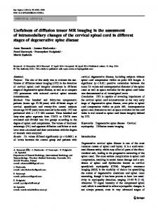
Diffusion tensor data were not normalized for purposes of manual ROI-based analysis. Using the directionally encoded color maps, a single experienced observer manually placed ROIs on the posterior limbs of the internal capsules (PLIC) two slices above the anterior commissure and on the callosal genu (CG) and callosal splenium (CS) (Fig. 1). The FA values of the selected areas of the brain were averaged over 6– 8 adjacent voxels for the CG and CS, and 12–15 voxels for the PLIC because of variable size of the selected regions of each infant. Placement of each ROI was repeated until the standard deviation (SD) of the FA for each measurement was