Original Paper Received: August 9, 2011 Returned for revision: September 27, 2011 Accepted after revision: October 7, 2011 Published online: December 16, 2011
Brain Behav Evol DOI: 10.1159/000334469
Distinct Development of Peripheral Trigeminal Pathways in the Platypus (Ornithorhynchus anatinus) and Short-Beaked Echidna (Tachyglossus aculeatus) Ken W.S. Ashwell a Craig D. Hardman a Peter Giere b a
Department of Anatomy, School of Medical Sciences, The University of New South Wales, Sydney, N.S.W., Australia; b Museum für Naturkunde, Leibnitz Institut für Evolutions- und Biodiversitätsforschung an der Humboldt-Universität zu Berlin, Berlin, Germany
Key Words Monotremes ⴢ Electroreception ⴢ Trigeminal ganglion ⴢ Somatosensory system
Abstract The extant monotremes (platypus and echidnas) are believed to all be capable of electroreception in the trigeminal pathways, although they differ significantly in the number and distribution of electroreceptors. It has been argued by some authors that electroreception was first developed in an aquatic environment and that echidnas are descended from a platypus-like ancestor that invaded an available terrestrial habitat. If this were the case, one would expect the developmental trajectories of the trigeminal pathways to be similar in the early stages of platypus and short-beaked echidna development, with structural divergence occurring later. We examined the development of the peripheral trigeminal pathway from snout skin to trigeminal ganglion in sectioned material in the Hill and Hubrecht collections to test for similarities and differences between the two during the development from egg to adulthood. Each monotreme showed a characteristic and different pattern of distribution of developing epidermal sensory gland specializations (electroreceptor primordia) from the time of hatching. The cross-
© 2011 S. Karger AG, Basel 0006–8977/11/0000–0000$38.00/0 Fax +41 61 306 12 34 E-Mail
[email protected] www.karger.com
Accessible online at: www.karger.com/bbe
sectional areas of the trigeminal divisions and the volume of the trigeminal ganglion itself were also very different between the two species at embryonic ages, and remained consistently different throughout post-hatching development. Our findings indicate that the trigeminal pathways in the short-beaked echidna and the platypus follow very different developmental trajectories from the earliest ages. These findings are more consistent with the notion that the platypus and echidna have both diverged from an ancestor with rudimentary electroreception and/or trigeminal specialization, rather than the contention that the echidna is derived from a platypus-like ancestor. Copyright © 2011 S. Karger AG, Basel
Introduction
The monotremes are a unique group of mammals confined to Australia and New Guinea. All the extant monotremes, platypus (Ornithorhynchus anatinus), shortbeaked echidna (Tachyglossus aculeatus) and long-beaked echidnas (species of the genus Zaglossus) may be capable of electroreception in the trigeminal pathway [Scheich et al., 1986; Gregory et al., 1987, 1988, 1989; Andres et al., 1991; Manger and Hughes, 1992; Manger and Pettigrew, Prof. Ken Ashwell Department of Anatomy, School of Medical Sciences The University of New South Wales Sydney, NSW 2052 (Australia) Tel. +61 2 9385 2482, E-Mail k.ashwell @ unsw.edu.au
1996; Proske et al., 1998]. All monotremes appear to possess electroreceptors in the beak/bill, but the distribution and mode of electroreception employed are quite different [Manger et al., 1997; Pettigrew, 1999]. The platypus is reported to have approximately 40,000 mucous electroreceptors distributed in longitudinal stripes on the entire dorsal surface of the upper bill and the entire ventral surface of the lower bill [Manger and Pettigrew, 1996; Pettigrew, 1999]. These might be used to locate food by swinging the head from side to side while swimming, so that electrical field lines generated by the muscles of its crustacean prey sweep across the bands of electroreceptors [Pettigrew, 1999]. The short-beaked echidna is reported to have fewer than 400 electroreceptors confined to a sensitive spot at the end and underside of the beak tip, whereas the long-beaked echidna is said to have 2,000 to 3,000 electroreceptors concentrated over the tip and extending no further than 40% of the distance up the shaft [Manger et al., 1997; Pettigrew, 1999]. These might be used as a thrust probe to detect electrical fields generated by prey by inserting the beak tip into termite mounds or leaf litter. Some authors have argued that electroreception originally arose in an aquatic environment, in an ancestral monotreme much like the modern platypus, and that the modern echidnas (both short- and long-beaked) are derived from relatively recent (i.e. 20 million years ago) descendants of that ancient ‘platypus’ that invaded a terrestrial niche [Pettigrew, 1999; Phillips et al., 2009]. This view is based on molecular analysis, the available (fragmentary) fossil records and several anatomical features of echidnas (e.g. dorsoventral flattening of the body and ‘front-wheel drive’ locomotion) that suggest aquatic ancestry, but this contention has not been supported by other paleontologists [Musser, 2003; Camens, 2010]. If echidnas were derived recently from a platypus-like ancestor, then one would expect a great similarity in the early embryonic development of the platypus and echidna electroreceptive/trigeminal pathway, with later divergence producing the characteristic, restricted adult pattern of electroreceptors in echidnas. On the other hand, if the platypus and echidna have diverged from a common ancestor with only rudimentary electroreception and limited trigeminal specialization, then one would expect the early development of electroreceptors and the trigeminal pathway in embryonic and early post-hatchling monotremes to follow structurally distinct paths, with each showing characteristic features of their genus from the outset.
2
Brain Behav Evol
The aim of this study was to follow the development of the trigeminal pathway and putative mechano/electroreceptors from embryonic age to adulthood in the platypus and short-beaked echidna. By studying this system we hoped to determine whether the developmental trajectories of the trigeminal pathway in these two species show early divergence with features characteristic of the respective adult, or whether there are early similarities (with a bias towards the modern platypus) followed by subsequent structural divergence. A previous study has examined the development of electroreceptors in posthatching platypus [Manger et al., 1998a], but no study has yet examined receptor or trigeminal pathway development in the embryos of either species or in post-hatching echidnas.
Materials and Methods This study was based on 22 platypus and 12 short-beaked echidna specimens held at the Museum für Naturkunde, Berlin (tables 1, 2) with additional data from tammar wallaby (Macropus eugenii; table 3) and common shrew (Sorex araneus; table 4) specimens as representatives of altricial marsupial and eutherian young, respectively. All of the monotreme material is archival and was collected during the late 19th and early 20th centuries. Most of the material is part of the Hubrecht and Hill embryological collections, but 7 platypus and 1 echidna specimen were originally from collections in American museums (AMNH and USNM numbers) and had been sectioned by J. Zeller [Zeller, 1989]. The embryonic monotreme material had been embedded in paraffin and sectioned at 8- or 10-m thickness, usually in the transverse (coronal) plane (except for M37Sag, cut in the sagittal plane), before being stained with haematoxylin, haematoxylin and eosin, or Alcian blue and nuclear red. Post-hatching platypus specimens had been embedded in paraffin (M44, M45, MO38 and AMNH201969) or celloidin (all others) and sectioned at thicknesses of 10 m (M44, AMNH 201969, M45 and MO38), 35 m (AMNH202030, MO39, AMNH201311 and AMNH201312), 35– 50 m (AMNH202002 and AMNH 202003) or 35–80 m (USNM 221112 and an adult platypus) and stained with azan. Post-hatching echidnas were embedded in paraffin or celloidin (adult) and sectioned at 15 or 35 m and stained with haematoxylin and eosin, carmine or azan. The tammar wallaby (M. eugenii) material has been used previously in publications by our group [Ashwell et al., 2010] and came from a breeding colony. All procedures for animal handling and experimentation with the wallabies conformed to Australian National Health and Medical Research Council guidelines for animal care and experimentation. The Animal Ethics Experimentation Committee of the Australian National University approved all experimental procedures. The ages of animals were determined directly by noting the elapsed time from the date of birth, which was designated P0. The pouch young at ages P0, P5 and P12 were anaesthetised with hypothermia and fixed by perfusion with Bouin’s fixative, embedded in paraffin, sec-
Ashwell /Hardman /Giere
Table 1. Summary of 5Gn and 5n dimensions for platypus
No.
CRLa mm
DCL mm
Head length mm
Estimated ageb
Stainc
5Gn volume mm3
Sum of 5n 5oph division area area mm2 2 mm
5max area mm2
5man area mm2
M43 M37Sag M37 M39 M39x M38 M40 M41 M07 M42 M44 AMNH 201969 M45 MO38 AMNH 202030 MO39 AMNH 202002 AMNH 202003 AMNH 201311 AMNH 201312 USNM 221112 Adult
6.5 8.5 8.5 8.5 8.5 9 9 9 9 10 16.75 29.3 33.0 44.0 65 70 81.3 87 n.a. n.a. 160 n.a.
n.a. n.a. n.a. n.a. n.a. n.a. n.a. n.a. n.a. n.a. 28.0 74.0 56.0 122 193 180 215 240 260 300 333 400
n.a. n.a. n.a. n.a. n.a. n.a. n.a. n.a. n.a. n.a. 6.0 14.2 n.a. 22.5 n.a. 31.0 n.a. 41.7 43.0 55.5 60 100
H-9 H-6.5 H-6.5 H-6.5 H-6.5 H-6 H-6 H-6 H-6 H-5 PH0 to PH2 PH6 PH7 PH11 PH40 PH42 PH45 PH49 PH52 PH110 PH140 Adult
HE HE Haem HE HE HE Haem HE AB&NR HE Haem Azan HE Azan Azan Azan Azan Azan Azan Azan Azan HE/Azan
0.032 0.184 0.180 0.232 0.165 0.205 0.124 0.137 0.183 0.123 0.321 0.310 0.591 0.589 4.362 n.a. 4.006 6.497 n.a. 14.33 n.a. 55.34
n.a. n.a. n.a. 0.028 0.021 0.043 0.015 0.026 n.a. 0.050 0.052 0.108 0.065 0.259 1.034 1.313 1.400 2.572 1.894 4.950 9.337 15.80
n.a. n.a. n.a. 0.014 0.009 0.021 0.007 0.011 n.a. 0.022 0.026 0.037 0.031 0.113 0.574 0.650 0.644 0.986 0.911 2.350 4.777 7.125
n.a. n.a. n.a. 0.006 0.008 0.010 0.004 0.008 n.a. 0.015 0.017 0.036 0.018 0.084 0.291 0.436 0.476 0.808 0.551 1.619 1.805 5.568
n.a. n.a. n.a. 0.008 0.004 0.012 0.004 0.007 n.a. 0.013 0.009 0.035 0.016 0.062 0.169 0.227 0.280 0.778 0.432 0.981 2.755 3.106
n.a. = Not available; 5Gn = trigeminal ganglion; 5n = trigeminal nerve. a For embryonic ages, this is listed as GL (greatest length) in museum records. b Estimated on the basis of 10 days between egg-laying and hatching for pre-hatching ages, e.g. H-1 = 1 day prior to hatching
and PH = post-hatching. For PH specimens, we used the tables in Manger et al. [1998b]. c AB&NR = Alcian blue and nuclear red; Haem = haematoxylin; HE = haematoxylin and eosin; HE/Azan = sections alternately stained.
tioned transversely at a thickness of 10 m and stained with haematoxylin and eosin. The common shrew (S. araneus) specimens had been used for a previous study [i.e. Smeele, 1989; Giere et al., 2010] but they were sectioned from material originally collected from the wild for the Hubrecht collection during the late 19th century. The specimens were embedded in paraffin (S. araneus 58d and S. araneus 168a) or Medimplast (Medim, Giessen Germany; S. araneus 81a), and subsequently sectioned transversally. S. araneus 81a was sectioned at a thickness of 6 m, S. araneus 58d at 10–15 m and S. araneus 168a at 24–30 m. Most of the sectioned material was photographed with the aid of either a Zeiss Axioplan2 fitted with an AxioCam MRc5 camera, or with a Leica M420 macroscope fitted with an Apozoom 1: 6 lens and Leica DFC490 camera. Part of S. araneus 168a was photographed using a Prog/Res/3012 camera by Kontron Elektronik. All images were calibrated by photographing a scale bar at the same magnification. The volumes of the trigeminal ganglion (5Gn) were calculated by summing its cross-sectional area (measured with the aid of
ImageJ 1.37v software) in regularly spaced sections (every 3rd section for embryos and early hatchlings, every 5th–10th section for later post-hatchlings, or every 20th section for adults) and multiplying by the interval between the sections measured (Cavalieri’s basic estimator). The cross-sectional areas of the divisions of the trigeminal nerve (ophthalmic = 5oph, maxillary = 5max and mandibular = 5man) were calculated using transverse sections at the level of the caudal edge of the eye. At this level, motor fibres to the muscles of mastication have already left 5man and fibres in the available cross section are exclusively sensory. The cross-sectional areas of the divisions were calculated by outlining the nerve division using ImageJ 1.37v and ImageJ 1.41o (for common shrews) software. All tissues had been fully dehydrated and embedded in paraffin or celloidin, so tissue shrinkage was inevitable. We did not correct for tissue shrinkage because there were insufficient data to make appropriate corrections to fresh volumes, but for embryonic material of this kind one should expect a reduction in volume of 35–42% [Santander et al., 1997].
Extant Monotremes: Distinct Peripheral Trigeminal Pathway Development
Brain Behav Evol
3
Table 2. Summary of 5Gn and 5n dimensions for short-beaked echidnas
No.
M155 MOF142c M157 MOF161 M153 M154 MO55 M158 M161 M162 W98 Adult
CRLa mm 5.5c 6c 6.5c 6.6 7.3 7.5c 8 12.5 24 25 98 400
Estimated ageb
Stain
5Gn volume mm3
Sum of 5n 5oph division area area mm2 mm2
5max area mm2
5man area mm2
H-10 H-9 H-9 H-9 H-8 H-7.5 H-7 H-2 PH5 PH6 PH45 to 50 Adult
HE HE HE HE HE HE HE HE HE Carmine Haematoxylin Azan
0.0008 0.0017 0.0038 0.0059 0.0022 0.0028 0.0305 0.0628 n.a. 0.1408 n.a. 9.0804
n.a. n.a. n.a. n.a. 0.005 n.a. 0.005 0.008 0.019 0.031 n.a. 0.866
n.a. n.a. n.a. n.a. 0.002 n.a. 0.003 0.003 0.009 0.015 n.a. 0.483
n.a. n.a. n.a. n.a. 0.002 n.a. 0.001 0.002 0.004 0.006 n.a. 0.206
n.a. n.a. n.a. n.a. 0.001 n.a. 0.001 0.003 0.006 0.010 n.a. 0.177
HE = Haematoxylin and eosin; n.a. = not available; 5Gn = trigeminal ganglion; 5n = trigeminal nerve. For embryonic ages, this is listed as GL (greatest length) in museum records. b Estimated on the basis of 10 days between egg-laying and hatching for pre-hatching ages, e.g. H-1 = 1 day prior to hatching. PH = post-hatching ages, estimated on the basis of the relationship between CRL and weight [Rismiller and McKelvey, 2003]. c Estimated from available sections. a
Table 3. Summary of 5Gn and 5n dimensions for pouch young tammar wallaby (Macropus eugenii)
No.
Head lengtha mm
CRL mm
Age
Stain
5Gn volume mm3
Sum of 5n division area mm2
5oph area mm2
5max area mm2
5man area mm2
WP0 WP5 WP12
7.1 9.5 11.5
15.2 20.3 24.6
P0 P5 P12
HE HE HE
0.128 0.119 0.152
0.0218 0.0247 0.0230
0.0023 0.0034 0.0029
0.0159 0.0169 0.0155
0.0036 0.0044 0.0046
HE = Haematoxylin and eosin; 5Gn = trigeminal ganglion; 5n = trigeminal nerve. a Values for head length of pouch young made available by Dr. L. Marotte. CRL is estimated on the basis of the ratio of CRL to head length for pouch young diprotodontids in the Hill and Hubrecht collection.
Table 4. Summary of 5Gn and 5n dimensions for common shrew (Sorex araneus)
No.
81a 58d 168a
Head lengtha mm 5.85 >6.4 20
CRL mm
Age
Stain
5Gn volume mm3
Sum of 5n division area mm2
5oph area mm2
5max area mm2
5man area mm2
14b 11c 40d
prenatal prenatal subadult
Azan HE Azan
0.0158 0.0600 0.3260
0.0086 n.a. 0.1137
0.0016 n.a. 0.0088
0.0056 n.a. 0.0895
0.0014 n.a. 0.0154
HE = Haematoxylin and eosin; n.a. = not available; 5Gn = trigeminal ganglion; 5n = trigeminal nerve. Values of head length derived from sections. b Derived from photograph of embryo. c Taken from Hubrecht notebook. d CRL taken from Hubrecht notebook. a
4
Brain Behav Evol
Ashwell /Hardman /Giere
b a
c
e
d
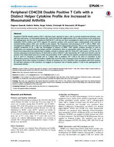
Fig. 1. Transverse sections showing epi-
dermal specializations in the developing bill of late embryonic and early post-hatching platypus. In the 10-mm CRL embryo (a), thickening of the epidermis begins in longitudinally oriented bands (arrows) on the dorsal surface of the upper bill. Shortly after hatching (b), ingrowth of the epidermis into the dermis is visible in longitudinally oriented ridges. These are adjacent to nerve branches (c), but do not appear to be innervated as yet. By the 3rd week after hatching, large numbers of epidermal specializations (arrows) are visible on the dorsal surface of the upper bill (d, e) and the ventral surface of the lower bill (f). Those on the dorsum are arranged in longitudinal rows (clusters of arrows in d), whereas those on the lower bill are less advanced structurally (arrow in f). The midline is to the left in photographs a, b, d, f. Md = Mandible; v = venous sinus.
f
Results
Early Development of Putative Electroreceptors in the Bill or Beak In the 10-mm crown to rump length (CRL) platypus embryo (M42), some thickening of the epidermis in longitudinal ridges along the dorsal surface of the upper bill was visible (fig. 1a). These thickenings are formed by palisades of darkly stained stratum germinativum cells and Extant Monotremes: Distinct Peripheral Trigeminal Pathway Development
are adjacent to fibres of 5max. Around the time of birth (16.75-mm CRL, M44; fig. 1b, c) the first definitive epidermal specializations begin to appear in the platypus bill. These are made up of longitudinal ridges of thickened epidermis, rather than individual pegs, and invade the dermis along the dorsolateral surface of the upper bill and the ventral surface of the lower bill. Over the subsequent week (i.e. by 33-mm CRL, M45; fig. 1d–f), these ridges break up into individual epidermal pegs penetratBrain Behav Evol
5
a
b
c
g
f
d
e
h
mm CRL post-hatching platypus as represented in transverse sections at intervals of 0.3 mm. Epidermal specializations are indicat-
ed by filled circles. Note the early presence of longitudinal zonation of putative electroreceptors. The midline is to the left in all diagrams. Md = Mandible; Mx = maxilla; VNO = vomeronasal organ.
ing 50–100 m into the dermis, predominantly along the dorsal surface of the upper bill, but also on the ventral surface of the lower bill. These are identical in appearance to those structures identified as the developing ducts of mucous electroreceptors [Manger et al., 1998a]. The specializations are loosely clustered into longitudinal domains in the 33-mm CRL platypus (fig. 2). One zone begins at the rostrocaudal level of the naris and extends dor-
sal to the nasal cavity to the large skin fold that marks the caudal edge of the beak. Another zone lies along the ventrolateral edge of the upper bill, beginning from immediately rostral to the naris, but becoming discontinuous towards the lateral angle of the oral cavity. It should be emphasized that although the early appearance of these epidermal specializations is broadly similar to developing hair follicles, at no time during the life of the platypus
Fig. 2. Distribution of epidermal specializations on the bill of a 33-
6
Brain Behav Evol
Ashwell /Hardman /Giere
a
c
b
d
Fig. 3. Transverse sections showing epidermal specializations in the developing beak of late embryonic and early posthatching short-beaked echidna. Immediately before hatching (a), punctate epidermal specializations begin to appear on the ventral surface of the upper beak (arrows), on each side of the egg tooth. At high power (b) of the structure marked by an asterisk in (a), these consist of epidermal plugs invading the dermis. Shortly after hatching, these structures are more advanced morphologically, with the development of a duct to the surface (c) and are distributed on the ventral surface of the upper beak and the tip of the lower beak (arrows in d). c Image of the structure marked with an asterisk in (d).
do hair follicles appear on this region of the body. The subsequent development of these specializations over the next 6 weeks involves a rapid increase in their number, their alignment into longitudinal stripes and their elongation/further penetration of the dermis as described by Manger et al. [1998a]. The development of epidermal specializations in the beak of the short-beaked echidna follows a rather different pattern from that seen in the platypus. Even before birth (12.5-mm CRL, immediately pre-hatching, M158; fig. 3a, b), the epidermal specializations in the echidna
beak appear as a few discrete epidermal pegs penetrating the dermis. Histologically, these appear to be more developmentally advanced than the early post-hatching platypus. Furthermore, in contrast to the platypus, they are located only on the underside of the upper beak at this early age. In subsequent ages (24-mm CRL, M161; fig. 3c, d), these epidermal specializations continue to be concentrated on the underside of the beak tip, with others appearing on the ventral surface of the lower beak (fig. 3d, 4a–e). Only a very few are seen on the dorsolateral surface of the end of the upper beak. By 98-mm CRL, the tip of
Extant Monotremes: Distinct Peripheral Trigeminal Pathway Development
Brain Behav Evol
7
Table 5 . Relationship between trigeminal pathway dimensions
and body length in platypus and short-beaked echidna Structure
No. measurements
ma
ba
5Gn platypus 5Gn echidna 5oph platypus 5oph echidna 5max platypus 5max echidna 5man platypus 5man echidna 5oph + 5max + 5man platypus 5oph + 5max + 5man echidna
20 10 18 6 18 6 18 6 18 6
+1.378 +2.012 +1.560 +1.285 +1.607 +1.364 +1.605 +1.270 +1.596 +1.313
–2.155 –4.020 –3.682 –4.015 –3.464 –3.862 –3.652 –4.029 –3.108 –3.469
a Regression line of the form: logX = b + m(log10 body length), where X is the volume of 5Gn in mm3 or the area of branches of 5n in mm2.
the upper beak and the distal ventral surface of the lower beak are covered by putative developing electroreceptors penetrating deeply into the dermis (fig. 4f–j), with a few epidermal specializations on the dorsal surface of the upper beak caudal to the naris. These specializations can be definitively identified as developing eccrine glands of the gland duct receptor system because of the presence of ducts (fig. 4i). As already observed for the platypus, we can exclude that these epidermal specializations are developing hair follicles because hairs never develop on the echidna beak. One feature that both species have in common is that the epidermal specializations appear first towards the tip of the bill or beak and, with subsequent development, progressively emerge more caudally along it. Development of the 5Gn and Divisions There is a substantial difference in the size of the platypus and echidna 5Gn even at the earliest ages that it can be recognized and its volume measured (i.e. 0.0317 vs. 0.00085 mm3 at 6.5-mm CRL in the platypus and at 5.5mm CRL in the short-beaked echidna, respectively). This difference in volume consistently remains at about 5-fold during pre-hatching and early post-hatching development (fig. 5; tables 1, 2). The regression lines for the two species (fig. 5; table 5) converge towards adulthood, but this may be an artefact of the few juvenile echidna specimens available for the analysis. The earliest outgrowth of branches of the trigeminal nerve (5n) into the maxillary and mandibular processes 8
Brain Behav Evol
occurs as soon as 5Gn is recognizable and discrete 5oph, 5max and 5man can be identified at 8.5-mm CRL (M39) in the platypus and at 7.3-mm CRL (M153) in the shortbeaked echidna. Even at this early age, there is a substantial difference between the two species in the cross-sectional areas of the divisions (tables 1, 2). This difference not only continues throughout development, but increases from an approximately 6-fold difference in total crosssectional area for all 5n divisions at hatching to more than 15-fold in adulthood [fig. 6; table 5: values of slope for the regression lines (‘m’ column)]. It should also be noted that the 5man tends to be of greater cross-sectional area than 5oph during post-hatching development in the platypus, whereas in the developing echidna they are roughly similar. These differences reflect the greater behavioural significance of the ventral surface of the lower bill in the platypus. In order to place these measurements in perspective, it is useful to compare the cross-sectional areas of 5n branches and 5Gn volume with those in a marsupial (tammar wallaby, M. eugenii) and a eutherian (common shrew, S. araneus), both of which are born in an altricial state. In addition, both exhibit (vibrissal) specialization of the trigeminal pathways. Tables 3 and 4 and figures 5 and 6 show values for these measurements for the newborn and early pouch young tammar and developing common shrew alongside the two monotremes. With respect to both the volume of 5Gn (fig. 5c) and the crosssectional area of 5max (fig. 6c), the tammar is intermediate between the two monotremes at a similar developmental stage. The common shrew has a similar 5Gn volume and 5oph and 5man cross-sectional area to both the wallaby and echidna (fig. 5c, 6a, c), but has a large 5max cross-sectional area due to a comparatively high number of vibrissae [Hyvärinen, 1972] which is reflected in a large area of the sensory part of 5n in soricine shrews [Hyde, 1957] (fig. 6b).
Discussion
Ethical and Technical Considerations: Benefits and Limitations of Archived Material Our findings are based on analysis of archived material collected in the late 19th and early 20th centuries. It would be impossible to make such a collection today, because of public concern over the conservation of monotremes and ethical limitations on experimentation with wild-caught animals, so the material in the Hill and Hubrecht collections is quite literally precious. Acquisition Ashwell /Hardman /Giere
a
b
c
f
d
e
h
g
i
j
Fig. 4. a–e Distribution of epidermal specializations on the beak of a 24-mm CRL echidna as represented on transverse sections at intervals of 0.3 mm. f, g Epidermal specializations (arrows) in the upper and lower snout tip of a 98-mm CRL echidna. h, i The epidermal specializations at higher power and with a narrow lumen.
j The concentric arrangement of epidermal cells around the pe-
Extant Monotremes: Distinct Peripheral Trigeminal Pathway Development
Brain Behav Evol
riphery of the duct primordium of the putative electroreceptor, with mitotic figures at the rim (arrow). Md = Mandible; Mx = maxilla; VNO = vomeronasal organ.
9
a
Fig. 5. 5Gn in the platypus (a) and echidna (b) embryos at the same developmental stage. Images were taken at the largest cross-sectional area and the midline is to the left in both photomicrographs. Note that the 5Gn is immediately adjacent to the pons (Pn) in the echidna, but its large size in the platypus pushes it rostrally and away from the pons. c Changes in volume of the 5Gn with body length (CRL for embryos and early post-hatching animals, DCL for subadults and adults) in platypus, echidna, tammar wallaby and common shrew. Note that both scales are logarithmic to accommodate the large changes in volume of the 5Gn during development, but that the 5Gn of the developing platypus is always 5! larger in volume than that of the echidna at a similar body length. Regression lines, correlation coefficients and p values are given for the short-beaked echidna and platypus (see table 5: regression line coefficients).
b
c
of the 34 embryos, juveniles and adults used in our study would require the capture, stress and/or death of many more than 34 adult female monotremes, a practice which would be unacceptable to the modern Australian public. The Hubrecht and Hill collections therefore provided a unique scientific opportunity to ask questions about monotreme development in the light of modern hypotheses concerning monotreme phylogeny, physiology and behaviour. It is inevitable that such material brings with it limitations. The staining of the material was actually still suitable for our analysis more than a century after preparation. But most of the embryonic material had been sectioned at 10-m thickness and the post-hatching material even thicker, so the quantitative assessment of the number of 5Gn neurons or the number of developmentally pyknotic ganglion cells was not possible. Nev10
Brain Behav Evol
ertheless, volumetric and areal analysis could still be accurately performed using calibrated photographs. All of the material, with the exception of S. araneus 81a, had been paraffin- or celloidin-embedded with consequent dehydration, so we did not attempt to apply any corrections back to fresh volume. In any event, the necessary calibration data to perform such a correction are not available. Developmental Ages of Monotremes A further limitation of working with archived material in these species is that it is difficult to assign precise developmental ages to the specimens. Information on the time course of the development of monotremes is patchy. It is not known exactly how many days pass between conception and egg-laying, but it is between 15 and 21 days Ashwell /Hardman /Giere
a
b
c
d
Fig. 6. Changes in the cross-sectional area of the 3 divisions of the trigeminal nerve, 5oph (a), 5max (b) and 5man (c) and their sum (d) during development of the platypus, echidna, tammar wallaby and common shrew. Note that both scales are logarithmic to accommodate the large changes in both nerve area and
body length during development, but that the trigeminal divisions of the platypus are always much larger than those of the echidna at a similar body length. Regression lines, correlation coefficients and p values are given for the short-beaked echidna and platypus.
for the platypus [Holland and Jackson, 2002; Hawkins and Battaglia, 2009] and 20 to 24 days for the shortbeaked echidna [Morrow et al., 2009]. The greater part of embryogenesis occurs in the period from egg-laying to hatching [Hughes and Hall, 1998; Werneburg and Sánchez-Villagra, 2011], which is believed to be approximately 10–11 days for both platypus and short-beaked echidnas [Renfree et al., 2009]. Semon [1894a, b] recognized 14 stages of development during the incubation of echidna pouch eggs (termed 40–53) and stages 46–53 in pouch young. Stage 40 (i.e. the newly laid egg) is an embryo 7 mm long with lobate forelimbs, buds
for hind limbs, prominent pharyngeal pouches, about 39 pairs of somites and a tail. Although the precise time sequence of embryological development is uncertain, the size of newly hatched monotremes is known with some confidence. Since the laid monotreme egg is approximately 16–17 mm at most in length, this sets an upper limit on the embryonic size of about 14–15 mm [similar to observed dimensions (14.7-mm CRL) in Griffiths et al., 1969; see discussion in Rismiller and McKelvey, 2003]. Body masses of newly hatched echidnas have been reported to be 0.303 g [Rismiller and McKelvey, 2003] or 0.378–0.380 g [Griffiths et al., 1969; Griffiths, 1978], re-
Extant Monotremes: Distinct Peripheral Trigeminal Pathway Development
Brain Behav Evol
11
spectively. So, when assigning pre-hatching ages, we assumed 10 days between laying and hatching with a steady 1 mm/day increase in length (tables 1, 2). We do not know precisely the full length of the embryonic period because the length of the period prior to egg-laying is uncertain, so we gave the estimated pre-hatching ages as days before hatching. Of course, all these estimates of age may be subject to change as better data become available. In this study we therefore relied mainly on body length measurements as an indicator of developmental age, because there remains some uncertainty as to how these can be translated into prenatal developmental age in both species, and postnatal ages in the echidna. Nevertheless, the observed differences between the species are sufficiently robust that small errors in assigning developmental age should have no impact on the interpretation of our findings. For embryonic material we used CRL, because that is usually the only measurement provided for the embryos and for early post-hatching animals and dorsal contour length (DCL) for older platypus and echidna (AMNH 201311, AMNH 201312, USNM 221112, adults) where the profound body curvature of early post-hatching young has been lost. Functional Anatomy of Electroreceptors in Monotremes: When Do Distinct Characters Emerge in Development? All of the extant monotremes are believed to use electroreception to find their prey, although this has been behaviourally tested only in the case of the platypus and short-beaked echidna; for the long-beaked echidna it is inferred from histology [Manger et al., 1997]. The platypus and echidna use electroreception in rather different ways and this is reflected in the distribution of electroreceptors on their bill or beak, respectively. The platypus actively sweeps its bill from side to side while swimming close to the river or lake bottom [Manger and Pettigrew, 1995; Pettigrew, 1999]. This sweeping movement carries electrical field lines generated by its crustacean prey across the longitudinally arranged rows of bill electroreceptors, giving rise to time-dependent changes in signal. Central processors then use these signal changes (perhaps in concert with mechanoreceptive input) to generate a map of electrical field sources in the immediate vicinity [Pettigrew et al., 1998]. When the prey is close enough, mechanoreceptors in the bill contribute by detecting pressure waves from its movement. As would be expected from this, mucous gland electroreceptors are found in longitudinal arrays over the entire cutaneous surface of the upper bill and the rostral two thirds of the 12
Brain Behav Evol
ventral cutaneous surface of the lower bill, with additional longitudinal alignments on the palatal surface of the upper bill and the lingual surface of the lower bill [Manger and Pettigrew, 1996]. By contrast, the short- and long-beaked echidnas use their electroreceptors to probe leaf litter and termite mound interiors. Consequently, the electroreceptors are concentrated in regions that would face arthropods in the leaf litter, i.e. on the tip or underside of the upper beak tip and the ventral surface of the lower beak tip [Andres et al., 1991; Manger and Hughes, 1992], forming an electroreceptive ‘hot spot’ rather than the longitudinal stripes of the platypus. More electroreceptors are reported to be present in the beak of Zaglossus than Tachyglossus [Manger et al., 1997], perhaps because the environment of Zaglossus contains abundant, moist leaf litter that would favour the use of electroreception. The number of electroreceptors is substantially greater in the platypus than the tachyglossids, and this is reflected in the much larger size of the trigeminal pathways (5Gn volume and cross-sectional areas of divisions) in the adult platypus compared to the echidnas. Our findings suggest that this very different distribution of electroreceptors in the platypus and echidna has its origins even before the time of hatching. Manger et al. [1998a] identified putative mucous electroreceptors in sections through the platypus bill at post-hatching day 28, but they did not examine any sectioned material from an age younger than this. However, they did claim to be able to identify putative mucous gland electroreceptors from post-hatching day 10 on the basis of external duct pits seen by stereomicroscopic examination of the bill surface in undissected specimens. In the sectioned material at the Museum für Naturkunde, Berlin, we saw epidermal specializations with very different topography in the two species from around hatching time. These structures develop ducts in both species and are identical in appearance to the developing mucous gland electroreceptors described in the platypus at post-hatching day 28 by Manger et al. [1998a]. We were not able to identify definitive unmyelinated axons surrounding the cuff area of the putative developing electroreceptor at the peri-hatching period, perhaps because of the thickness of the archived sections, but these epidermal specializations are usually surrounded by a pale area, much like that occupied by unmyelinated axons at post-hatching day 28 [Manger et al., 1998a]. Even at posthatching day 28, unmyelinated axons are very difficult to discern in the cuff region of the electroreceptor [Manger et al., 1998a, fig. 8a] and it is possible that they have Ashwell /Hardman /Giere
Table 6. Key differences in peripheral trigeminal pathway development between platypus and short-beaked echidna
Platypus
Short-beaked echidna
Site of first appearance of epidermal specializations on bill/beak
dorsal surface of upper bill
ventral surface of upper beak
Nature of earliest epidermal specialization
longitudinal ridge of epidermal thickening
focal epidermal invasion of dermis
Distribution of epidermal specializations
longitudinal ridges or stripes
focal or spot-like
Size of 5Gn
very large at all stages of development; approximately 5! that of the echidna
moderate in size throughout development; comparable in size to tammar wallaby and common shrew
Size of 5n components
very large at all stages of development; approximately 5! that of the echidna
moderate in size throughout development; comparable in size to tammar wallaby
Relative size of 5n divisions
5max > 5man > 5oph
5max > 5oph = 5man
yet to reach the developing electroreceptors at the time of hatching. It is possible that some of the structures are nonsensory mucous glands, but we believe that the bulk of them are sensory, for the following reasons. (1) In the adult platypus, sensory mucous glands are larger and more prominent than nonsensory ones, (2) they outnumber the nonsensory type by 3 to 2 in the adult platypus [Manger and Pettigrew, 1996] and (3) they appear to be the first to develop [Manger et al., 1998a]. In any event, the nonsensory glands of the adult and developing platypus have a linear arrangement like the sensory ones (only complementary in position) [Manger and Pettigrew, 1996], so the presence of linearly aligned mucous gland epidermal specializations (whether sensory or nonsensory) leads to the same conclusion, i.e. that the developing platypus bill is programmed to develop towards the adult structure from an early stage of development. In the platypus, the first specializations of the epidermis are seen in the last third of incubation (10-mm CRL) and consist of longitudinally running zones of epidermal thickening, which progress to ridge-like epidermal ingrowth (immediately post-hatching), which in turn develop into individual finger-like epidermal invasions of the dermis (end of 1st week of post-hatching life). All of these specializations have a longitudinal arrangement that closely matches the major arrays of electroreceptors reported for the late post-hatching and mature platypus bill [Manger and Pettigrew, 1996; Manger et al., 1998a], suggesting that the genetic control of the development of the distinctively platypus electroreceptor topography is active from late incubation.
In the case of the echidna, the first epidermal specializations were seen on the underside of the beak tip (late incubation, 12.5-mm CRL), exactly where the peak density of electroreceptors will be in the adult [Manger and Hughes, 1992]. At no stage was any longitudinal banding of epidermal specializations seen in the developing echidna beaks, suggesting that the distinctive ‘spot-like’ topography of electroreceptors seen in the adult echidna is established from the very earliest differentiation of electroreceptors. We did not see any putative pushrod mechanoreceptors in the embryonic or immediate post-hatching specimens. These mechanoreceptors have a distinctive appearance that cannot be confused with mucous gland electroreceptors, because pushrod receptors do not have a duct system that invades the dermis, and are characterized by central and peripheral vesicle chains through the core of the rod [Manger et al., 1998a]. These mechanoreceptors appear to develop towards the end of the 1st month of post-hatching life [Manger et al., 1998a].
Extant Monotremes: Distinct Peripheral Trigeminal Pathway Development
Brain Behav Evol
Phylogenetic Significance of the Developmental Patterns in the Trigeminal Pathways of the Platypus and Echidna We have observed that the peripheral trigeminal pathways of the platypus and short-beaked echidnas have distinct features from the earliest stages of development (summarized in table 6) and that these species-specific features are anatomically and functionally consistent with the role of the bill or beak in the respective adults. It is easier for a complex sensory apparatus like the electro13
receptive components of the trigeminal pathway to maintain functionality during evolution by confining evolutionary modifications to the later stages of development. Our findings of quite distinct anatomical features in the embryonic and early post-hatching trigeminal pathways of these two species are therefore consistent with the two lineages of modern monotremes (i.e. ornithorhynchids and tachyglossids) having pursued separate paths of sensory specialization for long periods of time. The early ontogeny of species-specific, spatially determined distributions of epidermal specializations is entirely consistent with developmental processes having been shaped differently from early embryonic stages in both lineages in order to produce the characteristic distribution pattern of electroreceptors in the adult. Our findings do not support the proposition that the modern echidna is derived from a platypus-like ancestor that left an aquatic environment to invade a land-based insectivorous niche, although we would agree that electroreception was present in the common ancestor of both and may have been used to search for invertebrates in moist rainforest leaf litter. We would suggest that the extreme trigeminal specializations of the platypus (enlarged 5Gn and 5n divisions, lon-
gitudinal zonation of electroreceptors) arose by the retention of 5Gn cells generated in excess during early embryonic life, in concert with the progressive shift of bill electroreceptors into longitudinal arrays. The most parsimonious explanation of our observations is that these changes emerged after the divergence of the ornithorhynchid and tachyglossid lineages, because no sign of 5Gn hyperplasia or longitudinal zonation of electroreceptors is seen at any stage in the developing echidna. Our conclusions are consistent with the proposal of Finlay et al. [2011] that the periphery of the nervous system is the principal locus for producing functional changes in neuroanatomy between species.
Acknowledgements We would like to thank the Alexander von Humboldt Foundation for their financial support of this project. We are very grateful to Professor Ulrich Zeller of the Museum für Naturkunde, Berlin, for kindly providing access to his collection of sectioned platypus and echidna heads and to Dr. Lauren Marotte of the Australian National University for wallaby head-length measurements.
References Andres KH, von Düring M, Iggo A, Proske U (1991): The anatomy and fine structure of the echidna Tachyglossus aculeatus snout with respect to its different trigeminal sensory receptors including the electroreceptors. Anat Embryol 184: 371–393. Ashwell K, Marotte LR, Mai JK (2010): Atlas of the brain of the developing tammar wallaby (Macropus eugenii); in Ashwell KWS (ed): The Neurobiology of Australian Marsupials. Cambridge, Cambridge University Press, pp 245–248. Camens, AB (2010) Were early tertiary monotremes really all aquatic? Inferring paleobiology and phylogeny from a depauperate fossil record. Proc Natl Acad Sci USA 107:E12. Finlay BL, Hinz F, Darlington RB (2011) Mapping behavioural evolution onto brain evolution: the strategic roles of conserved organization in individuals and species. Phil Trans Roy Soc 366:2111–2123. Giere, P, Möller L, Hilger A, Paulke A, Riesemeier H, Kuehbacher M (2010): Modern and classic approaches towards revealing the orbital mosaic in the common shrew Sorex araneus Linnaeus, 1758 (Soricidae, Lipotyphla, Mammalia). Zoosyst Evol 86:343–350. Gregory JE, Iggo A, McIntyre AK, Proske U (1987): Electroreceptors in the platypus. Nature 326:386–388.
14
Brain Behav Evol
Gregory JE, Iggo A, McIntyre AK, Proske U (1988): Receptors in the bill of the platypus. J Physiol 400:349–366. Gregory JE, Iggo A, McIntyre AK, Proske U (1989): Responses of electroreceptors in the snout of the echidna. J Physiol 414:521–538. Griffiths M (1978): The Biology of the Monotremes. New York, Academic Press. Griffiths M, McIntosh DL, Coles REA (1969): The mammary gland of the echidna, Tachyglossus aculeatus, with observations on the incubation of the egg and on the newly hatched young. J Zool (Lond) 158:371–386. Hawkins M, Battaglia A (2009): Breeding behaviour of the platypus (Ornithorhynchus anatinus) in captivity. Aust J Zool 57:283–293. Holland N, Jackson SM (2002): Reproductive behaviour and food consumption associated with the captive breeding of platypus (Ornithorhynchus anatinus). J Zool (Lond) 256: 279–288. Hughes RL, Hall LS (1998): Early development and embryology of the platypus. Phil Trans Roy Soc Lond B Biol Soc 353:1101–1114. Hyde JB (1957): A comparative study of certain trigeminal components in two soricid shrews, Blarina brevicauda and Sorex cinereus. J Comp Neurol 107:339–351. Hyvärinen H (1972): On the histology and histochemistry of the snout and vibrissae of the
common shrew (Sorex araneus L.). Z Zellforsch 124:445–453. Manger PR, Collins R, Pettigrew JD (1997): Histological observations on presumed electroreceptors and mechanoreceptors in the beak skin of the long-beaked echidna, Zaglossus bruijnii. Proc Roy Soc Lond B Biol Sci 264: 165–172. Manger PR, Collins R, Pettigrew JD (1998a): The development of the electroreceptors of the platypus (Ornithorhynchus anatinus). Phil Trans Roy Soc Lond B Biol Sci 353:1171–1186. Manger PR, Hall LS, Pettigrew JD (1998b): The development of the external features of the platypus (Ornithorhynchus anatinus). Phil Trans Roy Soc Lond B Biol Sci 353:1115–1125. Manger PR, Hughes RL (1992): Ultrastructure and distribution of epidermal sensory receptors in the beak of the echidna, Tachyglossus aculeatus. Brain Behav Evol 40:287–296. Manger PR, Pettigrew JD (1995): Electroreception and the feeding behaviour of platypus (Ornithorhynchus anatinus, Monotremata, Mammalia). Proc Roy Soc Lond B Biol Soc 347:359–381. Manger PR, Pettigrew JD (1996): Ultrastructure, number, distribution and innervation of electroreceptors and mechanoreceptors in the bill skin of the platypus, Ornithorhynchus anatinus. Brain Behav Evol 48:27–54.
Ashwell /Hardman /Giere
Morrow G, Andersen NA, Nicol SC (2009): Reproductive strategies of the short-beaked echidna – a review with new data from a long-term study on the Tasmanian subspecies (Tachyglossus aculeatus setosus). Aust J Zool 57:275–282. Musser AM (2003): Review of the monotreme fossil record and comparison of paleontological and molecular data. Comp Biochem Physiol A Molec Integr Physiol 136:927–942. Pettigrew JD (1999): Electroreception in monotremes. J Exp Biol 202:1447–1454. Pettigrew JD, Manger PR, Fine SLB (1998): The sensory world of the platypus. Phil Trans Roy Soc Lond B Biol Sci 353:1199–1210. Phillips MJ, Bennett TH, Lee MSY (2009): Molecules, morphology, and ecology indicate a recent, amphibious ancestry for echidnas. Proc Natl Acad Sci USA 106:17089–17094.
Extant Monotremes: Distinct Peripheral Trigeminal Pathway Development
Proske U, Gregory JE, Iggo A (1998): Sensory receptors in monotremes. Phil Trans Roy Soc Lond B Biol Sci 353:1187–1198. Renfree MB, Papenfuss AT, Shaw G, Pask AJ (2009): Eggs, embryos and the evolution of imprinting: insights from the platypus genome. Reprod Fertil Dev 21:935–942. Rismiller PD, McKelvey MW (2003): Body mass, age and sexual maturity in short-beaked echidnas, Tachyglossus aculeatus. Comp Biochem Physiol A Molec Integr Physiol 136: 851–865. Santander RG, Cuadrado GM, González-Santander M, Monteagudo M, Alonso FJM, Lobo MVT (1997): The use of different fixatives and hydrophilic embedding media (HistoresinTM and UnicrylTM) for the study of embryonic tissues. Microsc Res Tech 36:151– 158. Scheich H, Langner G, Tidemann C, Coles RB, Guppy A (1986): Electroreception and electrolocation in platypus. Nature 319:401–402.
Brain Behav Evol
Semon R (1894a): Die Embryonalhüllen der Monotremen und Marsupialier. Denkschriften Med-Naturwiss Ges Jena 5:19–58. Semon R (1894b): Zur Entwicklungsgeschichte der Monotremen. Denkschriften Med-Naturwiss Ges Jena 5:61–74. Smeele LE (1989): Ontogeny of relationship of middle ear and temporomandibular (squamomandibular) joint in mammals. II. Morphology and ontogeny in insectivores. Acta Anat (Basel) 134:62–66. Werneburg I, Sánchez-Villagra MR (2011): The early development of the echidna, Tachyglossus aculeatus (Mammalia: Monotremata), and patterns of mammalian development. Acta Zoologica 92:75–88. Zeller U (1989): Die Entwicklung und Morphologie des Schädels von Ornithorhynchus anatinus: (Mammalia, Prototheria, Monotremata). Abh Senckenberg Naturforsch Ges 544:1–188.
15