INTERNATIONAL TRENDS IN IMMUNITY ISSN 2326-3121 (Print) ISSN 2326-313X (Online)
VOL.1 NO.2 http://www.researchpub.org/journal/iti/iti.html
APRIL 2013
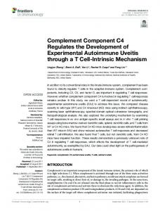
Does the Complement System Work for or Against the Host during Parasite Infections Hiro Goto and Maria Carmen Arroyo Sanchez
the intended function of parasite destruction. The reason for this failure is the development of efficient mechanisms for the parasites to evade complement attack or the subversion of this system (i.e., using components of the complement system to facilitate the infection process). In this review, we intend to convey the complexity of the interaction with the complement system, which can function to eliminate parasite function, to facilitate infection or to facilitate the pathogenesis of various disease manifestations. This review is not comprehensive; however, we emphasize the systems and components necessary for an understanding of complement system-parasite interactions, as exemplified by helminth and protozoan infections (see Boxes 1, 2 and 3 for information on the epidemiology, parasitic life cycle and clinical manifestations). Briefly, the complement system comprises more than 30 plasma and membrane-associated proteins that interact in a cascade through the sequential activation of many proenzymes that catalyze the activation of other enzymes (reviewed in [1]). Several complement activation pathways have been described (reviewed in [2]). The classical (CP), alternative (AP) and mannose-binding lectin (MBL) pathways (Fig. 1) lead to the formation of the C3 convertases, which cleave C3 into C3b and C3a [3]. The classical pathway begins with the binding of the C1 complex, specifically the C1q fraction, usually to an antibody in the immune complex. C1q then sequentially activates the serine proteases C1r and C1s, which cleave C4 and C2 to generate the C4a, C4b, C2a and C2b fragments. The C4b and C2a fragments then form the C4b2a complex, which constitutes the C3 convertase of the classical pathway. The alternative pathway is activated regardless of the presence of an antibody when C3 reacts with a hydroxyl or amino group on the surface of a pathogen. The resulting surface-bound C3b forms C3bB with factor B. Factor D then cleaves factor B into Ba and Bb, and the latter associates with C3b to form C3bBb, which constitutes the C3 convertase of the alternative pathway. The lectin pathway is activated when mannose-binding lectin (MBL) binds to mannose on the pathogen surface. This pathway is similar to the classical pathway but acts in the absence of antibody. MBL, or alternatively ficolin, activates MBL-associated serine proteases 1 and 2 (MASP-1 and MASP-2). These MASPs cleave C4 and C2 similarly to C1r and C1s in the classical pathway, leading to the formation of the C3 convertase C4b2a. C3 is the main component of the complement system, and all three pathways with their
Abstract—Studies regarding the role of the complement system in parasitic infections have demonstrated that the complement system often fails to achieve the intended task of destroying the parasite. The reason for this failure is the development of efficient mechanisms for the parasites to evade attack or the subversion of the complement system (i.e., using components of the complement system to facilitate the infection progress). In this review, we intend to demonstrate the complexity of the interaction between parasites and the complement system in schistosomiasis mansoni, leishmaniases and malaria. The complement system is activated through the classical, alternative or lectin pathways, but in many circumstances, it facilitates infection, as occurs in leishmaniases, or exerts almost no protective effects against the parasite, as observed in schistosomiasis mansoni, due to the evasion mechanisms and biological characteristics of the parasite. In malaria, the complement system participates in erythrocyte invasion and, depending on the degree of activation, in different pathogenic mechanisms related to the uncomplicated or severe manifestations of malaria. This review is not comprehensive; however, we emphasize the systems and components necessary for an understanding of complement system-parasite interactions. Index Terms— complement system, evasion mechanisms, innate immunity, leishmaniasis, malaria, parasite, schistosomiasis
I. INTRODUCTION
T
HE complement system is a major player in innate immunity, and because complement is present in tissue fluids and in the blood, it encounters parasites immediately upon introduction into the host organism. Most of the initial studies on the role of complement in parasite infections have approached this system as an innate host component for controlling infection by inducing parasite killing, as the system has been long perceived. This perception is changing in many fields and types of infections. The development of studies examining the role of complement in parasitic infections has shown that the complement system fails in many ways to exert Submitted Feb 18, 2013 and accepted March 15, 2013 Hiro Goto is with the Laboratorio de Soroepidemiologia e Imunobiologia, Instituto de Medicina Tropical de São Paulo, USP, São Paulo, Brazil and Departamento de Medicina Preventiva, Faculdade de Medicina da USP, São Paulo, Brazil. Address: Instituto de Medicina Tropical de São Paulo, Av. Dr. Enéas de Carvalho Aguiar 470, prédio II, 4°andar, 05403-000, São Paulo, SP, Brazil. Tel.: +55-11-30617023; Fax: +55-11-30618270 (e-mail:
[email protected]). Maria Carmen Arroyo Sanchez is with the Laboratorio de Soroepidemiologia e Imunobiologia, Instituto de Medicina Tropical de São Paulo, USP, São Paulo, Brazil (e-mail:
[email protected]).
11
INTERNATIONAL TRENDS IN IMMUNITY ISSN 2326-3121 (Print) ISSN 2326-313X (Online)
VOL.1 NO.2 http://www.researchpub.org/journal/iti/iti.html
respective C3 convertases converge to promote the cleavage of C3 into C3a (chemotactic factor) and C3b. C3b with C3 convertase then form the C5 convertase that cleaves C5, generating the C5a (chemotactic factor) and C5b fragments. Subsequently, the stepwise assembly of the components C5b, C6, C7, C8 and C9 is initiated, resulting in the formation of the membrane attack complex (MAC) C5b-C6-C7-C8-C9 [4]. The polymerized final components form a pore-like structure in the membrane, leading to the lysis of the targeted cells. There are two additional pathways that are independent of C3: the thrombin-mediated pathway, in which thrombin acts as a C5 convertase [5], and the pathway in which activated neutrophils and macrophages secrete serine proteases that cleave C5 [6].
APRIL 2013
The resultant fragments and the complex define the main functions of the complement system in the innate immune response, which are as follows [1]: 1) membrane lysis through the assembly of the membrane attack complex (MAC), which leads to the formation of membrane pores; 2) inflammation due to the generation of the potent pro-inflammatory anaphylatoxins C3a, C4a, and C5a, which bind to their respective receptors expressed on macrophages, neutrophils, basophils, eosinophils, mast cells and some dendritic cells; and 3) the opsonization and clearance of opsonized elements through the C4b, C3b, and C3bi fragments, which can bind to complement receptors on phagocytic cells. Due to the destructive potential of complement activation, the complement cascade is regulated by several proteins in the plasma and on the cell surface [7] to prevent autologous complement attacks [1]. The regulators include membrane cofactor protein (MCP or CD46), decay-accelerating factor (DAF or CD55), membrane inhibitor of reactive lysis (MIRL or CD59) and complement receptor 1 (CR1 or CD35) (reviewed in [8]). CR1, CD55, and MCP are members of the regulators of complement activation (RCA) protein family [8]. CR1, CD55, and CD59 are found on the erythrocyte surface and are responsible for the complement-regulatory properties of these cells [9]. Erythrocytes account for more than 85% of the CR1 in the blood [10]. In humans, the number of CR1 molecules expressed on erythrocytes varies between individuals within a range of 50–1200 molecules per cell [11]. Various single nucleotide polymorphisms (SNPs) of the CR1 gene are associated with variations in the expression levels of erythrocyte CR1 [12] [13]. CR1 plays essential roles in mammalian immunity. It is responsible for the complement-regulatory properties of the erythrocyte surface [9]. CR1 has decay-accelerating activity for C3- and C5-convertases [14], limiting the deposition of C3b and C4b and subsequent excessive complement activation [15]. CR1 acts as a cofactor in the factor I-mediated cleavage of C3b and C4b to iC3b and iC4b, respectively [16]. This activity is thought to play an important role in limiting the activation of the pathway. The expression of CR1 on human erythrocytes is believed to provide an important clearance function. On erythrocytes, CR1 is an immune-adherence receptor that binds to C3b/C4b-opsonized immune complexes and transports them to the spleen and liver for removal [17]. On phagocytic cells, the interaction between the complement receptor and C3bi, together with the binding of C3b to CR1, which is also present on phagocytes, promotes the adherence and phagocytosis of antigens in the liver and spleen [15]. When C3b is uniformly distributed over the erythrocyte surface, CR1 can diffuse through the erythrocyte membrane, interacting with deposited C3b; CR1 also diffuses on neutrophils and lymphocytes [18]. Another function of CR1 is the retention of opsonized antigens necessary to maintain immunological memory on follicular dendritic cells in germinal centers [15] [19]. CD55 and CD59 are erythrocyte surface proteins that also have important roles in protecting erythrocytes from
Fig. 1. The three Activation Pathways of Complement: the Classical, the Mannose-Binding Lectin, and the Alternative Pathways. The classical pathway begins with the binding of the C1 complex (which consists of C1q, two molecules of C1r, and two molecules of C1s) to antibodies (Ab) in the immune complex (the classical pathway may also be activated by apoptotic cells, certain viruses and gram-negative bacteria and C-reactive protein bound to ligand). C1s sequentially cleaves C4 and C2, leading to the formation of the C4b2a enzyme complex, the C3 convertase of the classical pathway. The lectin pathway is initiated by binding of the complex of mannose-binding lectin (MBL) and the serine proteases M–associated proteases 1 and 2 (MASP1 and MASP2, respectively) to mannose groups on the surface of a bacterial cell. MASPs cleave C4 and C2, leading to the formation of the C3 convertase enzyme C4b2a. The alternative pathway is activated by many bacteria, fungi, viruses and tumor cells. This pathway is activated by cleavage of C3 in plasma. The resulting C3b binds covalently to hydroxyl groups on cell-surface carbohydrates and proteins and then binds to factor B to form the C3bB complex. Factor D cleaves factor B bound to C3b to form the complex C3bBb, the alternative pathway C3 convertase. The three pathways lead to the formation of C3 convertase enzymes, which cleave C3 into C3b and C3a fragments. C3b binds to the C3 convertase enzymes, forming C5 convertase enzymes, which cleave C5, generating C5a and C5b fragments. The C5b fragment initiates the stepwise assembly of the components C6, C7, C8, and C9, resulting in the formation of the membrane-attack complex (MAC), C5b-C6C7C8C9n. (reviewed in [152]).
12
INTERNATIONAL TRENDS IN IMMUNITY ISSN 2326-3121 (Print) ISSN 2326-313X (Online)
VOL.1 NO.2 http://www.researchpub.org/journal/iti/iti.html
complement activation by promoting the inactivation of C3 convertases (CD55) and interfering with the assembly of the MAC C5b-9 (CD59) [20].
APRIL 2013
secretion known as membranocalyx [38] [39]. This change is important for evasion because the membranocalyx is weakly immunogenic, can avoid the exposure of parasite molecules present in the plasma membrane, and can take up various host molecules to disguise the parasite as ‘self’ within the host [40]. Membranocalyx was believed to undergo rapid turnover to avoid the fixation of the MAC of the complement system, but data that are more recent indicate that the turnover is not rapid enough to block fixation [40]. Possibly related to the characteristics of the adult worm membrane, which should be better characterized, an intriguing observation was that almost no inflammation was found around the parasite in vessels of CBA and BALB/c mouse strains and in samples from chimpanzees, although inflammation was evident in other tissues around the eggs [41]. Another constituent facilitating evasion is the presence of the Fc receptor detected on the surface of adult Schistosoma mansoni [42], which is similar to the paramyosin molecule. This receptor presumably captures immunoglobulin via the Fc domain, thus disabling the C1q binding [43]. Another molecule with similarity to paramyosin is schistosome complement inhibitory protein-1 (SCIP-1) [44], which is also similar to human complement inhibitor protein CD59, which can bind C8 and C9 and circumvent cell lysis [45]. The existence and localization of paramyosin in Schistosoma mansoni is controversial [35]; furthermore, a recent proteomic analysis did not confirm the presence of the paramyosin molecule on the Schistosoma mansoni surface [36]. Notably, however, a paucity of immunoglobulin deposits were detected on the parasite [42], which may indicate the scarcity of exposed parasite antigen in the tegument, seemingly situated at the plasma membrane [36]. This low amount of bound immunoglobulins may be more relevant for evasion than for the putative role of the Fc receptor and paramyosin in the inactivation of complement attack. Schistosomes have some regulatory molecules that inhibit the activation of the complement cascade. One known regulatory protein is CRIT (complement C2 receptor inhibiting trispanning), also known as Schistosoma trispanning orphan receptor [46], which functions as a receptor for the C2 protein and is shown to inhibit the activation of the classical pathway [47] [48]. In a proteomic analysis of parasite surface membranes, this molecule was not detected [36], but the SmTOR gene was identified in another study, and its structure was analyzed. It was suggested that the receptor is tetraspanning instead of trispanning (however, keeping the same abbreviation TOR), and the protein was detected in adult worm extracts but was mainly found on the cercariae surface [46]. A critical component of complement activation is the C3 protein, and in Schistosome, a C3-binding molecule was detected, and its production by the parasite confirmed [49]. This protein would interfere with complement activation, but its existence is controversial, and the molecule remains uncharacterized [40]. Schistosomes also acquire some molecules from the host that may contribute to evasion. One such protein is DAF, which
II. THE COMPLEMENT SYSTEM IN SCHISTOSOMIASIS MANSONI During the life cycle within the vertebrate host, Schistosoma mansoni induces a variety of innate and acquired immune responses. However, this parasite not only survives these numerous attacks but also lives in hostile environments for years and decades using different mechanisms to evade the immune response (reviewed in [21]). Because the cercariae are the first form that the host copes with, the innate elements are the main players of the immune response. Innate elements also affect other forms, but the acquired immune response is induced by schistosomula, adult worms and eggs in the tissues, which are targets of acquired humoral and cell-mediated mechanisms [22]. Antibody-dependent, cell-mediated cytotoxicity is considered an important defense mechanism against schistosomula, which involves IgG and IgE antibodies and various cell types, including T cells, eosinophils and neutrophils [23] [24]. The cell-mediated immune response is important against eggs in tissues, leading to granuloma formation and regulation [25]. Recently in the field of vaccine development, a discussion has been ongoing regarding the parasite life cycle phase that should be targeted, the vulnerable structures, and the identity of relevant immunogenic molecules; in this discussion, in vitro and in vivo data from different animal models are being critically considered [21]. One of the potent innate elements of the immune system that the Schistosoma must face is the complement system, which also functions as an effector of the acquired humoral immune response. Cercariae [26], schistosomula [27] [28] [29] [30], and adult worms [31] [32] activate the alternative pathway in vitro. MBL was shown to adhere to surface glycoproteins of cercariae and adult worms [33], but in MBL knock-out mice, the course of infection was not altered, raising questions about its biological relevance [34]. In terms of complement activation by the classical pathway, there are data demonstrating the presence of various immunoglobulin classes and subclasses on the Schistosoma surface that may activate the classical complement pathway [35]. Recently, a proteomic analysis confirmed the deposition of different host immunoglobulin classes and subclasses and the further presence of the complement C3 fraction fragments C3c/C3dg [36] [37], indicating the activation of complement at the parasite surface. To evade complement attack, Schistosoma mansoni presents various mechanisms that are persistent throughout the life cycle, in addition to the parasite constituents that are inherently appropriate for life in a hostile environment. An initial alteration that the cercariae manifest within the host during differentiation into the schistosomulum is the shedding of the surface membrane glycocalyx, which is a potent activator of the alternate complement pathway [28]. Concomitantly, the parasite acquires a tegument, which is a syncytium composed of a plasma membrane with an overlaid membrane-like 13
INTERNATIONAL TRENDS IN IMMUNITY ISSN 2326-3121 (Print) ISSN 2326-313X (Online)
VOL.1 NO.2 http://www.researchpub.org/journal/iti/iti.html
protects the schistosomula from antibody-mediated lysis [50]. However, this protein was not detected by proteomic analysis of the adult parasite extract [36]. Complement receptor-related protein y (Crry) was also identified, which inactivates the C3-derived fragments, by proteomic analysis of the adult schistosome extract [36]. It is worth mentioning that most of the data were derived from in vitro observations. In C3-deficient mice [51], no significant difference was observed compared with the wild-type controls. However, in C5-deficient mice, when cobra venom factor (CVF) was used to deplete complement, the worm burden was higher [52], suggesting in vivo a role for the alternative pathway in protection from pathogens. From the above-described data, we conclude that the Schistosoma uses so many tricks and constitutive components to evade complement attack that the presence or absence of complement activation appears to exert only a marginal effect on the parasite.
APRIL 2013
hindering the formation of MAC [70]. Another factor is protein kinase C, which exhibits increased expression in the metacyclic form and phosphorylates several components of the complement system, such as C3, C5, and C9 [71], to block the activation of the classical and alternative pathways. Most studies in the literature have used promastigotes, but L. donovani amastigotes were initially considered nonsusceptible to lysis by complement [55]; however, they have since been shown to be susceptible but to a much lesser extent [72]. Another consequence of complement activation is the generation of the chemotactic fragments C3a and C5a, which promote tissue inflammation by attracting polymorphonuclear neutrophils and monocytes/macrophages to the site of infection. Recently, another mechanism of the migration of effector macrophages to the L. major infection site was shown to be initiated by complement activation, which causes the binding of platelets to L. major, leading to the release of PDGF, which induces CCL2 (MCP-1). It was demonstrated that these effector macrophages efficiently kill the parasites at the early phase of infection [73]. Macrophages phagocytose Leishmanias that engage various membrane receptors, such as fibronectin, mannose, b-glucan, and complement receptor 1 (CR1) and 3 (CR3) [74]. The receptors involved in the interactions between macrophages and amastigotes have not been extensively studied, but the CR3 and Fc receptors are considered important [75] [76]. The parasite LPG, gp63, and advanced glycosylation products participate in this interaction. However, the opsonized parasites appear to play a predominant role in promoting phagocytosis, involving the binding of C3b to the CR1 and CR3 and the binding of iC3b to the CR3 of the macrophage [74]. These interactions contribute to the establishment of infection and the evasion of the immune response required for the survival of the parasite within the macrophages, which possess potent microbicidal mechanisms. Phagocytized Leishmania initially localizes in the parasitophorous vacuole surrounded by the membrane of the host cell, which then fuses with the lysosome to form the phagolysosome that contains the proteolytic enzymes capable of lysing the parasites. Furthermore, parasite phagocytosis generates the macrophage effector elements, reactive oxygen species (mainly hydrogen peroxide = H2O2), which involve NADPH oxidase and superoxide dismutase, and reactive nitrogen species (nitric oxide = NO), which involve inducible nitric oxide synthase (iNOS) [77] [78] [79] [80] [81] [82] [83]. Recently, we observed that the protein-disulfide isomerase –NADPH oxidase interaction influences the phagocytosis of promastigotes of Leishmania chagasi [84]. The major mechanisms of evasion by Leishmania for parasite survival are as follows. Phagosome and lysosome fusion is inhibited, and the differentiation of the promastigotes to amastigotes is favored by LPG [85], which also sequesters hydroxyl radicals and superoxide anions [86]. Parasite lysis by lysosomal enzymes is hindered by gp63 [87] [88], the secreted factors [89] [90] and the cysteine proteinases of Leishmania [91].
III. THE COMPLEMENT SYSTEM IN LEISHMANIASES As soon as the Leishmania promastigotes are injected by phlebotomine sandflies into the skin, the parasite comes into contact with the complement system. Most Leishmania species, such as Leishmania major, L. enrietti, L. mexicana, L. amazonensis, and L. braziliensis, activate the alternative complement pathway [53] [54]. In contrast, L. donovani appears to activate the classical pathway [54] [55], suggesting the participation of natural antibodies. However, because L. major, L. Mexicana, and L. braziliensis promastigotes bind to mannan [56] [57], the activation of the lectin pathway must also be considered [58] [59]. Nonetheless, in experiments under more physiological conditions using whole blood or high serum concentrations, the importance of natural antibodies and the classical pathway has been reconsidered, and the participation of classical pathway has also extended to other Leishmania species [60] [61]. Regardless of the pathway used, C3b deposition on the surface of the parasite constitutes a crucial step in complement activation that may proceed either with the formation of C5 convertase with the subsequent formation of membrane attack complex C5b-C9 implicated in parasite lysis or with its inactivation by generating iC3b. The most abundant molecules on the Leishmania surface, gp63 (also called membrane surface protease) [62] and lipophosphoglycan (LPG) [63], have been identified as C3 acceptors. These molecules have been much studied [64] [65] and has been shown to participate in different processes during the life cycle. Promastigotes become infectious within the insect when they reach the stationary phase of growth and turn into metacyclic forms [66] that are also more resistant to lysis by complement [67]. This resistance to lysis was attributed to the modification of LPG, which becomes longer at this stage [64] and blocks the binding of MAC to the parasite membrane [68]. The expression of gp63 is also increased in the metacyclic form [69], and as a protease, it accelerates the conversion of C3b to iC3b, 14
INTERNATIONAL TRENDS IN IMMUNITY ISSN 2326-3121 (Print) ISSN 2326-313X (Online)
VOL.1 NO.2 http://www.researchpub.org/journal/iti/iti.html
A decrease in the production of reactive oxygen species is desirable to the parasite survival, and this decrease is observed in Leishmania-infected macrophages [92] [93], which may involve the gp63 molecule [94]. Importantly, the opsonization mediated by the C3b and iC3b fragments and the related receptors CR1 and CR3 play an important role in these mechanisms, leading to significant decrease in the oxidative burst [95] [96]. Notably, the oxidative burst induced during the phagocytosis of amastigotes in macrophages is smaller than that induced by promastigotes [97]. Another leishmanicidal element, NO, is also modulated by infection, which may involve Leishmania glicoinositolphospholipid [98], the tyrosine phosphatase SHP-1 [99] or the exposure of phosphatidyl serine observed specifically in the tissue-derived Leishmania amazonensis amastigotes, mimicking cell apoptosis [100]. NO is mainly considered to have a role in specific immunity, but it is very important in the nonspecific response, which functions within the first 24 hours of infection, as observed in mice deficient in iNOS activity [101] [102]. There is no known involvement of complement in the modulation of NO, but we have observed the inhibition of NO production by another nonspecific factor, the insulin-like growth factor –I, which is constitutively present in the skin and within macrophages [103]. Most of data presented here are derived from in vitro studies, but some in vivo experiments reinforce the role of complement in the progression of infection and lesion development rather than parasite control. The most relevant studies focus on the C3 and C3R components. In hamsters transiently depleted of C3 by injection with cobra venom factor (CVF) and infected with L. chagasi, the inflammatory infiltrate and cell parasitism was analyzed during the initial 72 hours and at 240 days post-infection. The depletion of C3 led to a decrease in the amount of infected polymorphonuclear neutrophils (PMNs) and monocytes in the initial period and no detection of the whole parasite at 240 days in the liver or spleen, in contrast to patent infection in nondepleted animals [104]. In experimental cutaneous leishmaniasis, in BALB/c mice transiently depleted of C3 with CVF injection or permanently depleted of C3 with CVF gene transfection and infected with L. major, the lesion development was, respectively, considerably delayed or inhibited [105]. In the latter study, this outcome was attributed to the decreased migration of PMNs to the site of infection, but in the former study using hamsters, PMNs were detected at greater quantities after 2 hours of infection, which contradicts this supposition. The assumption of reduced PMN infiltration leading to lower infectivity is appropriate because in susceptible strains of mice, such as BALB/c, the PMNs are mainly necrotic and apoptotic and contribute to the development of the infection [106]. However, considering that the inhibition of the oxidative burst would occur when phagocytosis is mediated by CR1 and CR3 [95] [96], it may be more likely that in the absence of the engagement of CR1 and CR3 receptors during phagocytosis in C3-depleted animals, the process leads to the full activation of the oxidative burst with consequent parasite killing. The participation of CR3 was
APRIL 2013
analyzed in CR3-depleted (CD11b knock out) BALB/c mice, in which the degree of L. major infection reached an intermediate size between the susceptible wild-type BALB/c mice and resistant C57BL/6 mice [107]. This phenotype was likely due to the preservation of complement activation and the phagocytosis of opsonized parasites through other complement receptors, such as CR1, leading to the partial modulation of the oxidative burst. In human visceral leishmaniasis, higher serum MBL levels were observed in active disease when compared with asymptomatic infected individuals or healthy controls [108]. Furthermore, a gene polymorphism in MBL2 that encodes human MBL was addressed in a case-control study in a population from northeastern Brazil, and it was observed that genotypes related to high serum levels of MBL were present at a significantly greater proportion among patients with active disease and with signs of severe manifestations [109]. These results suggest that the high MBL levels favor the development of infection. In the pathogenesis of glomerular lesions in visceral leishmaniasis, immune complex deposition with the consequent activation of the complement cascade and the resulting inflammation has been the accepted mechanism [110] [111]. However, we have recently shown that T cell-mediated immune mechanisms are apparently more important without any clear participation from complement activation [112]. We suggest that in leishmaniases, when complement is activated, the massive destruction of extracellular parasites likely occurs during the early phase of infection. However, the components of the complement system functioning in parasite opsonization and in the host macrophage-parasite interaction seem to contribute more to evasion, thus favoring progression of the infection. Furthermore, the complement system has seemingly marginal or no participation in the pathogenesis of glomerulitis in visceral leishmaniasis. IV. THE COMPLEMENT SYSTEM IN MALARIA In the course of Plasmodium infection, malaria antigens, either on the infected erythrocytes, released from schizont rupture, or in immune complexes with specific antibodies, can activate complement [113]. The hemoglobin and hematin that result from the intravascular lysis of Plasmodium falciparum-infected erythrocytes can also activate complement [114] [115]. The digestive vacuole, a membrane-bound organelle containing the malaria pigment hemozoin, which is released after infected erythrocyte rupture, activates the alternative complement cascade and the intrinsic clotting pathway. Activation is initiated by the intact organelle but not by isolated hemozoin. This is a protective function that allows the host to rapidly remove foreign material [116] [117]. The complement system is also a first line of defense against invasion by Plasmodium [118], but excessive complement activation may have deleterious consequences for the host (reviewed in [2]). Here, we will review some of the mechanisms that account for the host defense and how they 15
INTERNATIONAL TRENDS IN IMMUNITY ISSN 2326-3121 (Print) ISSN 2326-313X (Online)
VOL.1 NO.2 http://www.researchpub.org/journal/iti/iti.html
may result in complications that can lead to death. One element that has diverse roles in the development of infection and in various clinical manifestations of malaria is CR1. The invasion of erythrocytes by P. falciparum is a complex multistep process that involves multiple ligands on the merozoite surface and multiple erythrocyte receptors [8]. The majority of P. falciparum strains invade erythrocytes through interactions with sialic acid on glycophorins [119]. Most recently, it was reported that CR1 is a sialic acid-independent invasion receptor of P. falciparum on human erythrocytes [120]. The ligand for CR1 on the erythrocyte surface is PfRh4, and the CR1-PfRh4 interaction mediates the functional sialic acid–independent pathway for the entry of P. falciparum into human erythrocytes [121]. Nevertheless, additional parasite ligands may exist [119]. The region within CR1 that is recognized by the P. falciparum adhesin PfRh4 is within the complement control protein modules 1 to 3 (CCP1-3) of CR1, and other regions of CR1 are unlikely to be involved. The binding of PfRh4 to CR1 does not affect its C3b/C4b binding capability but does inhibit the convertase decay-accelerating activity of CR1, whereas there is no effect on the factor H-mediated decay-accelerating activity [118]. CR1 and other complement control proteins, including CD55 and CD59, protect erythrocytes from hemolysis (reviewed in [18]). In vivo, the binding of circulating immune complexes to erythrocytes via CR1 does not induce erythrocyte destruction, whereas the formation of immune complexes at other sites promotes hemolysis and/or clearance. Erythrocytes are spared because of a process known as the transfer reaction, in which the engagement of CR1-bound immune complexes by macrophage Fc receptors leads to proteolytic cleavage of CR1, resulting in the loss of CR1 and the associated immune complexes from the surface of erythrocytes [122]. DAF or glycophorin A-bound immune complexes consisting of multiple antibodies, with or without complement, are also transferred from erythrocytes to model macrophages. DAF is lost from the erythrocyte during this transfer process. However, when immune complexes are bound to glycophorin A, either the transfer of immune complexes to macrophages or the phagocytosis of the erythrocyte by macrophages can occur [18]. In a study of infected Malian children [10], circulating immune complex levels correlated with malaria severity, indicating that the erythrocyte CR1 removal system was overburdened. Low levels of erythrocyte CR1 copy numbers were found in children with severe malaria. The circulating immune complexes remaining in the circulation became deposited in internal organs, causing renal failure or coma. Rosetting is an adhesion property in which the erythrocytes infected with late-stage trophozoites bind to uninfected erythrocytes to form clumps of cells [123], which can potentially obstruct small capillaries [8]. When P. falciparum merozoites invade erythrocytes, they make changes to the cytoskeleton and insert parasite-derived proteins into and onto the erythrocyte membrane. One such protein is the erythrocyte membrane protein PfEMP1, which is
APRIL 2013
encoded by a specific var gene [124] [125]). Rosetting is mediated through the interaction between PfEMP1 expressed on the surface of infected erythrocytes and several receptors on endothelial cells and uninfected erythrocytes, such as blood group antigens A and B, serum components, glycosaminoglycans and CR1 [12]. CR1 acts as the major rosetting receptor in P. falciparum infection [126]. The CR1 region required for rosetting is short consensus repeat modules (SCRs) 10 and 17 [121]. The regions that bind to PfEMP-1 and PfRh4 may be distinct within CR1 [127]. The interaction between PfEMP1 and CR1 likely provides an advantage to the parasite that remains sequestered in the microcapillaries, avoiding destruction in the spleen and liver [15]. The possible mechanisms by which rosetting is thought to contribute to severe malaria are increased microvascular obstruction to blood flow [128], impaired tissue perfusion [128], elevated parasite densities, favoring invasion of the merozoites [129] and immune evasion [130]. High levels of CR1 are more likely to form rosettes that contribute to sequestration in brain capillaries and other vital organs, leading to the blockage of blood flow and to cerebral malaria [124]. The greater the number of CR1 molecules per erythrocyte, the greater the tendency to form rosettes [124]. Although other erythrocyte molecules, such as blood group A, might be involved in rosetting [131], CR1 appears to be the major component [9]. High levels of CR1 also carry more immune complexes that can interact with monocytes and endothelial cells to produce pro-inflammatory mediators [9]. However, varied results have been obtained in population surveys on the association between malaria and genetic variants of CR1 that influence the levels of CR1 expression [12]. Low levels of erythrocyte CR1 are more likely to correspond to complement-mediated damage and the removal of erythrocytes in the spleen, leading to severe anemia [132]. The polymorphisms associated with erythrocyte CR1 deficiency confer protection against severe malaria. Studies in Papua New Guinea [11] showed that this deficiency occurs in up to 80% of healthy individuals from the malaria-endemic regions and that rosetting may be an important parasite virulence phenotype that should be a target for drug and vaccine development. Severe malaria-associated anemia (SMA), one of the complications of malaria, cannot be explained by the direct lysis of infected erythrocytes, as only a small proportion of erythrocytes become infected [18]. Additionally, patients treated for P. falciparum malaria continue to experience erythrocyte destruction after treatment [133]. Therefore, additional mechanisms act to destroy uninfected erythrocytes during infection [8]. C3 and IgG deposition on erythrocytes, reduced CR1 and DAF, and pronounced splenic enlargement were observed in children with severe P. falciparum anemia, suggesting that antibodies, complement, and phagocytosis may be involved in erythrocyte loss [18]. The binding of antibodies, immune complexes, and complement to erythrocytes may lead to the following: 16
INTERNATIONAL TRENDS IN IMMUNITY ISSN 2326-3121 (Print) ISSN 2326-313X (Online)
VOL.1 NO.2 http://www.researchpub.org/journal/iti/iti.html
hemolysis by MAC, erythrophagocytosis by fixed tissue macrophages in the liver and spleen, or removal of immune complexes or antibodies bound to erythrocytes by transfer to macrophages. Under normal conditions, erythrocytes are protected from hemolysis by the action of complement control proteins, such as CR1. The transfer reaction spares the erythrocytes from being removed and destroyed by fixed tissue macrophages [18]. Waitumbi et al. [134] have demonstrated increased levels of circulating immune complexes in patients with SMA compared with control subjects. Children with SMA manifest lower levels of CR1 and CD55 than children with uncomplicated malaria or uninfected children [8] [132] [134]. This decrease in the levels of complement regulatory proteins does not appear to have a genetic basis because after treatment for malaria and blood transfusions, these patients exhibited a normalization of the CR1 and CD55 levels [8] [132]. The decline in CR1 on erythrocyte surfaces has been shown to be associated with increased C3b deposition [2] [135] and is thought to result from the increased proteolysis of CR1-bound immune complexes (transfer reaction) by phagocytic cells during the process of removing immune complexes from circulation. Young children may be more susceptible to SMA because of their lower levels of erythrocyte complement regulatory proteins [134]. Individuals with low levels of CD55 and CD35 on erythrocytes, either as a result of repeated malaria infections or due to physiologic changes in expression with age, are at high risk for SMA due to the formation of immune complexes or complement activation during malaria infection (reviewed in Stoute [8]). When CR1 and CD55 deficiency reach a certain level, the erythrocytes lose their ability to control complement activation and remove immune complexes from circulation. In this way, C3b and immune complexes lead to the destruction of erythrocytes by either phagocytosis and/or complement-mediated lysis [132]. The pathogenesis of CM is complex, and it may be the result of the interplay between the sequestration of infected erythrocytes in the brain microvasculature, the pro-inflammatory response of the host, and the virulence of the parasite variant (reviewed in [125]). The role of CR1 in CM pathogenesis has also been investigated. A population survey of the association between CR1 genetic variants and malaria reported some conflicting findings [12]. In Papua New Guinea, Cockburn et al. [11] found that intermediate CR1 levels had a protective effect against severe malaria. In contrast, Nagayasu et al. [136] reported that deficiencies in CR1 levels were significantly more common in severe malaria. In Thailand, Teeranaipong et al. [126] studied 17 different polymorphisms and identified one in the promoter region of CR1 that was associated with protection against CM and increased erythrocyte CR1, whereas low levels of CR1 were not associated with protection against CM, pointing to a role for CR1 in CM. If erythrocytes with high CR1 levels can plug brain capillaries, the immune complexes carried by these erythrocyte could also interact with monocytes and endothelial cells, leading to the production of local inflammatory mediators,
APRIL 2013
which can cross the blood-brain barrier [8] [9]. In a hyperendemic region of India, Rout et al. [12] found that increased erythrocyte CR1 levels, as determined by genotyping, were associated with CM. CM and SMA are epidemiologically and clinically distinct; children with SMA are generally younger than children with CM [8]. Nevertheless, erythrocyte complement regulatory proteins have been implicated in the pathogenesis of both [134]. Patients with CM tend to have higher levels of erythrocyte complement regulatory proteins than patients with SMA: older children and adults may be at a higher risk of CM because of increasing levels of erythrocyte complement regulatory proteins with age, whereas young children may be more susceptible to SMA because of their lower levels of RBC complement regulatory proteins. The levels of expression of CR1 and CD55 on erythrocytes vary with age in both endemic and nonendemic countries. The levels are low in young children and increase into adulthood, and in regions with intense malaria transmission, compared to nonendemic areas, the changes in levels could help to explain the epidemiological differences between CM and SMA [9] [134]. Experimental CM in mice infected with P. berghei has been a model for studying the role of complement in the development and progression of malaria and CM. In this model, the resistance to CM development in naturally C5-deficient mice suggests the involvement of C5 [137] [138], but the complement anaphylatoxin receptor appears to not have a role in pathogenesis [139]. In fact, the critical role of C5 in ECM is apparently mediated at the level of C5b and MAC formation instead of complement anaphylatoxins. In another study, the deletion of C4, Factor B, or C3 was shown to have no effect on the susceptibility to experimental CM, suggesting no involvement of the classical or alternative pathways and C5 convertases in experimental CM development and progression [140]. It was suggested that the activation of C5 in experimental CM likely occurs via the coagulation enzymes of the extrinsic protease pathway, as factors Xa, IXa, and XIa can cleave both C3 and C5 to generate C3a and C5a [5] in the absence of C5 convertases [141]. During P. falciparum infection in pregnancy, parasitized erythrocytes bind to chondroitin sulfate A and sequester it in the placental intervillous space, resulting in placental malaria [142]. Women living in areas of stable transmission develop antibodies that block sequestration after successive pregnancies, and primigravidae or HIV-infected individuals are the most susceptible to adverse outcomes of placental malaria [143]. Placental malaria can lead to life-threatening consequences for both the mother and the fetus, such as severe maternal anemia, spontaneous abortion, stillbirth, and low birth weight at delivery [143]. During pregnancy, complement activation is augmented and is 50% more augmented in primigravidae women than in uninfected pregnant women [138]. This augmentation protects against infections and maintains a tolerant state towards the semi-allogeneic fetus [144]. The complement regulatory proteins, such as CD55, CD59, and CD46, expressed in the 17
INTERNATIONAL TRENDS IN IMMUNITY ISSN 2326-3121 (Print) ISSN 2326-313X (Online)
VOL.1 NO.2 http://www.researchpub.org/journal/iti/iti.html
placenta may be overwhelmed by the excess complement activation during PM, contributing to placental injury (reviewed in [2]). A model of immunopathogenesis during malaria in pregnancy has been suggested, in which infected erythrocytes present in the placental intervillous space contribute to the increased production of C5a and the immunologic activation of fetal trophoblasts and maternal mononuclear cells [143]. In this model, C5a exacerbates the inflammatory reaction of the placenta and deregulate angiogenesis, impairing the adequate vascularization of the placenta, via an inflammation-independent pathway. The complement factor C5a would therefore constitute a central initiator of poor birth outcomes associated with placental malaria. An association between reduced birth weight and higher levels of terminal complement complex was also found in placental blood, suggesting a role for complement activation as one of the underlying mechanisms. The release of anaphylatoxins C3a and C5a would activate monocytes and other inflammatory cells in the placenta, leading to the secretion of abnormal levels of proinflammatory cytokines [145]. The balance between the activation and regulation of the complement system may be an important determinant of the severity and outcome of malaria. When in excess, activation may be deleterious to the host. However, when the parasite evades or subverts the immune system, such as by inhibiting complement activation and effector functions or using receptors to facilitate entry into host cells, their ability to survive will increase.
APRIL 2013
fever and pulmonary symptoms during the period when the schistosomula circulate through the lung. The localization of the adult worms in the mesenteric vein initiates the intestinal phase, which is usually asymptomatic. Some of the eggs reach the liver, inducing the formation of granulomas and ensuing hepatointestinal schistosomiasis. Granulomas may affect the local circulation, which may result in portal hypertension, leading to the hepatosplenic phase. VI. BOX 2. LEISHMANIA SPP AND LEISHMANIASES Leishmaniases are diseases caused by various species of protozoan of the genus Leishmania that are transmitted by phlebotomine sandflies of the genus Lutzomyia and Phlebotomus to vertebrate hosts with the incidence of two million new cases per year. They are zoonotic diseases, and diverse mammalian species can be infected that constitute reservoirs of Leishmania. Humans become infected when they live in or visit areas that are endemic for the parasite [146]. Leishmania are present in phlebotomine sandflies as promastigotes (elongated forms of the protozoa with an external flagellum) and in the vertebrate host as amastigotes (round forms without any external flagellum) [147]. Visceral and tegumentary manifestations have been observed in humans; visceral leishmaniasis is caused by Leishmania (L.) donovani and L. (L.) infantum, and tegumentary leishmaniasis is caused by approximately 15 species of parasites, with L. (L.) major, L. (L.) aethiopica, L. (Viannia) braziliensis, L. (L.) amazonensis, and L. (V.) guyanensis being the more prevalent [148]. Patients with active visceral leishmaniasis present fever, hepatosplenomegaly, pancytopenia, and hypergammaglobulinemia. Currently, severe cases involving secondary infections, hemorrhage, pancytopenia, and liver dysfunction are increasing with consequently high lethality. The manifestations of tegumentary leishmaniasis are variable, from localized cutaneous to mucosal forms, and also present as disseminated or diffuse forms or as leishmaniasis recidiva cutis [148], and some forms correspond to particular species.
V. BOX 1. SCHISTOSOMA SPP AND SCHISTOSOMIASES Schistosomiases are diseases caused by various trematode species of the genus Schistosoma. Schistosoma mansoni and S. japonicum are most prevalent, but S. mansoni is the most widespread species infecting 83.3 million individuals worldwide [4], which we will consider in this review. Freshwater snails of the genus Biomphalaria function as the intermediary host. In humans, the definitive host, adult worms that live in the mesenteric veins lay their eggs, which reach the circulation and the intestinal lumen. When the eggs reach freshwater in the environment, they transform into miracidium that may infect the snails. The infected snails liberate the parasites in the form of cercariae that may penetrate the human skin that is exposed to contaminated water. Within the mammalian host, they transform in the skin into schistosomula that migrate to lung and liver. As sexually mature adult worms, the males and females form pairs and localize to the mesenteric veins around the intestinal wall, where they remain to lay eggs for years or decades. From the moment of penetration of the skin to the final localization in the mesenteric vein, the cycle takes approximately four to six weeks [21]. The symptoms in humans derive from the reactions to the various stages of the life cycle of the parasite [25]. The infected individuals may present skin erythema during the penetration of the cercariae,
VII. BOX 3. PLASMODIUM SPP AND MALARIA Malaria is a disease caused by various species of protozoan of the genus Plasmodium. In humans, the more prevalent species are Plasmodium falciparum and P. vivax and less frequently, P. malariae and P. ovale. Recently, a growing number of human infections with P. knowlesi, a species typically infecting Old World monkeys, have been reported [149] [150]. The World Health Organization (WHO) [151] estimates that 3.3 billion people worldwide were at risk of malaria in 2010, with between 154 and 289 million cases of clinical disease and between 490,000 and 836,000 deaths caused by malaria. The majority of deaths (86%) was in children less than 5 years old living in sub-Saharan Africa [151]. The life cycle is complex, and in man, it begins with the inoculation of sporozoites by the Anopheles mosquito during its blood meal. These sporozoites invade the hepatocytes and begin development by multiplying by schizogony (liver stage). After a period of 5 to 14 days, the schizonts develop into thousands of merozoites that are delivered into the bloodstream, 18
INTERNATIONAL TRENDS IN IMMUNITY ISSN 2326-3121 (Print) ISSN 2326-313X (Online)
VOL.1 NO.2 http://www.researchpub.org/journal/iti/iti.html
[9]
where they invade erythrocytes, initiating the blood stage. Within the erythrocytes, each merozoite differentiates into a trophozoite that matures and divides, generating a schizont that develops to up to 32 merozoites within 48 hours, which reinfect new erythrocytes to maintain the cycle. Some merozoites may develop into sexual forms, the male or female gametocytes. When a female Anopheles mosquito ingests the gametocytes during a blood meal, it becomes infected. Passing through fertilization within the insect, the sporozoites are generated that migrate to the salivary gland prepared to start a new infection in man when the mosquito bites the host. The initial symptoms may be nonspecific headache, fever, and shivering, but the characteristic feature of the subsequent phase is the paroxystic occurrence of sudden shivering/coldness followed by fever occurring every two or three days depending on the parasite species.
[10]
[11]
[12]
[13]
[14]
The most severe clinical forms of malaria are caused by P. falciparum, especially in nonimmune adults, children, and pregnant women. The WHO estimates that in 2010, there were between 490,000 and 836,000 malaria-related deaths [151]. The overwhelming majority of malaria-associated mortality is caused by P. falciparum, and most deaths occur because of complications, such as cerebral malaria (CM), severe malarial anemia (SMA) and placental malaria (PM) (reviewed in [2]). SMA is usually observed in children under 5 years of age living in areas with intense transmission in Africa (reviewed in [8]). In holoendemic areas, most cases of SMA occur a few months after birth, when the transplacental immunity begins to wane, up to 24 months of age [134]. CM has a fatality rate of 15–30%, and more than 10% of the children who recover from CM have ongoing neurological complications, such as cognitive impairment [125] [140]. In holoendemic areas, SMA is more common than CM. However, in low transmission areas, SMA is less common [134]. In stable transmission areas, PM is most common in primigravidae or pregnant women with impaired acquired immunity [143].
[15]
[16]
[17]
[18]
[19]
[20] [21]
[22]
[23]
REFERENCES [1]
[2]
[3]
[4] [5]
[6]
[7] [8]
J. R. Dunkelberger and W. C. Song, “Complement and its role in innate and adaptive immune responses,” Cell. Res., vol. 20, no. 1, pp. 34-50, Jan. 2010. K. L. Silver, S. J. Higgins, C. R. McDonald, and K. C. Kain, “Complement driven innate immune response to malaria: fuelling severe malarial diseases,” Cell. Microbiol., vol. 12, no. 8, pp. 1036-1045, Aug. 2010. P. Gros, F. J. Milder, and B. J. Janssen, “Complement driven by conformational changes,” Nat. Rev. Immunol., vol. 8, no. 1, pp. 48-58, Jan. 2008. D. W. Crompton, “How much human helminthiasis is there in the world?,” J. Parasitol., vol. 85, no. 3, pp. 397-403, Jun. 1999. M. Huber-Lang, J. V. Sarma, F. S. Zetoune, D. Rittirsch, T. A. Neff, S. R. McGuire, J. D. Lambris, R. L. Warner, M. A. Flierl, L. M. Hoesel, F. Gebhard, J. G. Younger, S. M. Drouin, R. A. Wetsel, and P. A. Ward, “Generation of C5a in the absence of C3: a new complement activation pathway,” Nat. Med., vol. 12, no. 6, pp. 682-687, Jun. 2006. M. Huber-Lang, E. M. Younkin, J. V. Sarma, N. Riedemann, S. R. McGuire, K. T. Lu, R. Kunkel, J. G. Younger, F. S. Zetoune, and P. A. Ward, “Generation of C5a by phagocytic cells,” Am. J. Pathol., vol. 161, no. 5, pp. 1849-1859, Nov. 2002. B. P. Morgan, “Regulation of the complement membrane attack pathway,” Crit. Rev. Immunol., vol. 19, no. 3, pp. 173-198, Jul. 1999. J. A. Stoute, “Complement receptor 1 and malaria,” Cell. Microbiol., vol. 13, no. 10, pp. 1441-1450, Oct. 2011.
[24]
[25]
[26]
[27]
[28]
[29]
19
APRIL 2013
J. A. Stoute, “Complement-regulatory proteins in severe malaria: too little or too much of a good thing?,” Trends Parasitol., vol. 21, no. 5, pp. 218-223, May 2005. B. N. Thomas, D. A. Diallo, G. T. Noumsi, and J. M. Moulds, “Circulating Immune Complex Levels are Associated with Disease Severity and Seasonality in Children with Malaria from Mali,” Biomark. Insights, vol. 7, pp. 81-86, Jul. 2012. I. A. Cockburn, M. J. Mackinnon, A. O'Donnell, S. J. Allen, J. M. Moulds, M. Baisor, B. Bockarie, J. C. Reeder, and J. A. Rowe, “A human complement receptor 1 polymorphism that reduces Plasmodium falciparum rosetting confers protection against severe malaria,” Proc Natl. Acad. Sci. U S A, vol. 101, no. 1, pp. 272-277, Jan. 2004. R. Rout, G. Dhangadamajhi, B. N. Mohapatra, S. K. Kar, and M. Ranjit, “High CR1 level and related polymorphic variants are associated with cerebral malaria in eastern-India,” Infect. Genet. Evol., vol. 11, no. 1, pp. 139-144, Jan. 2011. A. K. Panda, M. Panda, R. Tripathy, S. S. Pattanaik, B. Ravindran, and B. K. Das, “Complement receptor 1 variants confer protection from severe malaria in Odisha, India,” PLoS One, vol. 7, no. 11, pp. e494202012, Nov. 2012. M. Krych-Goldberg and J. P. Atkinson, “Structure-function relationships of complement receptor type 1,” Immunol. Rev., vol. 180, pp. 112-122, Apr. 2001. M. Krych-Goldberg, J. M. Moulds, and J. P. Atkinson, “Human complement receptor type 1 (CR1) binds to a major malarial adhesion,” Trends Mol. Med., vol. 8, no. 11, pp. 531-537, Nov. 2002. J. M. Ahearn and D. T. Fearon, “Structure and function of the complement receptors, CR1 (CD35) and CR2 (CD21),” Adv. Immunol., vol. 46, pp. 183-219, Jan. 1989. D. J. Birmingham and L. A. Hebert, “CR1 and CR1-like: the primate immune adherence receptors,” Immunol. Rev., vol. 180, pp. 100-111, Apr. 2001. M. L. Craig, J. N. Waitumbi, and R. P. Taylor, “Processing of C3b-opsonized immune complexes bound to non-complement receptor 1 (CR1) sites on red cells: phagocytosis, transfer, and associations with CR1,” J. Immunol., vol. 174, no. 5, pp. 3059-3066, Mar. 2005. R. Roozendaal and M. C. Carroll, “Complement receptors CD21 and CD35 in humoral immunity,” Immunol. Rev., vol. 219, pp. 157-166. Oct. 2007. D. V. Devine, “The regulation of complement on cell surfaces,” Transfus. Med. Rev., vol. 5, no. 2, pp. 123-131, Apr. 1991. R. A. Wilson and P. S. Coulson, “Immune effector mechanisms against schistosomiasis: looking for a chink in the parasite's armour,” Trends Parasitol., vol. 25, no. 9, pp. 423-431. Sep. 2009. A. E. Butterworth, D. W. Taylor, M. C. Veith, M. A. Vadas, A. Dessein, R. F. Sturrock, and E. Wells, “Studies on the mechanisms of immunity in human schistosomiasis,” Immunol. Rev., vol. 61, pp. 5-39, 1982. M. Capron and A. Capron, “Effector functions of eosinophils in schistosomiasis,” Mem. Inst. Oswaldo Cruz, vol. 87, Suppl. 4, pp. 167-170, 1992. A. Dessein, A. E. Butterworth, M. A. Vadas, and J. R. David, “Maturation in vivo of Schistosoma mansoni schistosomula after culture in vitro with granulocytes and antibody,” Infect. Immun., vol. 39, no. 1, pp. 225-232, Jan. 1983. S. M. Philips, “Mechanisms of Immunopathology in parasitic infections”, in Immunology and molecular biology of parasitic infections, K.s. Warren, Ed. Boston: Blackwell Scientific Publications, 1993, pp. 52-63. J. C. Samuelson and J. P. Caulfield, “Cercarial glycocalyx of Schistosoma mansoni activates human complement,” Infect. Immun., vol. 51, no. 1, pp. 181-186, Jan. 1986. M. Marikovsky, M. Parizade, R. Arnon, and Z. Fishelson, “Complement regulation on the surface of cultured schistosomula and adult worms of Schistosoma mansoni,” Eur. J. Immunol., vol. 20, no. 1, pp. 221-227, Jan. 1990. W. Dias Da Silva and M. D. Kasatchkine, “Schistosoma mansoni: activation of the alternative pathway of human complement by schistosomula,” Exp. Parasitol., vol. 50, no. 2, pp. 278-286, Oct. 1980. E. J. Pearce, B. F. Hall, and A. Sher, “Host-specific evasion of the alternative complement pathway by schistosomes correlates with the presence of a phospholipase C-sensitive surface molecule resembling human decay accelerating factor,” J. Immunol., vol. 144, no. 7, pp. 2751-2756, Apr. 1990.
INTERNATIONAL TRENDS IN IMMUNITY ISSN 2326-3121 (Print) ISSN 2326-313X (Online)
VOL.1 NO.2 http://www.researchpub.org/journal/iti/iti.html
[30] A. Ruppel, D. J. McLaren, H. J. Diesfeld, and U. Rother, “Schistosoma mansoni: escape from complement-mediated parasiticidal mechanisms following percutaneous primary infection,” Eur. J. Immunol., vol. 14, no. 8, pp. 702-708, Aug. 1984. [31] E. Linder and G. Huldt, “Antibody-independent binding and activation of complement by Schistosoma mansoni adult worms,” Parasite Immunol., vol. 5, no. 2, pp. 183-194, Mar. 1983. [32] K. R. Rasmussen and W. M. Kemp, “Schistosoma mansoni: interactions of adult parasites with the complement system,” Parasite Immunol., vol. 9, no. 2, pp. 235-248, Mar. 1987. [33] J. Klabunde, J. Berger, J. C. Jensenius, M. Q. Klinkert, U. E. Zelck, P. G. Kremsner, and J. F. Kun, “Schistosoma mansoni: adhesion of mannan-binding lectin to surface glycoproteins of cercariae and adult worms,” Exp. Parasitol., vol. 95, no. 4, pp. 231-239, Aug. 2000. [34] R. A. Lawrence, T. Carter, L. V. Bell, K. J. Else, J. Summerfield, and Q. Bickle, “Altered antibody responses in mannose-binding lectin-A deficient mice do not affect Trichuris muris or Schistosoma mansoni infections,” Parasite Immunol., vol. 31, no. 2, pp. 104-109, Feb. 2009. [35] P. J. Skelly, Intravascular schistosomes and complement,” Trends Parasitol., vol. 20, no. 8, pp. 370-374, Aug. 2004. [36] S. Braschi and R. A. Wilson, “Proteins exposed at the adult schistosome surface revealed by biotinylation,” Mol. Cell. Proteomics, vol. 5, no. 2, pp. 347-356, Feb. 2006. [37] S. Braschi, W. C. Borges, and R. A. Wilson, “Proteomic analysis of the schistosome tegument and its surface membranes,” Mem. Inst. Oswaldo Cruz, vol. 101, Suppl. 1, pp. 205-212, Sep. 2006. [38] R. A. Wilson and P. E. Barnes, “The tegument of Schistosoma mansoni: observations on the formation, structure and composition of cytoplasmic inclusions in relation to tegument function,” Parasitology, vol. 68, no. 2, pp. 239-258, Apr.1974. [39] D. J. Hockley and D. J. McLaren, “Schistosoma mansoni: changes in the outer membrane of the tegument during development from cercaria to adult worm,” Int. J. Parasitol., vol. 3, no. 1, pp. 13-25, Jan. 1973. [40] P. J. Skelly and R. A. Wilson, “Making sense of the schistosome surface,” Adv. Parasitol., vol. 63, pp. 185-284, 2006. [41] J. H. Keating, R. A. Wilson, and P. J. Skelly, “No overt cellular inflammation around intravascular schistosomes in vivo,” J. Parasitol., vol. 92, no. 6, pp. 1365-1369, Dec. 2006. [42] R. L. Tarleton and W. M. Kemp, “Demonstration of IgG-Fc and C3 receptors on adult Schistosoma mansoni,” J. Immunol., vol. 126, no. 1, pp. 379-384, Jan. 1981. [43] A. Loukas, M. K. Jones, L. T. King, P. J. Brindley, and D. P. McManus, “Receptor for Fc on the surfaces of schistosomes,” Infect. Immun., vol. 69, no. 6, pp. 3646-3651, Jun. 2001. [44] J. Deng, D. Gold, P. T. LoVerde, and Z. Fishelson, “Inhibition of the complement membrane attack complex by Schistosoma mansoni paramyosin,” Infect. Immun., vol. 71, no. 11, pp. 6402-6410, Nov. 2003. [45] M. Parizade, R. Arnon, P. J. Lachmann, and Z. Fishelson, “Functional and antigenic similarities between a 94-kD protein of Schistosoma mansoni (SCIP-1) and human CD59,” J. Exp. Med., vo. 179, no. 5, pp. 1625-1636, May 1994. [46] C. Lochmatter, J. A. Schifferli, and P. J. Martin, “Schistosoma mansoni TOR is a tetraspanning orphan receptor on the parasite surface,” Parasitology, vol. 136, no. 5, pp. 487-498, Apr. 2009. [47] J. M. Inal, “Complement C2 receptor inhibitor trispanning: from man to schistosome,” Springer Semin. Immunopathol., vol. 27, no. 3, pp. 320-331, Nov. 2005. [48] J. M. Inal, K. M. Hui, S. Miot, S. Lange, M. I. Ramirez, B. Schneider, G. Krueger, and J. A. Schifferli, “Complement C2 receptor inhibitor trispanning: a novel human complement inhibitory receptor,” J. Immunol., vol., 174, no. 1, pp. 356-366, Jan. 2005. [49] E. E. Silva, M. W. Clarke, and R. B. Podesta, “Characterization of a C3 receptor on the envelope of Schistosoma mansoni,” J. Immunol., vol. 151, no. 12, pp. 7057-7066, Dec. 1993. [50] M. F. Horta and F. J. Ramalho-Pinto, “Role of human decay-accelerating factor in the evasion of Schistosoma mansoni from the complement-mediated killing in vitro,” J. Exp. Med., vol. 174, no. 6, pp. 1399-1406, Dec. 1991. [51] A. C. La Flamme, A. S. MacDonald, C. R. Huxtable, M. Carroll, and E. J. Pearce, “Lack of C3 affects Th2 response development and the sequelae of chemotherapy in schistosomiasis,” J. Immunol., vol. 170, no. 1, pp. 470-476, Jan. 2003.
APRIL 2013
[52] F. Santoro, B. Vandemeulebroucke, M. C. Liebart, and A. Capron, “Schistosoma mansoni: role in vivo of complement in primary infection of mice,” Exp. Parasitol., vol. 54, no. 1, pp. 40-46, Aug. 1982. [53] D. M. Mosser and P. J. Edelson, “Activation of the alternative complement pathway by Leishmania promastigotes: parasite lysis and attachment to macrophages,” J. Immunol., vol. 132, no. 3, pp. 1501-1505, Mar. 1984. [54] D. M. Mosser, S. K. Burke, E. E. Coutavas, J. F. Wedgwood, and P. J. Edelson, “Leishmania species: mechanisms of complement activation by five strains of promastigotes,” Exp. Parasitol., vol. 62, no. 3, pp. 394-404, Dec. 1986. [55] R. D. Pearson and R. T. Steigbigel, “Mechanism of lethal effect of human serum upon Leishmania donovani,” J. Immunol., vol. 125, no. 5, pp. 2195-2201, Nov. 1980. [56] A. R. Ambrosio and I. J. De Messias-Reason, “Leishmania (Viannia) braziliensis: interaction of mannose-binding lectin with surface glycoconjugates and complement activation. An antibody-independent defence mechanism,” Parasite Immunol., vol. 27, no. 9, pp. 333-340, Sep. 2005. [57] P. J. Green, T. Feizi, M. S. Stoll, S. Thiel, A. Prescott, and M. J. McConville. “Recognition of the major cell surface glycoconjugates of Leishmania parasites by the human serum mannan-binding protein,” Mol. Biochem. Parasitol., vol. 66, no. 2, pp. 319-328, Aug. 1994 [58] D. R. Claus, J. Siegel, K. Petras, A. P. Osmand, and H. Gewurz, “Interactions of C-reactive protein with the first component of human complement,” J. Immunol., vol. 119, no. 1, pp. 187-192, Jul. 1977. [59] K. Ikeda, T. Sannoh, N. Kawasaki, T. Kawasaki, and I. Yamashina, “Serum lectin with known structure activates complement through the classical pathway,” J. Biol. Chem., vol. 262, no. 16, pp. 7451-7454, Jun. 1987. [60] M. Domínguez, I. Moreno, C. Aizpurua, and A. Toraño, “Early mechanisms of Leishmania infection in human blood,” Microbes Infect., vol. 5, no. 6, pp. 507-513, May 2003. [61] M. Domínguez, I. Moreno, M. López-Trascasa, and A. Toraño, “Complement interaction with trypanosomatid promastigotes in normal human serum,” J. Exp. Med., vol. 195, no. 4, pp. 451-459, Feb. 2002. [62] C. Yao, “Major surface protease of trypanosomatids: one size fits all?,” Infect. Immun., vol. 78, no. 1, pp. 22-31, Jan. 2010. [63] D. L. Sacks, “The structure and function of the surface lipophosphoglycan on different developmental stages of Leishmania promastigotes,” Infect. Agents Dis., vol. 1, no. 4, pp. 200-206, Aug. 1992. [64] S. M. Puentes, D. L. Sacks, R. P. da Silva, and K. A. Joiner, “Complement binding by two developmental stages of Leishmania major promastigotes varying in expression of a surface lipophosphoglycan,” J. Exp. Med., vol. 167, no. 3, pp. 887-902, Mar. 1988. [65] D. G. Russell, “The macrophage-attachment glycoprotein gp63 is the predominant C3-acceptor site on Leishmania mexicana promastigotes,” Eur. J. Biochem., vol. 164, no. 1, pp. 213-221, Apr. 1987. [66] D. L. Sacks, “Metacyclogenesis in Leishmania promastigotes,” Exp. Parasitol., vol. 69, no. 1, pp. 100-103, Jul. 1989. [67] E. D. Franke, P. B. McGreevy, S. P. Katz, and D. L. Sacks. “Growth cycle-dependent generation of complement-resistant Leishmania promastigotes,” J. Immunol., vol. 134, no. 4, pp. 2713-2718, Apr. 1985. [68] S. M. Puentes, R. P. Da Silva, D. L. Sacks, C. H. Hammer, and K. A. Joiner, “Serum resistance of metacyclic stage Leishmania major promastigotes is due to release of C5b-9,” J. Immunol., vol. 145, no. 12, pp. 4311-4316, Dec. 1990. [69] M. Kweider, J. L. Lemesre, F. Santoro, J. P. Kusnierz, and M. Sadigursky, A. Capron, “Development of metacyclic Leishmania promastigotes is associated with the increasing expression of GP65, the major surface antigen,” Parasite Immunol., vol. 11, no. 3, pp. 197-209, May 1989. [70] A. Brittingham, C. J. Morrison, W. R. McMaster, B. S. McGwire, K. P. Chang, and D. M. Mosser, “Role of the Leishmania surface protease gp63 in complement fixation, cell adhesion, and resistance to complement-mediated lysis,” J. Immunol., vol. 155, no. 6, pp. 3102-3111, Sep. 1995. [71] T. Hermoso, Z. Fishelson, S. I. Becker, K. Hirschberg, and C. L. Jaffe, “Leishmanial protein kinases phosphorylate components of the complement system,” EMBO J., vol. 10, no. 13, pp. 4061-4067, Dec. 1991. [72] D. L. Hoover, M. Berger, C. A. Nacy, W. T. Hockmeyer, and M. S. Meltzer, “Killing of Leishmania tropica amastigotes by factors in normal human serum,” J. Immunol., vol. 132, no. 2, pp. 893-897, Feb. 1984.
20
INTERNATIONAL TRENDS IN IMMUNITY ISSN 2326-3121 (Print) ISSN 2326-313X (Online)
VOL.1 NO.2 http://www.researchpub.org/journal/iti/iti.html
APRIL 2013
[92] Y. Buchmuller-Rouiller and J. Mauel, “Impairment of the oxidative metabolism of mouse peritoneal macrophages by intracellular Leishmania spp,” Infect. Immun., vol. 55, no. 3, pp. 587-593, Mar. 1987. [93] J. H. Passwell, R. Shor, J. Smolen, and C. L. Jaffe, “Infection of human monocytes by Leishmania results in a defective oxidative burst,” Int. J. Exp. Pathol., vol. 75, no. 4, pp. 277-284, Aug. 1994. [94] A. L. Sorensen, A. S. Hey, and A. Kharazmi, “Leishmania major surface protease Gp63 interferes with the function of human monocytes and neutrophils in vitro,” APMIS, vol. 102, no. 4, pp. 265-271, Apr. 1994. [95] J. M. Blackwell, “Receptors and recognition mechanisms of Leishmania species,” Trans. R. Soc. Trop. Med. Hyg., vol. 79, no. 5, pp. 606-612, 1985. [96] S. D. Wright and S. C. Silverstein, “Receptors for C3b and C3bi promote phagocytosis but not the release of toxic oxygen from human phagocytes,” J. Exp. Med., vol. 158, no. 6, pp. 2016-2023, Dec. 1983. [97] J. Y. Channon, M. B. Roberts, and J. M. Blackwell, “A study of the differential respiratory burst activity elicited by promastigotes and amastigotes of Leishmania donovani in murine resident peritoneal macrophages,” Immunology, vol. 53, no. 2, pp. 345-355, Oct. 1984. [98] L. Proudfoot, C. A. O'Donnell, and F. Y. Liew, “Glycoinositolphospholipids of Leishmania major inhibit nitric oxide synthesis and reduce leishmanicidal activity in murine macrophages,” Eur. J. Immunol., vol. 25, no. 3, pp. 745-750, Mar. 1995. [99] G. Forget, K. A. Siminovitch, S. Brochu, S. Rivest, D. Radzioch, and M. Olivier, “Role of host phosphotyrosine phosphatase SHP-1 in the development of murine leishmaniasis,” Eur. J. Immunol., vol. 31, no. 11, pp. 3185-3196, Nov. 2001. [100] J. M. de Freitas Balanco, M. E. Moreira, A. Bonomo, P. T. Bozza, G. Amarante-Mendes, C. Pirmez, and M. A. Barcinski, “Apoptotic mimicry by an obligate intracellular parasite downregulates macrophage microbicidal activity,” Curr. Biol., vol. 11, no. 23, pp. 1870-1873, Nov. 2001. [101] C. Bogdan, M. Rollinghoff, and A. Diefenbach, “The role of nitric oxide in innate immunity,” Immunol. Rev., vol. 173, pp. 17-26, Feb. 2000. [102] A. Diefenbach, H. Schindler, N. Donhauser, E. Lorenz, T. Laskay, J. MacMicking, M. Röllinghoff, I. Gresser, and C. Bogdan, “Type 1 interferon (IFNalpha/beta) and type 2 nitric oxide synthase regulate the innate immune response to a protozoan parasite,” Immunity, vol. 8, no. 1, pp. 77-87, Jan. 1998. [103] C. M. Vendrame, M. D. Carvalho, F. J. Rios, E. R. Manuli, F. Petitto-Assis, and H. Goto, “Effect of insulin-like growth factor-I on Leishmania amazonensis promastigote arginase activation and reciprocal inhibition of NOS2 pathway in macrophage in vitro,” Scand. J. Immunol., vol. 66, no. 2-3, pp. 287-296, Aug-Sep. 2007. [104] M. D. Laurenti, C. E. Corbett, M. N. Sotto, I. L. Sinhorini, and H. Goto. “The role of complement in the acute inflammatory process in the skin and in host-parasite interaction in hamsters inoculated with Leishmania (Leishmania) chagasi,” Int. J. Exp. Pathol., vol. 77, no. 1, pp. 15-24, Feb. 1996. [105] T. Jacobs, J. Andrä, I. Gaworski, S. Graefe, K. Mellenthin, M. Krömer, R. Halter, J. Borlak, and J. Clos, “Complement C3 is required for the progression of cutaneous lesions and neutrophil attraction in Leishmania major infection,” Med. Microbiol. Immunol., vol. 194, no. 3, pp. 143-149, May 2005. [106] A. A. Filardy, D. R. Pires, and G. A. Dos Reis, “Macrophages and neutrophils cooperate in immune responses to Leishmania infection,” Cell. Mol. Life Sci., vol. 68, no. 11, pp. 1863-1870, Jun. 2011. [107] C. R. Carter, J. P. Whitcomb, J. A. Campbell, R. M. Mukbel, and M. A. McDowell, “Complement receptor 3 deficiency influences lesion progression during Leishmania major infection in BALB/c mice,” Infect. Immun., vol. 77, no. 12, pp. 5668-5675, Dec. 2009. [108] I. K. Santos, C. H. Costa, H. Krieger, M. F. Feitosa, D. Zurakowski, B. Fardin, R. B. Gomes, D. L. Weiner, D. A. Harn, R. A. Ezekowitz, and J. E. Epstein, “Mannan-binding lectin enhances susceptibility to visceral leishmaniasis,” Infect. Immun., vol. 69, no. 8, pp. 5212-5215, Aug. 2001. [109] D. P. Alonso, A. F. Ferreira, P. E. Ribolla, I. K. de Miranda Santos, M. do Socorro Pires e Cruz, F. Aécio de Carvalho, A. R. Abatepaulo, D. Lamounier Costa, G. L. Werneck, T. J. Farias, M. J. Soares, and C. H. Costa, “Genotypes of the mannan-binding lectin gene and susceptibility to visceral leishmaniasis and clinical complications,” J. Infect. Dis., vol. 195, no. 8, pp. 1212-1217, Apr. 2007. [110] T. de Brito, S. Hoshino-Shimizu, V. A. Neto, I. S. Duarte, and D. O. Penna, “Glomerular involvement in human kala-azar. A light,
[73] R. Goncalves, X. Zhang, H. Cohen, A. Debrabant, and D. M. Mosser, “Platelet activation attracts a subpopulation of effector monocytes to sites of Leishmania major infection,” J. Exp. Med., Vol. 208, no. 6, pp. 1253-1265, Jun. 2011. [74] D. M. Mosser and A. Brittingham, “Leishmania, macrophages and complement: a tale of subversion and exploitation,” Parasitology, vol. 115, Suppl., pp. S9-S23, 1997. [75] R. A. Guy and M. Belosevic, “Comparison of receptors required for entry of Leishmania major amastigotes into macrophages,” Infect. Immun., vol. 61, no. 4, pp. 1553-1558, Apr. 1993. [76] C. Peters, T. Aebischer, Y. D. Stierhof, M. Fuchs, and P. Overath, “The role of macrophage receptors in adhesion and uptake of Leishmania mexicana amastigotes,” J. Cell. Sci., vol. 108, no. 12, pp. 3715-3724, Dec. 1995. [77] S. J. Green, M. S. Meltzer, J. B. Jr. Hibbs, and C. A. Nacy, “Activated macrophages destroy intracellular Leishmania major amastigotes by an L-arginine-dependent killing mechanism,” J. Immunol., vol. 144, no. 1, pp. 278-283, Jan. 1990. [78] C. G. Haidaris and P. F. Bonventre, “A role for oxygen-dependent mechanisms in killing of Leishmania donovani tissue forms by activated macrophages,” J. Immunol., vol. 129, no. 2, pp. 850-855, Aug. 1982. [79] F. Y. Liew, S. Millott, C. Parkinson, R. M. Palmer, and S. Moncada, “Macrophage killing of Leishmania parasite in vivo is mediated by nitric oxide from L-arginine,” J. Immunol., vol. 144, no. 12, pp. 4794-4797, Jun. 1990. [80] H. W. Murray, “Susceptibility of Leishmania to oxygen intermediates and killing by normal macrophages,” J. Exp. Med., vol. 153, no. 5, pp. 1302-1315, May 1981. [81] S. Stenger, H. Thüring, M. Röllinghoff, and C. Bogdan, “Tissue expression of inducible nitric oxide synthase is closely associated with resistance to Leishmania major,” J. Exp. Med., vol. 180, no. 3, pp. 783-793, Sep. 1994. [82] X. Q. Wei, I. G. Charles, A. Smith, J. Ure, G. J. Feng, F. P. Huang, D. Xu, W. Muller, S. Moncada, and F. Y. Liew, “Altered immune responses in mice lacking inducible nitric oxide synthase,” Nature, vol. 375, no. 6530, pp. 408-411, Jun. 1995. [83] J. H. Zarley, B. E. Britigan, and M. E. Wilson, “Hydrogen peroxide-mediated toxicity for Leishmania donovani chagasi promastigotes. Role of hydroxyl radical and protection by heat shock,” J. Clin. Invest., vol. 88, no. 5, pp. 1511-1521, Nov. 1991. [84] C. X. Santos, B. S. Stolf, P. V. Takemoto, A. M. Amanso, L. R. Lopes, E. B. Souza, H. Goto, and F. R. Laurindo, “Protein disulfide isomerase (PDI) associates with NADPH oxidase and is required for phagocytosis of Leishmania chagasi promastigotes by macrophages,” J. Leukoc. Biol., vol. 86, no. 4, pp. 989-998, Oct. 2009. [85] M. Desjardins and A. Descoteaux, “Inhibition of phagolysosomal biogenesis by the Leishmania lipophosphoglycan,” J. Exp. Med., vol. 185, no. 12, pp. 2061-2068, Jun. 1997. [86] J. Chan, T. Fujiwara, P. Brennan, M. McNeil, S. J. Turco, J. C. Sibille, M. Snapper, P. Aisen, and B. R. Bloom, “Microbial glycolipids: possible virulence factors that scavenge oxygen radicals,” Proc. Natl. Acad. Sci. U S A, vol. 86, no. 7, pp. 2453-2457, Apr. 1989 [87] G. Chaudhuri, M. Chaudhuri, A. Pan, and K. P. Chang, “Surface acid proteinase (gp63) of Leishmania mexicana. A metalloenzyme capable of protecting liposome-encapsulated proteins from phagolysosomal degradation by macrophages,” J. Biol. Chem., vol. 264, no. 13, pp. 7483-7489, May 1989. [88] M. B. Seay, P. L. Heard, and G. Chaudhuri, “Surface Zn-proteinase as a molecule for defense of Leishmania mexicana amazonensis promastigotes against cytolysis inside macrophage phagolysosomes,” Infect. Immun., vol. 64, no. 12, pp. 5129-5137, Dec. 1996. [89] Y. Eilam, J. El-On, and D. T. Spira, “Leishmania major: excreted factor, calcium ions, and the survival of amastigotes,” Exp. Parasitol., vol. 59, no. 2, pp. 161-168, Apr. 1985. [90] E. Handman, L. F. Schnur, T. W. Spithill, and G. F. Mitchell, “Passive transfer of Leishmania lipopolysaccharide confers parasite survival in macrophages,” J. Immunol., vol. 137, no. 11, pp. 3608-3613, Dec. 1986. [91] J. C. Mottram, A. E. Souza, J. E. Hutchison, R. Carter, M. J. Frame, and G. H. Coombs, “Evidence from disruption of the lmcpb gene array of Leishmania mexicana that cysteine proteinases are virulence factors,” Proc. Natl. Acad. Sci. U S A, vol. 93, no. 12, pp. 6008-6013, Jun. 1996.
21
INTERNATIONAL TRENDS IN IMMUNITY ISSN 2326-3121 (Print) ISSN 2326-313X (Online)
VOL.1 NO.2 http://www.researchpub.org/journal/iti/iti.html
APRIL 2013
[128] G. B. Nash, B. M. Cooke, J. Carlson, and M. Wahlgren, “Rheological properties of rosettes formed by red blood cells parasitized by Plasmodium falciparum,” Br. J. Haematol., vol. 82, no. 4, pp. 757-763, Dec. 1992. [129] J. A. Rowe, J. Obiero, K. Marsh, and A. Raza, “Short report: Positive correlation between rosetting and parasitemia in Plasmodium falciparum clinical isolates,” Am. J. Trop. Med. Hyg., vol. 66, no. 5, pp. 458-460, May 2002. [130] A. M. Deans and J. A. Rowe, “Plasmodium falciparum: Rosettes do not protect merozoites from invasion-inhibitory antibodies,” Exp. Parasitol., vol. 112, no. 4, pp. 269-273, Apr. 2006. [131] A. Barragan, P. G. Kremsner, M. Wahlgren, and J. Carlson, “Blood group A antigen is a coreceptor in Plasmodium falciparum resetting,” Infect. Immun., vol. 68, no. 5, pp. 2971-2975, May 2000. [132] J. A. Stoute, A. O. Odindo, B. O. Owuor, E. K. Mibei, M. O. Opollo, and J. N. Waitumbi, “Loss of red blood cell-complement regulatory proteins and increased levels of circulating immune complexes are associated with severe malarial anemia,” J. Infect. Dis., vol. 187, no. 3, pp. 522-525, Feb. 2003. [133] L. H. Camacho, V. R. Gordeuk, P. Wilairatana, P. Pootrakul, G. M. Brittenham, and S. Looareesuwan, “The course of anaemia after the treatment of acute, falciparum malaria,” Ann. Trop. Med. Parasitol., vol. 92, no. 5, pp. 525-537, Jul. 1998. [134] J. N. Waitumbi, B. Donvito, A. Kisserli, J. H. Cohen, and J. A. Stoute, “Age-related changes in red blood cell complement regulatory proteins and susceptibility to severe malaria,” J. Infect. Dis., vol. 190, no. 6, pp. 1183-1191, Sep. 2004. [135] W. Ansar, S. K. Habib, S. Roy, C. Mandal, and C. Mandal, “Unraveling the C-reactive protein complement-cascade in destruction of red blood cells: potential pathological implications in Plasmodium falciparum malaria,” Cell. Physiol. Biochem., vol. 23, no. 1-3, pp. 175-190, Feb. 2009. [136] E. Nagayasu, M. Ito, M. Akaki, Y. Nakano, M. Kimura, S. Looareesuwan, and M. Aikawa, “CR1 density polymorphism on erythrocytes of falciparum malaria patients in Thailand,” Am. J. Trop. Med. Hyg., vol. 64, no. 1-2, pp. 1-5, Jan-Feb. 2001. [137] S. N. Patel, J. Berghout, F. E. Lovegrove, K. Ayi, A. Conroy, L. Serghides, G. Min-oo, D. C. Gowda, J. V. Sarma, D. Rittirsch, P. A. Ward, W. C. Liles, P. Gros, and K. C. Kain, “C5 deficiency and C5a or C5aR blockade protects against cerebral malaria,” J. Exp. Med., vol. 205, no. 5, pp. 1133-1143, May 2008. [138] A. Conroy, L. Serghides, C. Finney, S. O. Owino, S. Kumar, D. C. Gowda, W. C. Liles, J. M. Moore, and K. C. Kain, “C5a enhances dysregulated inflammatory and angiogenic responses to malaria in vitro: potential implications for placental malaria,” PLoS One, vol. 4, no. 3, pp. e4953, Mar. 2009. [139] T. N. Ramos, M. M. Darley, X. Hu, O. Billker, J. C. Rayner, M. Ahras, J. E. Wohler, and S. R. Barnum, “Cutting edge: the membrane attack complex of complement is required for the development of murine experimental cerebral malaria,” J. Immunol., vol. 186, no. 12, pp. 6657-6660, Jun. 2011. [140] T. N. Ramos, M. M. Darley, S. Weckbach, P. F. Stahel, S. Tomlinson, and S. R. Barnum, “The C5 convertase is not required for activation of the terminal complement pathway in murine experimental cerebral malaria,” J. Biol. Chem., vol. 287, no. 29, pp. 24734-24738, Jul. 2012. [141] U. Amara, M. A. Flierl, D. Rittirsch, A. Klos, H. Chen, B. Acker, U. B. Brückner, B. Nilsson, F. Gebhard, J. D. Lambris, and M. Huber-Lang, “Molecular intercommunication between the complement and coagulation systems,” J. Immunol., vol. 185, no. 9, pp. 5628-5636, Nov. 2010. [142] M. Fried and P. E. Duffy, “Adherence of Plasmodium falciparum to chondroitin sulfate A in the human placenta,” Science, vol. 272, no. 5267, pp. 1502-1504, Jun. 1996. [143] A. L. Conroy, C. R. McDonald, K. L. Silver, W. C. Liles, and K. C. Kain, “Complement activation: a critical mediator of adverse fetal outcomes in placental malaria?,” Trends Parasitol., vol. 27, no. 7, pp. 294-299, Jul. 2011. [144] K. Richani, E. Soto, R. Romero, J. Espinoza, T. Chaiworapongsa, J. K. Nien, S. Edwin, Y. M. Kim, J. S. Hong, and M. Mazor, “Normal pregnancy is characterized by systemic activation of the complement system,” J. Matern. Fetal Neonatal Med., vol. 17, no. 4, pp. 239-245, Apr. 2005.
immunofluorescent, and electron microscopic study based on kidney biopsies,” Am. J. Trop. Med. Hyg., vol. 24, no. 1, pp. 9-18, Jan. 1975. [111] J. R. Weisinger, A. Pinto, G. A. Velazquez, I. Bronstein, J. J. Dessene, J. F. Duque, J. Montenegro, F. Tapanes, and A. R. de Rousse, “Clinical and histological kidney involvement in human kala-azar,” Am. J. Trop. Med. Hyg., vol. 27, no. 2 Pt 1, pp. 357-359, Mar. 1978. [112] F. A. Costa, M. G. Prianti, T. C. Silva, S. M. Silva, J. L. Guerra, and H. Goto, “T cells, adhesion molecules and modulation of apoptosis in visceral leishmaniasis glomerulonephritis,” BMC Infect. Dis., vol. 10, pp. 112, May 2010. [113] C. Wenisch, S. Spitzauer, K. Florris-Linau, H. Rumpold, S. Vannaphan, B. Parschalk, W. Graninger, and S. Looareesuwan, “Complement activation in severe Plasmodium falciparum malaria,” Clin. Immunol. Immunopathol., vol. 85, no. 2, pp. 166-171, Nov. 1997. [114] W. Kaca and R. Roth, “Activation of complement by human hemoglobin and by mixtures of hemoglobin and bacterial endotoxin,” Biochim. Biophys. Acta, vol. 1245, no. 1, pp. 49-56, Aug. 1995. [115] A. W. Pawluczkowycz, M. A. Lindorfer, J. N. Waitumbi, and R. P. Taylor, “Hematin promotes complement alternative pathway-mediated deposition of C3 activation fragments on human erythrocytes: potential implications for the pathogenesis of anemia in malaria,” J. Immunol., vol. 179, no. 8, pp. 5543-5552, Oct. 2007. [116] P. Dasari, S. D. Heber, M. Beisele, M. Torzewski, K. Reifenberg, C. Orning, A. Fries, A. L. Zapf, S. Baumeister, K. Lingelbach, R. Udomsangpetch, S. C. Bhakdi, K. Reiss, and S. Bhakdi, “Digestive vacuole of Plasmodium falciparum released during erythrocyte rupture dually activates complement and coagulation,” Blood, vol. 119, no. 18, pp. 4301-4310, May 2012. [117] P. Dasari and S. Bhakdi, “Pathogenesis of malaria revisited,” Med. Microbiol. Immunol., vol. 201, no. 4, pp. 599-604, Nov. 2012. [118] W. H. Tham, C. Q. Schmidt, R. E. Hauhart, M. Guariento, P. B. Tetteh-Quarcoo, S. Lopaticki, J. P. Atkinson, P. N. Barlow, and A. F. Cowman, “Plasmodium falciparum uses a key functional site in complement receptor type-1 for invasion of human erythrocytes,” Blood, vol. 118, no. 7, pp. 1923-1933, Aug. 2011. [119] G. A. Awandare, C. Spadafora, J. K. Moch, S. Dutta, J. D. Haynes, and J. A. Stoute. “Plasmodium falciparum field isolates use complement receptor 1 (CR1) as a receptor for invasion of erythrocytes,” Mol. Biochem. Parasitol., vol. 177, no. 1, pp. 57-60, May 2011. [120] C. Spadafora, G. A. Awandare, K. M. Kopydlowski, J. Czege, J. K. Moch, R. W. Finberg, G. C. Tsokos, and J. A. Stoute, “Complement receptor 1 is a sialic acid-independent erythrocyte receptor of Plasmodium falciparum,” PLoS Pathog., vol. 6, no. 6, pp. e1000968, Jun. 2010. [121] W. H. Tham, D. W. Wilson, S. Lopaticki, C. Q. Schmidt, P. B. Tetteh-Quarcoo, P. N. Barlow, D. Richard, J. E. Corbin, J. G. Beeson, and A. F. Cowman, “Complement receptor 1 is the host erythrocyte receptor for Plasmodium falciparum PfRh4 invasion ligand,” Proc. Natl. Acad. Sci. U S A, vol. 107, no. 40, pp. 17327-17332, Oct. 2010. [122] M. L. Reinagel and R. P. Taylor, “Transfer of immune complexes from erythrocyte CR1 to mouse macrophages,” J. Immunol., vol. 164, no. 4, pp. 1977-1985, Feb. 2000. [123] P. H. David, S. M. Handunnetti, J. H. Leech, P. Gamage, and K. N. Mendis, “Rosetting: a new cytoadherence property of malaria-infected erythrocytes,” Am. J. Trop. Med. Hyg., vol. 38, no. 2, pp. 289-297, Mar. 1988. [124] J. A. Rowe, J. M. Moulds, C. I. Newbold, and L. H. Miller, “P. falciparum rosetting mediated by a parasite-variant erythrocyte membrane protein and complement-receptor 1,” Nature, vol. 388, no. 6639, pp. 292-295, Jul. 1997. [125] G. E. Grau and A. G. Craig, “Cerebral malaria pathogenesis: revisiting parasite and host contributions,” Future Microbiol., vol. 7, no. 2, pp. 291-302, Feb. 2012. [126] P. Teeranaipong, J. Ohashi, J. Patarapotikul, R. Kimura, P. Nuchnoi, H. Hananantachai, I. Naka, C. Putaporntip, S. Jongwutiwes, and K. Tokunaga, “A functional single-nucleotide polymorphism in the CR1 promoter region contributes to protection against cerebral malaria,” J. Infect. Dis., vol. 198, no. 12, pp. 1880-1891, Dec. 2008. [127] J. A. Rowe, S. J. Rogerson, A. Raza, J. M. Moulds, M. D. Kazatchkine, K. Marsh, C. I. Newbold, J. P. Atkinson, and L. H. Miller, “Mapping of the region of complement receptor (CR) 1 required for Plasmodium falciparum rosetting and demonstration of the importance of CR1 in rosetting in field isolates,” J. Immunol., vol. 165, no. 11, pp. 6341-6346, Dec. 2000.
22
INTERNATIONAL TRENDS IN IMMUNITY ISSN 2326-3121 (Print) ISSN 2326-313X (Online)
VOL.1 NO.2 http://www.researchpub.org/journal/iti/iti.html
[145] A. Khattab, P .G. Kremsner, and S. Meri, “Complement activation in primiparous women from a malaria endemic area is associated with reduced birthweight,” Placenta, vol. 34, no. 2, pp. 162-167, Feb. 2013. [146] WHO, “Control of Leishmaniases. Report of a meeting of the WHO Expert Committee,” WHO Technical Report Series, vol. 949, pp. 1-186, Mar. 2010. [147] A. L. Banuls, M. Hide, and F. Prugnolle, “Leishmania and the leishmaniases: a parasite genetic update and advances in taxonomy, epidemiology and pathogenicity in humans,” Adv. Parasitol., vol. 64, pp. 1-109, 2007. [148] H. Goto and J. A. Lindoso, “Current diagnosis and treatment of cutaneous and mucocutaneous leishmaniasis,” Expert Rev. Anti Infect. Ther., vol. 8, no. 4, pp. 419-433, Apr. 2010.
APRIL 2013
[149] J. Cox-Singh, T. M. Davis, K. S. Lee, S. S. Shamsul, A. Matusop, S. Ratnam, H. A. Rahman, D. J. Conway, and B. Singh, “Plasmodium knowlesi malaria in humans is widely distributed and potentially life threatening,” Clin. Infect. Dis., vol. 46, no. 2, pp. 165-171, Jan. 2008. [150] N. J. White, “Plasmodium knowlesi: the fifth human malaria parasite,” Clin. Infect. Dis., vol. 46, no. 2, pp. 172-173, Jan. 2008. [151] WHO, “World Malaria Report 2012,” World Health Organization: Switzerland. pp. 195, 2012. [152] M. J. Walport, “Complement. First of two parts,” N. Engl. J. Med., vol. 344, no. 14, pp. 1058-1066, Apr. 2001.
23