Online Submissions: wjg.wjgnet.com
[email protected] doi:10.3748/wjg.14.6786
World J Gastroenterol 2008 November 28; 14(44): 6786-6801 World Journal of Gastroenterology ISSN 1007-9327 © 2008 The WJG Press. All rights reserved.
TOPIC HIGHLIGHT Carlos J Pirola, PhD, FAHA, Series Editor
Dynamic localization of hepatocellular transporters in health and disease
Marcelo G Roma, Fernando A Crocenzi, Aldo D Mottino Marcelo G Roma, Fernando A Crocenzi, Aldo D Mottino, Instituto de Fisiología Experimental (IFISE)-Facultad de Ciencias Bioquímicasy Farmacéuticas (CONICET-U.N.R.), S2002LRL, Rosario, Argentina Author contributions: All authors made an equal intellectual contribution to this review. Supported by Grants from CONICET (PIP 6442) and Agencia Nacional de Promoción Científica y Tecnológica (ANPCyT; PICT 05-26115 and 05-26306), Argentina Correspondence to: Dr. Marcelo G Roma, Instituto de Fisiología Experimental (IFISE), Facultad de Ciencias Bioquímicas y Farmacéuticas, Suipacha 570, S2002LRL, Rosario, Argentina.
[email protected] Telephone: +54-341-4305799 Fax: +54-341-4399473 Received: August 21, 2008 Revised: October 28, 2008 Accepted: November 4, 2008 Published online: November 28, 2008
cholestatic liver disease are reviewed. In addition, the causes explaining the pathological condition (e.g. disorganization of actin or actin-transporter linkers) and the mediators involved (e.g. activation of cholestatic signaling transduction pathways) are also discussed. Finally, several experimental therapeutic approaches based upon the administration of compounds known to stimulate exocytic insertion of canalicular transporters (e.g. cAMP, tauroursodeoxycholate) are described. © 2008 The WJG Press. All rights reserved.
Key words: Hepatocellular transporters; Cholestasis; cAMP; Bile salts; Vesicular trafficking; Endocytosis; Signaling pathways Peer reviewers: Serhan Karvar, MD, Keck School of
Abstract Vesicle-based trafficking of hepatocellular transporters involves delivery of the newly-synthesized carriers from the rough endoplasmic reticulum to either the plasma membrane domain or to an endosomal, submembrane compartment, followed by exocytic targeting to the plasma membrane. Once delivered to the plasma membrane, the transporters usually undergo recycling between the plasma membrane and the endosomal compartment, which usually serves as a reservoir of pre-existing transporters available on demand. The balance between exocytic targeting and endocytic internalization from/to this recycling compartment is therefore a chief determinant of the overall capability of the liver epithelium to secrete bile and to detoxify endo and xenobiotics. Hence, it is a highly regulated process. Impaired regulation of this balance may lead to abnormal localization of these transporters, which results in bile secretory failure due to endocytic internalization of key transporters involved in bile formation. This occurs in several experimental models of hepatocellular cholestasis, and in most human cholestatic liver diseases. This review describes the molecular bases involved in the biology of the dynamic localization of hepatocellular transporters and its regulation, with a focus on the involvement of signaling pathways in this process. Their alterations in different experimental models of cholestasis and in human
www.wjgnet.com
Medicine, Division of Gastroin, University of Southern California, Los Angeles, United States; Silvana Zanlungo, Professor, Department of Gastroenterology, Pontificia Universidad Católica de Chile, Santiago, Chile Roma MG, Crocenzi FA, Mottino AD. Dynamic localization of hepatocellular transporters in health and disease. World J Gastroenterol 2008; 14(44): 6786-6801 Available from: URL: http://www.wjgnet.com/1007-9327/14/6786.asp DOI: http:// dx.doi.org/10.3748/wjg.14.6786
INTRODUCTION Bile secretion is a highly-regulated process. Such regulation is aimed at coping with the physiological demand for hepatocellular transport of endo- and xenobiotics. This is achieved by modulation of the constitutive expression, dynamic localization or intrinsic activity of relevant transport systems located at the sinusoidal (basolateral) and canalicular (apical) membranes of the hepatocyte. Modulation of carrier transport activity may occur at different time scales. Long-term regulations occur by changes in carrier turnover, which leads to modification of the synthesis-degradation balance. Altered synthesis rate involves transcriptional or translational changes in carrier expression. On the other hand, modification of the carrier degradation rate is a post-translational
Roma MG et al . Trafficking of hepatocellular transporters
process. This latter event may involve, as an initiating step, sustained internalization of the carrier protein from its plasma membrane domain, followed by lysosomal breakdown. In contrast to this irreversible fate, transitory, reversible changes in transporter localization by vesicle-mediated insertion/internalization from/to an endosomal recycling compartment may occur as part of a short-ter m, physiological mechanism aimed at quickly modulating carrier density at the plasma membrane. This is a tightly regulated process, and the signaling mediators involved are being actively characterized. Apart from its role in biliary physiology, changes in the proper localization of hepatocellular carriers also occur in a number of pathological conditions, and they may partly explain the cholestatic manifestations in these liver diseases. This has encouraged investigators to better understand the mechanisms involved in this particular pathomechanism at a molecular level, and to envisage and test in experimental models of cholestasis new therapeutic approaches based upon its prevention. This article aims to give an overview of this subject, by summarizing the current information available in the literature on physiological regulation and cholestatic changes in hepatocellular carrier dynamic localization, as well as its beneficial modulation by therapeutic agents.
HEPATOCELLULAR TRANSPORT SYSTEMS The hepatocyte is a polarized cell that expresses differential transport systems in its plasma membrane domains. These transporters play a key role in the vectorial transfer of solutes and water from sinusoidal blood into bile, thus contributing to bile formation and the biliary excretion of many xenobiotics. Most of these transport proteins have been identified by molecular cloning, and their transport properties characterized by functional studies. Their localization and transport function are shown in Figure 1. Sinusoidal solute uptake transporters Liver sinusoids possess a specific architecture that allows passage of organic compounds bound to albumin through endothelial fenestrae into the space of Disse, from where they can be taken up by the sinusoidal transport systems of the hepatocytes[1]. Basolateral uptake transporters can be divided into Na +-dependent and Na +-independent systems. Na +dependent uptake involves co-transport of solutes with Na+, and is driven by the electrochemical Na+ gradient generated and maintained by the Na+/K+-ATPase, which is strategically localized at the sinusoidal membrane. The Na+-independent transport of organic anions is driven primarily by anion exchange. Bile salts are the predominant organic solutes in bile, and the main determinants of bile flow [2]. Bile salts are mainly taken up by the Na+/taurocholate cotransporting polypeptide (NTCP/Ntcp for humans
6787
and rodents, respectively; also known as SLC10A1/ Slc10a1)[3]. A remaining fraction is taken up by a Na+independent transport system mediated by the organic anion-transporting polypeptide (OATP/Oatp) family of transporters [4,5] . In addition to conjugated and unconjugated bile salts, Oatps/OATPs accept other cholephilic compounds, including glucuronidated (and maybe unconjugated) bilirubin, exogenous organic anions (e.g. sulphobromophthalein), leukotrienes, estrogen-conjugates (e.g. estrone-3-sulfate or estradiol17- β -d-glucuronide), thyroid hormones, mycotoxins, and numerous xenobiotics [3,6-8] . Four OATPs have been cloned and characterized from human liver, namely: OATP1A2 (SLCO1A2/SLC21A3; formerly, OATP-A), OATP1B1 (SLC21A6; formerly, OATP-C or LST-1), OATP1B3 (SLC21A8; formerly, OATP-8) and OATP2B1 (SLC21A9; formerly, OATP-B). There are three Oatps identified in rats, namely: Oatp1a1 (Slc21a1; formerly, Oatp1), Oatp1a4 (Slc21a5; formerly, Oatp2) and Oatp1b2 (Slc21a10; formerly, Oatp4 or Lst-1). Oatp1b2 is the rodent ortholog of both OATP1B1 and OATP1B3[9]. Hepatocellular uptake of organic cations is mediated by two separate transport systems, which depends on the substrate molecular size[10]. Thus, small (type Ⅰ) organic cations are taken up by the organic cation transporter, OCT1/Oct1 (SLC22A1/Slc22a1), which is electrogenic in nature. On the other hand, human OATP-A (but not the remaining members of the OATP family) and rat Oatp2 mediate the uptake of bulky (type Ⅱ) organic cations. Canalicular solute export transporters After traversing the cell by Fick’s diffusion, mostly bound to high-affinity cytosolic proteins, cholephilic compounds are excreted into bile mainly by ATPdependent pumps of the superfamily of ATP-binding cassette (ABC) transporters, in particular those belonging to the family of multidrug-resistance proteins, MDR/ Mdr, or to the family of multidrug-resistance-associated proteins, MRP/Mrp. MDRs/Mdrs are members of the ABC superfamily that were originally described in cancer cell lines, where they confer resistance to therapeutic agents. Three gene products were identified in rodents, Mdr1a (Abcb1a), Mdr1b (Abcb1b) and Mdr2 (Abcb4), and two in humans, MDR1 (ABCB1) and MDR 3 (ABCB4). MDR1/Mdr1 functions as an efflux pump for a wide range of amphiphilic, bulky type Ⅱ cationic drugs, together with other hydrophobic compounds, such as endogenous and exogenous metabolites or toxins, steroid hormones, hydrophobic peptides and even glycolipids [8] . Two closely related but functionally distinct Mdr1 isoforms, mdr1a and mdr1b are present in the murine but not in the human phenotype[11]. MDR3/Mdr2 functions as a flippase, which translocates phosphatidylcholine (PC) from the inner to the outer leaflet of the canalicular membrane, followed by release of PC-containing vesicles from the outer leaflet into bile, a process facilitated by the detergent properties of luminal bile salts[12]. www.wjgnet.com
6788
ISSN 1007-9327
CN 14-1219/R
A
World J Gastroenterol
November 28, 2008
Volume 14
Number 44
Human liver -
+
HCO3 + H
H2O
Na CO2
AQP?
NHE
Hepatocyte -
+
HCO3 + H
H2O
+
3 Na
Cl
+
+
2K
+ -
OA , BGs, GSH, MRP2 divalent BS
NTCP BS
HCO3
AE2
-
AQP?
+
Na /K ATPase
-
Na
+
-
BS
-
OA , Br, BS
-
binding to cytosolic proteins
Bile canaliculus
BSEP
OATPs
monovalent BS
MDR3
MDR1
-
?
B
PC
"Type Ⅱ" OC, other hydrophobic compounds
Rat liver -
+
HCO3 + H
H2O
Na CO2
AQP9
NHE
Hepatocyte -
-
AE2
-
AQP8
-
OA , BGs, GSH, Mrp2 divalent BS
Ntcp BS
Cl
+
-
BS
-
OA , Br, BS
Oatps
-
Bile canaliculus
Mdr2
Mdr1a
binding to cytosolic proteins
Bsep Mdr1b monovalent BS
+
HCO3 + H
H2O
Na
+
-
?
HCO3
+
3 Na
+
+
Na /K ATPase
+
2K
PC
"Type Ⅱ" OC, other hydrophobic compounds
"Type Ⅱ" OC, other hydrophobic compounds
Figure 1 Localization and function of sinusoidal and canalicular hepatocellular transporters. A: humans; B: rodents. The Na+-dependent sinusoidal uptake of bile salts is mediated by NTCP (human)/Ntcp (rat). The Na+-independent hepatic uptake of organic anions (OA-), Bile salts and type Ⅱ organic cations (OC+) is mediated by members of the OATP/Oatp family. Sinusoidal uptake of type Ⅰ OC+ is mediated by OCT1/Oct1. Transport across the canalicular membrane is driven mainly by ATP-dependent export pumps (black circles). MDR1/Mdr1a, Mdr1b mediates canalicular excretion of amphiphilic type Ⅱ OC+ and other hydrophobic compounds. MDR3/Mdr2 functions as a phosphatidylcholine (PC) flippase. BSEP/Bsep mediates apical excretion of BSs. MRP2/Mrp2 transports non-bile-salt organic anions, such as bilirubin glucuronides, GSH, and sulfated/glucuronidated bile salts. Canalicular transport of HCO3- is mediated by the Cl-/HCO3- exchanger AE2/Ae2. Aquaporins AQP9 and AQP8 are involved in the transport of water across the rat sinusoidal and the canalicular membrane, respectively. The nature of the water channels in human liver has yet to be characterized.
Monoanionic bile salts are excreted in the canalicular pole by the bile salt export pump (BSEP/Bsep; ABCB11/ abcb11), another member of the MDR family [13]. In contrast, canalicular efflux of divalent, bipolar sulfated or glucuronidated bile salts is mediated by the multidrugresistance-associated protein 2 (MRP2/Mrp2; ABCC2/ Abcc2)[4,14]. This carrier is also engaged in the biliary excretion of many other organic anions, including glutathione S-conjugates (e.g. of leukotriene C4 or www.wjgnet.com
sulphobromophthalein, among others), glucuronides (e.g. of bilirubin and estrogens), and reduced (GSH) and oxidized glutathione (GSSG), the former with low affinity [15,16] . Both GSSG and GSH are major determinants of the so-called “canalicular bile-saltindependent bile flow”[17]. The canalicular membrane domain also contains the electroneutral anion exchanger 2 (AE2/Ae2; SLC4A2/ slc4a2), which extrudes HCO3- by exchanging the anion
Roma MG et al . Trafficking of hepatocellular transporters
for biliary Cl-[18]. It functions to regulate intracellular pH when hepatocytes are exposed to an alkaline load[18]. In addition, AE2/Ae2 plays a role in bile flow generation, since HCO3- excretion is thought to be an additional primary driving force of the canalicular bilesalt-independent bile flow [18,19]. Both in humans and rats, three transcript variants of AE2/Ae2 have been described, namely the full-length transcript AE2a/ Ae2a, expressed from the upstream promoter in most tissues, and the alternative transcripts AE2b1/Ae2b 1 and AE2b2/Ae2b2, expressed in a more tissue-restricted fashion (mainly in liver and kidney). AE2b1/2/Ae2b1/2 transcription is driven from overlapping promoter sequences within intron 2, which result in AE2/Ae2 protein isoforms with short N-terminal differences[20,21]. Water transporters For a solute to drive blood-to-bile vectorial water transport primarily, resultant osmotic forces need to be associated with aquaporin (AQP)-mediated transcellular movement of water molecules from plasma to the bile canaliculus. Both immunochemical and functional studies have demonstrated the constitutive expression of the water channel AQP9 at the basolateral membrane of rat hepatocytes, and the regulated expression of the water channel AQP8 at the hepatocellular canalicular membrane domain[22-24]. As a result of it being inserted in the canalicular membrane on demand, AQP8 is suggested to play a role in bile formation, facilitating the osmotic movement of water under a choleretic stimulus [23,24]. AQP isoforms that mediate polarized water transport in human hepatocytes, if any, remain to be identified.
MECHANISMS OF NORMAL TRAFFICKING OF HEPATOCELLULAR TRANSPORTERS AND ITS REGULATION BY SIGNALING PATHWAYS Basolateral transporters N TC P / N t c p : B a s o l a t e r a l t a r g e t i n g o f N T C P is mediated by a sor ting pathway that involves translocation of the protein from the endoplasmic reticulum (ER) to the Golgi apparatus, and from there to the plasma membrane, by a trans-Golgi-networkindependent pathway[25]. The process may also involve microtubular and microfilamental motor proteins. A role for the cytoskeleton in NTCP translocation has been studied in detail using green fluorescent protein (GFP)-tagged NTCP expressed in the HepG2 cell line[26]. This study showed that targeting of NTCP to the plasma membrane consists of two steps: (1) delivery of NTCP to the region of the plasma membrane via microtubules, and (2) insertion of NTCP into the plasma membrane, by a microfilament-mediated mechanism; this actin requirement was also observed in isolated rat hepatocytes[27]. The latter step more likely involves targeting of NTCP from an early (recycling) endosomal
6789
cPKC AMPc PKA? +
CaM
+
2+
Ca
Ntcp recycling pool
2+
Ca -CaM
P13K PP2B PKCζ
PDK2
PDK1
PKB PKCδ
Figure 2 Signaling pathways that regulate the cAMP-induced exocytic insertion of Ntcp into the basolateral membrane. cAMP stimulatory effect involves elevations in cytosolic Ca2+ and activation of PI3K-dependent pathway, probably via protein kinase A (PKA). CaM complex activates phosphatase 2B (PP2B), which promotes insertion of Ntcp by dephosphorylation. This pathway is counter-regulated by cPKC. cAMP also stimulates Ntcp targeting by PI3Kdependent activation of PDK1 and subsequent PKB activation. Alternatively, PKB is activated by the concerted action of the atypical PKCζ and PDK2. Finally, cAMP/PI3K signaling stimulatory pathway may involve PKCδ.
compartment[28]. These NTCP/Ntcp-containing vesicles also express the microtubule-based motor proteins dynein and kinesin, and the actin-based motor myosin [28] Ⅱa . This compartment may serve as a reservoir of transporters for their rapid insertion into the sinusoidal membrane under a physiological stimulus that requires their function. It is therefore not surprising that recycling of NTCP/Ntcp from this compartment is a highly regulated process. The cAMP-elevating hormone glucagon and the permeant cAMP analog dibutyryl cAMP stimulate hepatocyte Ntcp maximal transport in rats by insertional exocytosis from intracellular vesicles that contain the transporter[29]. The signaling pathways evoked by cAMP that account for this stimulatory effect are depicted in Figure 2. Protein kinase A (PKA) activation [30] , phosphatidylinositol 3-kinase (PI3K) activation[27,31] and elevations of cytosolic Ca 2+[30] all mediate the cAMP effect. Although the mechanism of PI3K activation by cAMP has not been elucidated as yet, there is evidence in other cell lines that the cAMP-dependent PKA can activate PI3K by phosphorylation of the PI3K regulatory subunit, p85[32]; if this applies to hepatocytes, this would explain the dual mediation of PKA and PI3K in the cAMP-stimulatory effect. The downstream mediators of the cAMP-PI3K signaling pathway are under debate, and may be multifactorial. The PI3K downstream enzyme, protein kinase B (PKB, also known as Akt), has been implicated[27,31]. Coincidently, hepatocellular swelling, which also evokes the PI3K/ www.wjgnet.com
6790
ISSN 1007-9327
CN 14-1219/R
World J Gastroenterol
PKB signaling pathway, favors Ntcp translocation to the plasma membrane as well[31,33]. The effect of PI3K/ PKB on Ntcp translocation seems to be mediated by the PI3K-dependent activation of atypical protein kinase C zeta (PKCζ)[34]. PKCζ is downstream of PI3K, since PI3K products activate this PKC isoform [35,36]. The requirement of PKCζ for the PKB effect can be explained by PKCζ modulation of activators upstream of PKB. Activation of PKB requires phosphorylation by 3-phosphoinositide phosphate-dependent kinase 1 (PDK1), followed by phosphorylation by a second kinase, PDK2; this latter kinase phosphor ylates and activates PKB fully only when associated with PKC ζ [36,37]. In addition, a direct, non-PKB-mediated stimulatory role for PKCζ on Ntcp translocation has been suggested[34]. Apart from PKCζ, cAMP-stimulated PI3K phosphorylates the novel protein kinase C delta (PKCδ) at Thr-505, and the resulting activation seems to be involved in Ntcp membrane translocation as well[38]. The molecular target/s phosphorylated by PKB, PKCζ and PKCδ that ultimately account for the translocation of Ntcp are unknown. Ntcp itself seems not to be a target, since cAMP may promote dephosphorylation rather than phosphorylation of the carrier[39-41]. However, studies in transfected COS-7 and Madin-Darby canine kidney (MDCK) cells using GFP-fused Ntcp constructs that lack the cytoplasmic Ntcp tail, which serves as a signal for basolateral sorting, have demonstrated that this moiety has regulatory phosphorylation sites that are essential for cAMP-induced stimulation of Ntcp translocation[42]. The relevance of this finding needs to be tested in a more physiological context. Other possible phosphorylation targets, at least of PKCζ, are the microtubule motors that drive movement of Ntcpcontaining vesicles. A majority (75%) of intracellular vesicles containing Ntcp were found to co-localize with PKCζ in rat hepatocytes, and the motility of these vesicles on microtubules, when assessed using an in vitro motility assay, was impaired by both PI3K and PKCζ inhibitors, and stimulated by PI3K products[28]. Apart from activating PKA and PI3K, cAMP induces elevations of cytosolic Ca2+ in hepatocytes[43,44]. The subsequent formation of the Ca2+-calmodulin (CaM) complex influences Ntcp localization by activating the Ca 2+/CaM-dependent serine–threonine phosphatase PP2B (also known as calcineurin)[39]. cAMP promotes both serine and threonine dephosphorylation of Ntcp via PP2B[39-41], and dephosphorylated Ntcp is located preferentially in the plasma membrane[45]. Phosphorylated Ser-226 in the third cytoplasmic loop of Ntcp may be the target for cAMP-stimulated dephosphorylation[45]. This cAMP-dependent, Ca 2+-mediated pathway may be counter-regulated by activation of “classical” (Ca2+dependent) PKC (cPKC), since pan-specific activation of PKC with phorbol esters counteracts the cAMPstimulatory effect[30]. OATP/Oatp: Unlike Ntcp, this family of transporters is not stored in intracellular vesicular compartments, and therefore regulation by trafficking is limited to www.wjgnet.com
November 28, 2008
Volume 14
Number 44
Subapical recycling compartment (Bsep pool)
Targeting through recycling compartment (Bsep)
Nucleus
RER
Golgi complex
Direct targeting (Mdr1/Mdr2)
Subapical recycling compartment (Mdr1/Mdr2 pool)
Figure 3 Routes involved in trafficking of canalicular transporters. The trafficking of vesicles delivering Bsep (gray vesicles) or Mdr1/Mdr2 (white vesicles) from the site of synthesis to the canalicular domain is distinct. Mdr1 and Mdr2 are directly targeted to the canalicular membrane, whereas Bsep is indirectly targeted via a subapical, endosomal compartment, which allows the recycling of transporters (exocytic insertion/endocytic internalization). Once targeted, Mdr1 and Mdr2 are also able to recycle between the subapical compartment and the canalicular membrane.
modulation of its transfer from synthesis sites. Sorting of human OATP-C to the basolateral membrane is mediated by both the Golgi complex- and the vacuolar H + -ATPase vesicle-mediated membrane sor ting pathways, and cAMP positively regulates the first sorting mechanism via activation of PKA[46]. Canalicular transporters ABC canalicular transporters: Vesicle-based trafficking steps of canalicular export pumps are depicted in Figure 3. Once synthesized by the rough ER, de novo ABC canalicular transporters belonging to either the MRP or the MDR family traffic via the Golgi complex directly to the apical membrane[47-49]. Pulse-chase studies using 35S-methionine followed by immunoprecipitation of the ABC transporters from subcellular fractions have revealed that these transporters are targeted directly to the canalicular membrane, as at no time between passage through Golgi and arrival at the canalicular membrane are the ABC transporters localized at the sinusoidal membrane [49] . However, the post-Golgi trafficking differs among the ABC transporters studied. Mdr1 and Mdr2 are fully delivered to the canalicular membrane 30 min after 35S-methionine administration [49]. This finding was confirmed for Mdr1 in WIF-B cells, a hybrid of rat hepatoma cells and human fibroblasts that has functional bile canaliculi[50]. Contrarily, Bsep only reaches
Roma MG et al . Trafficking of hepatocellular transporters
the canalicular membrane after 2 h, which suggests that, unlike Mdr1/2, Bsep is retained in an intracellular endosomal pool prior to delivery to the canalicular membrane [48]. This intrahepatic, large vesicular pool also serves as a reservoir of ABC transporters, which can be quickly recruited to the canalicular membrane on physiological demand that requires their function (e.g. increased biliary excretion of bile salts for lipid digestion/absorption during the post-prandial period). The recycling process involves exocytic insertion, followed by endocytic internalization once demand is satisfied[47,48]. Compelling evidence in the literature further supports the existence of this recycling compartment for canalicular hepatocellular transporters. Immunogold electron microscopy studies of rat hepatocytes have revealed that distribution of Bsep is not restricted to the canalicular membrane, but is also detected in electrontranslucent vacuolar structures close to the apical, but not the basolateral membrane [51]. Pericanalicular localization of Mrp2, Bsep and Mdr1 has also been demonstrated by immunofluorescent staining in isolated rat hepatocyte couplets[52]. Finally, direct visualization of the recycling between the canalicular membrane and subapical endosomes has been observed for BsepGDP chimeras in WIF-B cells stably transfected with adenoviral Bsep-GFP constructs[53]. Chimeric Bsep colocalizes with the marker of recycling endosomes Rab11, and its recycling was microtubule- and microfilamentdependent in both ways[53]. On the contrary, and unlike the de novo transporter pathway, this recycling does not involve the Golgi complex, since it is unaffected by brefeldin A. This suggests that recycling represents an independent step in the whole trafficking of de novo ABC transporters to the canalicular membrane, and that only replenishment of this recycling compartment with newly-synthesized transporters is Golgi-dependent. T his larg e-rang e, Golgi-de pendent vesicular trafficking of ABC transporters has been characterized by our group and others using the couplet model. Sorting of Mrp2 to the apical membrane has been analyzed by studying the spontaneous retargeting of the transporter after Mrp2 internalization that occurs during the isolation process[54,55]; this vesicle-based trafficking shares the route of newly-synthesized, apicallydirected proteins, since it is sensitive to disruption of the Golgi complex function with brefeldin A [55]. Inhibitors of microtubule polymerization diminish, but do not completely block, the restoration of Mrp2 localization[54,55]. Re-establishment of hepatocyte couplet secretory polarity is instead strikingly dependent on microfilament organization [55]. A similar differential cytoskeletal dependency has been suggested to occur for Bsep, as inferred by functional studies upon restoration of the hepatocyte couplet capability to secrete apically the Bsep substrate, cholyl-lysylfluorescein (CLF), and also for Ca 2+/Mg 2+-ATPase, another canalicular transporter [56] . The vesicle motor protein myosinⅡ may be crucially involved in the actin-dependent
6791
targeting of Bsep. Co-immunoprecipitation studies have identified myosin-Ⅱ regulatory light chain as a binding partner of BSEP, and reduced expression of this protein in dominant negative mutant MDCK cells reduces apical membrane BSEP levels[57]. Furthermore, pharmacological inhibition of myosin Ⅱ impedes delivery of newly synthesized transporter to the apical membrane in these cells[57]. These findings suggest that myosin-Ⅱ is required for BSEP trafficking to the apical membrane in polarized epithelial cells. Trafficking of ABC transporters from their place of synthesis to the canalicular membrane is under signaling modulation. Studies using the re-polarization approach in hepatocyte couplets described above have shown that the spontaneous canalicular targeting of Mrp2 after isolation and culture is Ca2+- but not PKA-dependent[55]. The Ca2+-elevating compound thapsigargin (an inhibitor of the ER Ca 2+ -ATPase) accelerates, whereas the intracellular Ca2+ chelator BAPTA/AM and the CaM inhibitor W7 greatly inhibit this process, which suggests Ca2+-CaM dependency. On the other hand, the PKCdependent signaling pathway is inhibitory in nature, since the PKC activator phorbol 12,13-dibutyrate inhibits this process, whereas both the pan-specific PKC inhibitor staurosporine and the specific inhibitor cPKC Gö6976 accelerate this process. This indicates that, under basal conditions, cPKC exerts an inhibitory effect on longrange trafficking of ABC transporters to the canalicular pole and that the stimulation induced by Ca2+ elevations may generate its own counter-regulatory mechanism, by activating cPKC. In this connection, selective activation of cPKC by administration of thymeleatoxin is associated with retrieval of Bsep and loss of bile salt secretory function in isolated rat perfused liver[58]. Both Roelofsen et al [54] and our g roup [55] have analyzed the influence of cAMP on the time-dependent re-targeting of Mrp2 after isolation-induced Mrp2 internalization. cAMP stimulates this process. This phenomenon is partially inhibited by inhibitors of microtubule polymerization. We have further examined this phenomenon by analyzing the involvement of signaling molecules downstream of cAMP, the cross talk with other signaling pathways, and the dependency of cAMP stimulus on cytoskeleton organization [55] (Figure 4). The cAMP-sensitive stimulatory pathway shares most downstream signaling constituents with the basal, spontaneous pathway described above, i.e. it is not PKA-dependent, but Ca2+-dependent, via Ca2+CaM complex for mation. This cAMP-dependent pathway is also counter-regulated by activation of cPKC[55]. Interestingly, a similar counter-regulatory crosstalk between cAMP- and PKC-dependent signaling pathways applies to the trafficking of other transporters, including Ae2 [59] and Ntcp [30]. Another candidate to mediate cAMP-stimulatory effects is PI3K. Studies in vivo have revealed that cAMP-mediated stimulation of ABC transporter insertion is inhibited by the PI3K inhibitor wortmannin, and restored by phosphoinositide PI3K products[60]. PKCδ has been identified recently www.wjgnet.com
6792
ISSN 1007-9327
TC
TUDC +
CAMP
+
CN 14-1219/R
+
World J Gastroenterol
November 28, 2008
Volume 14
Number 44
PKC
2+
Ca
+ CaM
P13K
2+
Ca -CaM
cPKC
?
AE2
PKCδ
Pas/Raf
Subapical recycling pool
MEK
AQP8 +
Erk1/Erk2 MAPKs
?
cAMP MAPK
p38
+ ?
P13K
+
PKA
glucagon receptor
TUDC
Glucagon
Figure 4 Signaling pathways involved in the exocytic insertion of canalicular transporters promoted by cAMP and by TC and TUDC. cAMP effect involves elevation in cytosolic Ca2+ and activation of the PI3K-dependent pathway. Formation of the CaM complex promotes apical insertion of transporters via unidentified mediators, and is counter-regulated by activation of cPKC. PI3K promotes exocytic insertion of canalicular transporters by activation of PKCδ and Erk-1 and Erk-2 of MAPK, via the Ras/Raf- MAPK kinase (MEK)Erk-1/2 pathway. TC and TUDC also evoke the PI3K-dependent signaling pathway and promote insertion of canalicular transporters via the Ras/RafMEK-Erk-1/2 pathway. TUDC also stimulates canalicular carrier insertion by activation of MAPKs of the p38MAPK type, by an unknown mechanism.
as a possible effector of the cAMP-dependent, PI3Kmediated pathways that leads to Mrp2 insertion[38]. The endogenous bile salt taurocholate (TC), which, as does cAMP, evokes the PI3K-dependent signaling pathway[61] and activates PKCδ[62], also promotes insertion of ABC transporters into the canalicular membrane in a PI3Ksensitive manner[61]. Another bile salt that stimulates exocytic insertion of canalicular transporters is tauroursodeoxycholate (TUDC)[63], but its action mechanism seems to involve another set of signaling molecules (Figure 4). TUDC activates within minutes mitogen-activated protein kinases (MAPKs) of both the p38 MAPK type [63] and of the extracellular signal-regulated kinase (Erk) type (Erk-1 and Erk-2)[64]. These effects are causally linked to increased biliary excretion of bile salts and canalicular inser tion of Bse p; the latter event having been demonstrated only for p38MAPK[63]. The stimulus induced by TUDC on Erk-1/2, but not on p38MAPK, is dependent on the sequential activation of PI3K and Ras/Raf[65]. The two MAPK-dependent pathways seem to act in parallel, and dual activation is required [63]. Studies in human hepatoblastoma HepG2 cells and in rat hepatocytes have shown that TUDC-stimulated insertion www.wjgnet.com
Figure 5 Signaling pathways involved in the co-stimulation of the canalicular targeting of AE2 and AQP8 by cAMP. AE2 and AQP8 are colocalized in the same population of pericanalicular vesicles, thus explaining common signaling modulation. cAMP stimulates AE2 and AQP8 targeting via activation of PKA. The PI3K pathway mediates the cAMP-stimulated, PKAdependent targeting of AQP8, and probably that of AE2. cAMP effect on both transporters is counteracted by activation of PKC.
of BSEP involves not only increased targeting from the subapical compartment to the canalicular membrane, but also enhanced trafficking from the Golgi complex to the subapical compartment, and that p38MAPK may be a key signaling molecule in mediating this latter effect[66]. Coincidently, hypo-osmotic cell swelling, which shares with TUDC several downstream signaling effectors, also stimulates bile salt excretion by activation of Erk-1/2 and p38MAPK[67], and both types of MAPKs are involved in hypotonicity-stimulated, microtubule-sensitive bile salt excretion[68,69]. AE2/Ae2:Apart from its functional localization in the canalicular membrane, the canalicular Cl-/HCO3exchanger AE2/Ae2 is present in pericanalicular vesicles[21,70], which migrate to the canalicular membrane on demand (Figure 5). The sorting in polarized liver cells of the three Ae2 variants, Ae2a, Ae2b1 and Ae2b2, has been studied using collagen-sandwiched primary rat hepatocytes[21]. After 72-96 h, GFP constructs from each recombinant Ae2 isoform co-localize in the canalicular membrane and in subapical, vesicular structures, and no signal is detected at the basolateral pole. This shared sorting of Ae2 isoforms is sensitive to the microtubuledisrupting agent colchicine, which suggests microtubuledependent vesicular transport and exocytotic insertion of these transporter isoforms in the canalicular membrane.
Roma MG et al . Trafficking of hepatocellular transporters
Microtubule-dependence of Ae2 trafficking has been confirmed by functional studies. Ae2-mediated Cl -/ HCO3- exchange is increased in rat hepatocytes exposed to a bicarbonate-containing medium or in response to cAMP, and this increased activity is blocked with colchicine[59]. The cAMP-elevating hormone glucagon also stimulates this activity through a microtubule- and a cAMP-dependent, PKA-mediated mechanism [71] . The stimulation of Cl -/HCO 3- exchange activity by cAMP or glucagon is inhibited by PKC agonists[59,71], which suggests the existence of a counter-regulatory mechanism similar to that occurring for the targeting of Ntcp and ABC canalicular transporters (see above). AQP8: This water canalicular channel is largely localized in intracellular vesicles in hepatocytes, as demonstrated by both subcellular fractionation [23] , confocal immunofluorescence[23] and immunoelectron microscopy studies[72]. As a result of this property, it can be quickly inserted in the canalicular membrane on demand[24,73]. The cell-permeable cAMP analog dibutyryl cAMP induces redistribution of AQP8 to the canalicular membrane, and increases hepatocyte membrane water permeability in a microtubule-dependent manner[22,23]. Further studies in isolated rat hepatocytes[74] have shown that, as with AE2, AQP8 is inserted in the canalicular membrane by the cAMP-elevating hormone glucagon, by a process that involves both PKA and PI3K activation[75]., Immunofluorescent co-staining studies in WIF-B cells have shown intracellular co-localization of AQP8 and AE2, which suggests that these transporters are expressed in the same population of pericanalicular vesicles[76] (Figure 5). This explains the similar behavior of both transporters in response to a similar regulatory stimulus. Thus, apart from modulating the biliary secretion of osmotically-active solutes to the bile canaliculus via exocytic insertion of relevant carriers (e.g., BSEP, MRP2, AE2), hepatocytes can also modulate their canalicular membrane water permeability by inserting AQP8, thus facilitating the osmotic movement of water under choleretic stimulus.
ALTERATIONS OF THE DYNAMIC LOCALIZATION OF TRANSPORTERS IN LIVER DISEASE Endocytic internalization of hepatocellular transporters is a common feature in liver disease. This applies mainly to those liver diseases that involve primary impairment in the capability of hepatocytes to produce bile (hepatocellular cholestasis). In these cases, changes in transpor ter localization may become a major pathomechanism that explains the secretory failure. Alternatively, changes in carrier localization can occur as a secondary consequence of a cholestatic manifestation caused by mechanical impediments to deliver bile to the duodenum (obstructive cholestasis). In this case, transporter mis-localization may aggravate/perpetuate the primary secretory halt. We summarize here the current evidence in the literature that alterations in the dynamic
6793
localization of transporters occur in experimental and human cholestatic liver disease. Endocytic internalization of transporters in animal models of cholestasis Endocytic internalization of the main canalicular transporters was first described in experimental models of cholestasis in rodents. Internalization of Mrp2 and Bsep into intracellular vesicles, mainly at the pericanalicular domain, has been shown to occur in experimental models of both obstr uctive and hepatocellular cholestasis. Bile duct ligation (BDL): Experimental ligation of the common bile duct in the rat is an accepted model of obstructive cholestasis. BDL leads to a marked alteration in the pattern of staining of both Mrp2 and Bsep, as detected by indirect immunofluorescence microscopy. Paulusma et al[77] have found that, 48 h after BDL in rats, immunostaining of these transporters at the canalicular level becomes fuzzy, contrasting with the well-delimited detection in sham-operated controls. The authors have assumed that this represents mislocalization of the transporters to intracellular vesicles at a subapical compartment, next to the canaliculus. These alterations are accompanied by a severe impairment of the biliary excretion of model solutes. For example, Mrp2-mediated transport of the model substrate dinitrophenyl glutathione is substantially impaired in isolated hepatocytes from rats with BDL[77]. Endocytic internalization seems not to be circumscribed to Mrp2 or Bsep, as a similar phenomenon was observed for the canalicular enzymes dipeptidyl peptidase Ⅳ[78] and Ca 2+/Mg 2+-ATPase [79]. Altered localization of Mrp2 and Bsep may represent aggravation of the secretory dysfunction caused by the parallel decrease in the hepatocellular content of the carriers that also occurs in this disease[80,81], or even to be a causal factor of this reduction[77,82-85]. Indeed, Paulusma et al[77] have also found that, in contrast to that which is observed for Mrp2 protein content, mRNA levels are preserved after BDL, which suggests post-transcriptional downregulation of Mrp2 expression. They have postulated that endocytic internalization may represent the primary step toward enhanced breakdown of the endocytosed carriers. If maintained with time in chronic cholestatic conditions, this may cause redirection of the protein to the lysosomal compartment, followed by degradation. The events leading to endocytic internalization of Mrp2 and Bsep in BDL rats remain uncertain. It is likely that accumulation of bile salts or other endogenous, potentially toxic compounds in the liver represents a causal factor. Bile salts are able to trigger oxidative stress [86,87], which in turn may explain the release of pro-inflammatory cytokines in BDL rats [88] . Both events have been involved in canalicular transporter internalization, as described below. We have found that the alteration in the normal pattern of localization of Mrp2, and that of the tight-junctional protein occludin, does not occur until 4 h after BDL in rats[78], in contrast www.wjgnet.com
6794
ISSN 1007-9327
CN 14-1219/R
World J Gastroenterol
to the immediate response observed in drug-induced cholestasis (see next section). This suggests that BDL alterations are secondary to intracellular accumulation of deleterious endogenous compounds. Dr ug-induced cholestasis: Administration to laboratory animals of drugs known to induce functional, hepatocellular cholestasis, or administration of endogenous compounds thought to be the etiological factors of human cholestatic liver diseases, has been used as an experimental tool to study the mechanisms of the disease. Administration of the cholestatic, naturally-occurring estrogen estradiol-17β-d-glucuronide (E 217G) [89,90], the cholestatic monohydroxylated bile salt taurolithocholate (TLC) [91,92] and the cholestatic immunosuppressor drug cyclosporine A[93] all induce cholestasis in a short-term fashion, accompanied by endocytic internalization of Mrp2 and Bsep. We have characterized in detail the mechanisms of transporter internalization in E 2 -17G-induced cholestasis, an experimental model that reproduces in part pregnancy-induced cholestasis. After a single, i.v. administration of this compound, bile flow decreases in a dose-dependent fashion with a nadir at 20 min, and spontaneously recovers to normality by 2 h postinjection [94]. The cholestatic phase is associated with endocytic internalization of Mrp2 and Bsep, whereas the recovery phase occurs in parallel with the spontaneous re-insertion of subapical vesicles into the canalicular membrane [89,90]. While the internalization process is microtubule-independent, re-insertion is microtubuledependent, and stimulated by cAMP [95] . We also found that repeated administration of E2-17G to rats leads to both a deeper internalization of Mrp2 and an abnormal localization of a small fraction to the lateral membrane [78] . The latter phenomenon likely reflects loss of the fence between apical and basolateral domains caused by the simultaneous alteration of the tight-junctional structures [95,96]. Unlike Mrp2 and Bsep, AQP8 has a preserved localization in E 2-17Ginduced cholestasis, and, like Mrp2 and Bsep, this water channel has a dual (intracellular plus plasma membrane) localization[97]. Lipopolysaccharide (LPS)-induced cholestasis: LPS is an endotoxin localized in the outer membrane of Gram-negative bacteria. The toxin induces cholestasis mainly by the release of pro-inflammatory cytokines, such as tumor necrosis factor-α and interleukin-1 by monocytes/macrophages and, in the liver, Kupffer cells[98]. Administration of LPS to laboratory animals represents, therefore, a good experimental model of inflammatory cholestatic diseases, not only of those caused by endotoxemia, but also those related to hepatitis caused by alcohol, autoimmune disease or drug intake. LPS administration leads to endocytic internalization of Mrp2 and Bsep, which relocalizes in intracellular vesicular structures [99-101] . The time-dependency of the effect of LPS on Mrp2 internalization has been www.wjgnet.com
November 28, 2008
Volume 14
Number 44
characterized by Kubitz et al [101] . T hese authors have found that, 3 h after LPS treatment, Mrp2 is found in intracellular vesicles in the vicinity of the canalicular membrane, and that these vesicles are deeply internalized after 6-12 h treatment. Endocytic inter nalization of ABC canalicular transpor ters seems to be specific, as localization of the canalicular enzyme dipeptidyl peptidase IV is not affected by the treatment. Mrp2 internalization is reversed by perfusing the liver with a hypo-osmotic buffer, a maneuver known to stimulate exocytic insertion of canalicular transporters under normal conditions[99,102]. However, this rescue of transporters occurs within 3 h of LPS administration, but not later on. It is possible that reversibility of the endocytic process depends on the degree of internalization of Mrp2, and that sustained internalization leads to delivery of the protein to the lysosomal compartment, followed by degradation. LPS effects can be prevented by administration of glucocorticoids[101] or by heat stress[103,104], two maneuvers that cause a decrease in synthesis and/or release of proinflammatory cytokines. Oxidative-stress-induced cholestasis: Oxidative stress is a common feature in most liver diseases [105]. Radical oxygen species induce biliary secretory failure and cholestasis, even at low, pre-necrotic levels[106], and endocytic internalization of canalicular transporters may play a key role. We have shown that Bsep undergoes endocytic internalization into intracellular vesicles in isolated rat hepatocyte couplets when exposed to low levels of the pro-oxidizing compound tertbuthylhydroperoxyde (tBOOH)[107]. This is accompanied by a reduced capability to accumulate the fluorescent bile salt analogue CLF in their canalicular vacuoles. A similar phenomenon has been described for Mrp2 after exposure of isolated perfused rat livers to the prooxidant agents tBOOH[108], chloro-dinitrobenzene[108] and ethacrynic acid [109,110], or after hepatic ischemiareperfusion[111]. Endocytic internalization of transporters in human cholestatic liver disease Changes in canalicular export pumps have been shown to occur in many human cholestatic liver diseases. Unlike the situation in rodents, downregulation of the expression of these transporters in human cholestatic disease is mostly post-transcriptional in nature, therefore, internalization of these transporters followed by degradation may represent a crucial mechanism to explain the disease in humans. Internalization of canalicular export pumps has been observed in virtually all kinds of human cholestasis, including: (1) obstructive extrahepatic cholestasis[112,113]; (2) inflammatory cholestasis associated with autoimmune hepatitis[113]; (3) mixed (obstructive plus inflammatory) cholestatic disease, such as primary biliary cirrhosis[114] and primary sclerosing cholangitis [113]; and (4) acute cholestasis induced by drugs, such as that triggered by
Roma MG et al . Trafficking of hepatocellular transporters
antibiotics, tiopronin, chlorpromazine and non-steroidal anti-inflammatory drugs[113,115]. Patients with obstructive cholestasis that are subjected to percutaneous transhepatic biliary drainage show different degrees of transporter dyslocalization, depending on the efficacy of the biliary drainage[112,113], which points to a central role for retained endogenous compounds in this pathomechanism. Mechanisms of endocytic internalization in cholestasis: role of signaling pathways The mechanisms by which endocytosis of canalicular transporters occurs in cholestasis remains poorly understood. At least in part, this may be because they are multifactorial. Alterations of actin-cytoskeletal integ rity by administration of the F-actin poison phalloidin [116], or secondary to the administration of pro-oxidant compounds, such as tBOOH[107] or the hydrophobic bile salts taurochenodeoxycholate[117], triggers canalicular transporter endocytosis. This may be related to the fact that actin cytoskeleton is involved in transcytosis processes by operating as a bridge between microtubules and the apical membrane itself, in a coordinated action of the microtubule- and the F-actin-based motor proteins, kinesin and myosin, respectively[118]. However, internalization of canalicular transporters also occurs with preserved actin organization, e.g. in E2-17G-[89,90] or TLC-[92]induced cholestasis. In these cases, components of the microfilament network other than actin, but associated with it, may be independently affected. Actin can interact with, and possibly regulate, transmembrane proteins via binding to plasma membrane actin crosslinking proteins, such as the ezrin-radixin-moesin (ERM) family of proteins, or by binding to interactingpartner proteins, such as PDZK1 and HAX-1. These cytoskeleton-associated proteins are required for the biosynthetic targeting of transmembrane proteins from the trans-Golgi network to the proper membrane domain, and for their further cell-surface retention[119-121]. Mice that lack radixin, the main ERM protein in liver, develop conjugated hyperbilirubinemia associated with retrieval of Mrp2[122]. Furthermore, downregulation of radixin using interfering RNA technology in collagensandwich-cultured rat hepatocytes disturbed the normal development of canalicular structures, and dissociated canalicular export pumps from their normal location at the apical membrane. Inside the cell, the transporters are found to be largely associated with Rab11-containing endosomes[123]. Furthermore, a disturbed co-localization of MRP2/Mrp2 and radixin associated with endocytic internalization of the carrier is apparent in obstructive and estrogen-induced cholestasis in rats [124], and in several cholestatic liver diseases in humans, including primar y biliar y cir rhosis stage Ⅲ, dr ug-induced liver injury, obstructive jaundice, primary sclerosing cholangitis and autoimmune hepatitis [113,114]. On the contrary, alteration in cholestasis of the localization/ function of interacting-partner proteins, such as PDZK1 (for Mrp2) and HAX-1 (for Bsep, Mdr2 Mrp1) remains
6795
cAMP
TUDC
silibinin
2+
Ca
MAPKS PKCα/PKA MAPK (Erks, p38 )
lysosomes SAC
?
PKCα E217G
PKCε
P13K
TLC
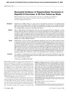
Figure 6 Endocytic internalization of canalicular transporters in E217G and in TLC-induced cholestasis. Protection from these cholestatic agents by the anticholestatic agents cAMP and TUDC is also shown. E217G and TLC induce endocytic internalization of canalicular transporters into the subapical compartment (SAC); this may lead to delivery to the lysosomal compartment, followed by degradation. E217G-induced activation of PKCα and TLC-induced, phosphatidylinositol 3-kinase (PI3K)-dependent activation of PKCε have been proposed to mediate this retrieval. Elevation of intracellular cAMP levels induced by administration of the permeant cAMP analogue DBcAMP, or by the phosphodiesterase inhibitor silibinin, prevents internalization, and accelerates re-insertion, via cytosolic Ca2+ elevations. On the other hand, TUDC prevents transporter endocytosis probably via co-stimulation of PKCα- and PKAdependent pathways.
to be confirmed. This possibility however exists, since retention of Mrp2 [122] and Oatp1a1 [125] in the apical and the basolateral membranes, respectively, requires interaction with the PDZ-domain protein, PDZ1. In addition, there is evidence that HAX-1 participates in clathrin-mediated Bsep endocytosis from the canalicular plasma membrane[126]. Accumulating evidence indicates that changes in canalicular transporter localization that occur in cholestasis also depend on activation of critical intracellular signaling pathways (Figure 6). Representative examples are cPKC (mainly, PKC α in hepatocytes). Selective activation of cPKC induces endocytic internalization of Bsep from the canalicular membrane and cholestasis in the isolated perfused rat liver [58]. Coincidently, pan-specific activation of PKC also induces redistribution of MRP2 from the canalicular to the basolateral membrane in HepG2 cells[127]. A critical participation of cPKC in the endocytic internalization of Bsep and the associated bile-salt secretory failure has recently been demonstrated by our g roup in E2-17G-induced cholestasis in rats[128]. A similar role for cPKC has also been reported in cholestasis associated with tBOOH-induced oxidative stress [107] (Figure 7). However, under oxidative stress, the type of canalicular protein that is internalized and the signaling molecule involved seem to depend on the magnitude of the oxidative challenge. Low concentrations of the oxidizing www.wjgnet.com
6796
ISSN 1007-9327
CN 14-1219/R
World J Gastroenterol
November 28, 2008
Volume 14
Number 44
Blebbing 2+
Ca ROS
BS BS GSH GSSG
F-actin
2+
Ca
Mrp2
iNOS cPKC activation
Bsep
t BOOH Ethacrynic acid
GSH GSSG
nPKC activation
iNO GC activation cGMP
Figure 7 Endocytic internalization of canalicular transporters under oxidative stress. In normal cells, the pericanalicular arrangement of F-actin allows for the appropriate insertion of the canalicular transporters in their membrane domain. Reactive oxygen species produced by the administration of oxidizing compounds, such as tBOOH or ethacrynic acid, induces mobilization of Ca2+ across the plasma membrane and membranes of the calciosome (smooth ER and mitochondria), and the subsequent activation of cPKC. cPKC activation induces blebbing and redistribution of F-actin from the pericanalicular region to the cell body. This rearrangement, in turn, leads to canalicular transporter internalization. Moderate Ca2+ elevations may also activate iNOS, which induces NO-mediated guanylate cyclase activation and further cGMP-mediated activation of nPKC, which may internalize selectively Mrp2.
compound, ethacr ynic acid, does not translocate cPKC, but novel PKC isoforms (nPKC). Under these conditions, the compound internalizes selectively Mrp2 without affecting Bsep, by a mechanism that probably involves Ca 2+ -dependent activation of inducible nitric oxide (NO) synthase (iNOS), followed by NOmediated cGMP increase, and further cGMP-activation of nPKC [110] . However, higher doses of ethacrynic acid, sufficient to activate cPKC isoforms, induce internalization of Bsep and Mrp2[110]. The nPKC isofor m PKC ε is also activated in TLC-induced cholestasis, and has been suggested to be involved in the TLC cholestatic effect [129] . This phenomenon occurs in a PI3K-dependent manner, which is consistent with the finding that PI3K products are potent activators of PKCε[130] (Figure 6). Since PI3K has been also shown to have pro-insertion properties (see above), this may be regarded as paradoxical. However, pro-exocytic and pro-endocytic effects of PI3K have been inferred by using pan-specific inhibitors of PI3K, and different isoforms of this kinase may have accounted for by these different effects. Anticholestatic therapeutic approaches based upon modulation of dynamic carrier localization As illustrated above for E 217G-induced cholestasis, internalization of hepatocellular transporters in cholestasis is spontaneously reversed if the cholestatic insult is transient. This spontaneous recovery occurs by a microtubule-dependent re-targeting of the endocytosed www.wjgnet.com
transporters to the canalicular membrane [95] . Some experimental therapeutic approaches have been designed to prevent transporter internalization and/or to accelerate this re-insertion, so as to avoid irreversible consequences of sustained internalization (Figure 6). The therapeutic agents studied include the following. cAMP: This second messenger partially prevents the impairment of bile flow and internalization of ABC transporters in experimental cholestasis, consistent with its capability to stimulate vesicle-mediated targeting of canalicular transporters[54,55,60]. The drop in bile flow and transport activity of Bsep[90] and Mrp2[89] in the acute phase of E 217G-induced cholestasis can be partially prevented by cAMP. More significantly, cAMP shortens spontaneous recovery to normality of bile flow, Mrp2 function and Mrp2 localization[89]. A similar acceleration of the re-insertion of endocytosed transporters has been described by our group for Bsep in TLC-induced cholestasis [92] . In isolated rat hepatocyte couplets, a preventive effect of cAMP has been obser ved in E 2 17G- [90,131] and TLC [92,131] -induced Bsep mislocalization. In this case, however, prevention by cAMP is complete. This protective effect is significantly blocked by the Ca2+ chelator, BAPTA/AM, but not by the PKA inhibitor, KT5720, which suggests involvement of Ca2+dependent signaling pathways. A similar anticholestatic mechanism in ter ms of the signaling modulators involved is afforded by silibinin, the active component of the hepatoprotector silymarin[131]. This most likely
Roma MG et al . Trafficking of hepatocellular transporters
results from the capability of silibinin to inhibit cAMP phosphodiesterase, thus increasing endogenous cAMP intracellular levels[131]. TUDC: This taurine-conjugate bile salt stimulates exocytic insertion of canalicular export pumps as part of its choleretic effect [63], and counteracts endocytic internalization of Bsep[134] and Mrp2[91] in TLC-induced cholestasis (Figure 6). The Ca2+-sensitive, PKC isoform, PKCα, has been proposed to mediate its anticholestatic effect [91] , via a cooperative PKC α /PKA-dependent mechanism[133]. This is in apparent contradiction with more recent findings that PKCα is cholestatic rather than hepatoprotective [58] . However, the biological response evoked by the interplay between different protein kinases (PKCα/PKA) may be different from that evoked by just one of them (PKCα). Furthermore, TUDC activates Erk [64] and p38 MAPK[63] , and the cholestatic effect of PKCα may be overridden by the choleretic effects of these signal transduction pathways. 4-Phenylbutyrate (4-PBA): This compound has been shown to restore the reduced cell surface expression of cystic fibrosis transmembrane conductance regulator in cystic fibrosis patients, who have mutated forms of the protein, which suggests improved targeting of the transporter to its membrane domain. When the 4PBA-proinserting property was tested for Bsep in normal rats, it was observed that canalicular expression and bile-salt transport function were improved by this compound[134]. A possible mechanism that 4PBA treatment increases the cell-surface-resident Bsep is the interruption of the internalization process from the cell surface to the intracellular compartment, or promotion of recycling from the intracellular compartment back to the cell surface [134]. Stabilization of Bsep in the membrane by 4PBA has also been confirmed in MDCK cells for wild-type Bsep and Bsep with E297G and D482G mutations, which occurs in progressive familiar intrahepatic cholestasis type 2 (PFIC2). Since trafficking of these Bsep-mutated proteins is impaired in PFIC2[135], this agent may be a potential candidate to halt the progression of this genetic disease. Its efficacy in acquired cholestatic diseases remains to be ascertained.
FUTURE DIRECTIONS The overwhelming progress in molecular biolog y techniques and the availability of in vitro, polarized cell models for the study of hepatobiliary function has greatly facilitated the characterization at a molecular level of the mechanism involved in the sorting of hepatobiliary transport systems from their sites of synthesis, and their recycling from/to endosomal compartments available on demand. However, the increasing number of new cytoskeletal, motor and signaling proteins that are being discovered as a result of these technological developments makes the characterization of their role in transporter trafficking an endless challenge.
6797
Advances in the molecular field have promoted a parallel prog ress in the understanding of the consequences that the alterations in the mechanisms of trafficking have in liver disease. It is becoming increasingly evident that impairment in the dynamic localization of hepatocellular transporters is a common feature in hepatocellular cholestasis. However, the characterization of the molecular mechanisms that underlie this alteration is in its infancy. Many crucial questions remain to be answered, for example: (1) which are the signaling mediators that trigger endocytosis of canalicular transporters in each kind of cholestasis; (2) which are the molecular targets of these cholestatic mediators that ultimately govern carrier internalization?; and (3) can changes in localization of these transporters be not only prevented but, what is more important from the therapeutic point of view, reversed by factors that counteract these dysfunctions? Satisfactory answers to these questions would allow the design of new therapeutic strategies in cholestatic liver diseases to assure proper localization of transporters in an attempt to prevent their accelerated degradation. We hope that progress in experimental therapeutics based on this current information encourages clinical researchers to apply this knowledge to envisage better, innovative therapeutic alternatives for the treatment of human cholestatic liver disease.
REFERENCES 1
Reichen J. The Role of the Sinusoidal Endothelium in Liver Function. News Physiol Sci 1999; 14: 117-121 2 Hofmann AF. Bile Acids: The Good, the Bad, and the Ugly. News Physiol Sci 1999; 14: 24-29 3 Bohan A, Boyer JL. Mechanisms of hepatic transport of drugs: implications for cholestatic drug reactions. Semin Liver Dis 2002; 22: 123-136 4 Kullak-Ublick GA, Stieger B, Hagenbuch B, Meier PJ. Hepatic transport of bile salts. Semin Liver Dis 2000; 20: 273-292 Pellicoro A, Faber KN. Review article: The function and 5 regulation of proteins involved in bile salt biosynthesis and transport. Aliment Pharmacol Ther 2007; 26 Suppl 2: 149-160 Ito K, Suzuki H, Horie T, Sugiyama Y. Apical/basolateral 6 surface expression of drug transporters and its role in vectorial drug transport. Pharm Res 2005; 22: 1559-1577 7 Niemi M. Role of OATP transporters in the disposition of drugs. Pharmacogenomics 2007; 8: 787-802 Müller M, Jansen PL. Molecular aspects of hepatobiliary 8 transport. Am J Physiol 1997; 272: G1285-G1303 Trauner M, Boyer JL. Bile salt transporters: molecular 9 characterization, function, and regulation. Physiol Rev 2003; 83: 633-671 10 van Montfoort JE, Müller M, Groothuis GM, Meijer DK, Koepsell H, Meier PJ. Comparison of "type I" and "type II" organic cation transport by organic cation transporters and organic anion-transporting polypeptides. J Pharmacol Exp Ther 2001; 298: 110-115 11 Silverman JA, Raunio H, Gant TW, Thorgeirsson SS. Cloning and characterization of a member of the rat multidrug resistance (mdr) gene family. Gene 1991; 106: 229-236 12 Crawford AR, Smith AJ, Hatch VC, Oude Elferink RP, Borst P, Crawford JM. Hepatic secretion of phospholipid vesicles in the mouse critically depends on mdr2 or MDR3
www.wjgnet.com
6798
13 14
15
16
17 18 19 20
21
22
23
24 25
26
27 28
29
30
31
ISSN 1007-9327
CN 14-1219/R
World J Gastroenterol
P-glycoprotein expression. Visualization by electron microscopy. J Clin Invest 1997; 100: 2562-2567 Suchy FJ, Ananthanarayanan M. Bile salt excretory pump: biology and pathobiology. J Pediatr Gastroenterol Nutr 2006; 43 Suppl 1: S10-S16 Akita H, Suzuki H, Ito K, Kinoshita S, Sato N, Takikawa H, Sugiyama Y. Characterization of bile acid transport mediated by multidrug resistance associated protein 2 and bile salt export pump. Biochim Biophys Acta 2001; 1511: 7-16 Paulusma CC, van Geer MA, Evers R, Heijn M, Ottenhoff R, Borst P, Oude Elferink RP. Canalicular multispecific organic anion transporter/multidrug resistance protein 2 mediates low-affinity transport of reduced glutathione. Biochem J 1999; 338 ( Pt 2): 393-401 Yang B, Hill CE. Nifedipine modulation of biliary GSH and GSSG/ conjugate efflux in normal and regenerating rat liver. Am J Physiol Gastrointest Liver Physiol 2001; 281: G85-G94 Ballatori N, Truong AT. Glutathione as a primary osmotic driving force in hepatic bile formation. Am J Physiol 1992; 263: G617-G624 Banales JM, Prieto J, Medina JF. Cholangiocyte anion exchange and biliary bicarbonate excretion. World J Gastroenterol 2006; 12: 3496-3511 Hardison WG, Wood CA. Importance of bicarbonate in bile salt independent fraction of bile flow. Am J Physiol 1978; 235: E158-E164 Medina JF, Lecanda J, Acín A, Ciesielczyk P, Prieto J. Tissue-specific N-terminal isoforms from overlapping alternate promoters of the human AE2 anion exchanger gene. Biochem Biophys Res Commun 2000; 267: 228-235 Aranda V, Martínez I, Melero S, Lecanda J, Banales JM, Prieto J, Medina JF. Shared apical sorting of anion exchanger isoforms AE2a, AE2b1, and AE2b2 in primary hepatocytes. Biochem Biophys Res Commun 2004; 319: 1040-1046 Huebert RC, Splinter PL, Garcia F, Marinelli RA, LaRusso NF. Expression and localization of aquaporin water channels in rat hepatocytes. Evidence for a role in canalicular bile secretion. J Biol Chem 2002; 277: 22710-22717 García F, Kierbel A, Larocca MC, Gradilone SA, Splinter P, LaRusso NF, Marinelli RA. The water channel aquaporin-8 is mainly intracellular in rat hepatocytes, and its plasma membrane insertion is stimulated by cyclic AMP. J Biol Chem 2001; 276: 12147-12152 Marinelli RA, Gradilone SA, Carreras FI, Calamita G, Lehmann GL. Liver aquaporins: significance in canalicular and ductal bile formation. Ann Hepatol 2004; 3: 130-136 Sun AQ, Swaby I, Xu S, Suchy FJ. Cell-specific basolateral membrane sorting of the human liver Na(+)-dependent bile acid cotransporter. Am J Physiol Gastrointest Liver Physiol 2001; 280: G1305-G1313 Dranoff JA, McClure M, Burgstahler AD, Denson LA, Crawford AR, Crawford JM, Karpen SJ, Nathanson MH. Short-term regulation of bile acid uptake by microfilamentdependent translocation of rat ntcp to the plasma membrane. Hepatology 1999; 30: 223-229 Webster CR, Anwer MS. Role of the PI3K/PKB signaling pathway in cAMP-mediated translocation of rat liver Ntcp. Am J Physiol 1999; 277: G1165-G1172 Sarkar S, Bananis E, Nath S, Anwer MS, Wolkoff AW, Murray JW. PKCzeta is required for microtubule-based motility of vesicles containing the ntcp transporter. Traffic 2006; 7: 1078-1091 Mukhopadhayay S, Ananthanarayanan M, Stieger B, Meier PJ, Suchy FJ, Anwer MS. cAMP increases liver Na+taurocholate cotransport by translocating transporter to plasma membranes. Am J Physiol 1997; 273: G842-G848 Grüne S, Engelking LR, Anwer MS. Role of intracellular calcium and protein kinases in the activation of hepatic Na+/taurocholate cotransport by cyclic AMP. J Biol Chem 1993; 268: 17734-17741 Webster CR, Srinivasulu U, Ananthanarayanan M, Suchy
www.wjgnet.com
32
33
34
35 36
37 38
39 40
41
42
43 44
45
46 47 48 49
November 28, 2008
Volume 14
Number 44
FJ, Anwer MS. Protein kinase B/Akt mediates cAMP- and cell swelling-stimulated Na+/taurocholate cotransport and Ntcp translocation. J Biol Chem 2002; 277: 28578-28583 Cosentino C, Di Domenico M, Porcellini A, Cuozzo C, De Gregorio G, Santillo MR, Agnese S, Di Stasio R, Feliciello A, Migliaccio A, Avvedimento EV. p85 regulatory subunit of PI3K mediates cAMP-PKA and estrogens biological effects on growth and survival. Oncogene 2007; 26: 2095-2103 Webster CR, Blanch CJ, Phillips J, Anwer MS. Cell swelling-induced translocation of rat liver Na(+)/ taurocholate cotransport polypeptide is mediated via the phosphoinositide 3-kinase signaling pathway. J Biol Chem 2000; 275: 29754-29760 McConkey M, Gillin H, Webster CR, Anwer MS. Crosstalk between protein kinases Czeta and B in cyclic AMPmediated sodium taurocholate co-transporting polypeptide translocation in hepatocytes. J Biol Chem 2004; 279: 20882-20888 Nakanishi H, Brewer KA, Exton JH. Activation of the zeta isozyme of protein kinase C by phosphatidylinositol 3,4,5-trisphosphate. J Biol Chem 1993; 268: 13-16 Miguel BG, Calcerrada MC, Mata F, Aller P, Clemente R, Catalán RE, Martínez AM. Differential redistribution of protein kinase C isoforms by cyclic AMP in HL60 cells. Biochem Biophys Res Commun 2000; 274: 596-602 Newton AC. Regulation of the ABC kinases by phosphorylation: protein kinase C as a paradigm. Biochem J 2003; 370: 361-371 Schonhoff CM, Gillin H, Webster CR, Anwer MS. Protein kinase Cdelta mediates cyclic adenosine monophosphatestimulated translocation of sodium taurocholate cotransporting polypeptide and multidrug resistant associated protein 2 in rat hepatocytes. Hepatology 2008; 47: 1309-1316 Webster CR, Blanch C, Anwer MS. Role of PP2B in cAMPinduced dephosphorylation and translocation of NTCP. Am J Physiol Gastrointest Liver Physiol 2002; 283: G44-G50 Mukhopadhyay S, Webster CR, Anwer MS. Role of protein phosphatases in cyclic AMP-mediated stimulation of hepatic Na+/taurocholate cotransport. J Biol Chem 1998; 273: 30039-30045 Mukhopadhyay S, Ananthanarayanan M, Stieger B, Meier PJ, Suchy FJ, Anwer MS. Sodium taurocholate cotransporting polypeptide is a serine, threonine phosphoprotein and is dephosphorylated by cyclic adenosine monophosphate. Hepatology 1998; 28: 1629-1636 Sun AQ, Arrese MA, Zeng L, Swaby I, Zhou MM, Suchy FJ. The rat liver Na(+)/bile acid cotransporter. Importance of the cytoplasmic tail to function and plasma membrane targeting. J Biol Chem 2001; 276: 6825-6833 Exton JH. Role of phosphoinositides in the regulation of liver function. Hepatology 1988; 8: 152-166 Staddon JM, Hansford RG. Evidence indicating that the glucagon-induced increase in cytoplasmic free Ca2+ concentration in hepatocytes is mediated by an increase in cyclic AMP concentration. Eur J Biochem 1989; 179: 47-52 Anwer MS, Gillin H, Mukhopadhyay S, Balasubramaniyan N, Suchy FJ, Ananthanarayanan M. Dephosphorylation of Ser-226 facilitates plasma membrane retention of Ntcp. J Biol Chem 2005; 280: 33687-33692 Sun AQ, Ponamgi VM, Boyer JL, Suchy FJ. Membrane trafficking of the human organic anion-transporting polypeptide C (hOATPC). Pharm Res 2008; 25: 463-474 Kipp H, Arias IM. Trafficking of canalicular ABC transporters in hepatocytes. Annu Rev Physiol 2002; 64: 595-608 Kipp H, Pichetshote N, Arias IM. Transporters on demand: intrahepatic pools of canalicular ATP binding cassette transporters in rat liver. J Biol Chem 2001; 276: 7218-7224 Kipp H, Arias IM. Newly synthesized canalicular ABC transporters are directly targeted from the Golgi to the hepatocyte apical domain in rat liver. J Biol Chem 2000; 275:
Roma MG et al . Trafficking of hepatocellular transporters
50
51
52
53
54
55
56 57
58
59 60 61
62
63
64
65
66
67 68
15917-15925 Sai Y, Nies AT, Arias IM. Bile acid secretion and direct targeting of mdr1-green fluorescent protein from Golgi to the canalicular membrane in polarized WIF-B cells. J Cell Sci 1999; 112 ( Pt 24): 4535-4545 Gerloff T, Stieger B, Hagenbuch B, Madon J, Landmann L, Roth J, Hofmann AF, Meier PJ. The sister of P-glycoprotein represents the canalicular bile salt export pump of mammalian liver. J Biol Chem 1998; 273: 10046-10050 Soroka CJ, Pate MK, Boyer JL. Canalicular export pumps traffic with polymeric immunoglobulin A receptor on the same microtubule-associated vesicle in rat liver. J Biol Chem 1999; 274: 26416-26424 Wakabayashi Y, Lippincott-Schwartz J, Arias IM. Intracellular trafficking of bile salt export pump (ABCB11) in polarized hepatic cells: constitutive cycling between the canalicular membrane and rab11-positive endosomes. Mol Biol Cell 2004; 15: 3485-3496 Roelofsen H, Soroka CJ, Keppler D, Boyer JL. Cyclic AMP stimulates sorting of the canalicular organic anion transporter (Mrp2/cMoat) to the apical domain in hepatocyte couplets. J Cell Sci 1998; 111 ( Pt 8): 1137-1145 Roma MG, Milkiewicz P, Elias E, Coleman R. Control by signaling modulators of the sorting of canalicular transporters in rat hepatocyte couplets: role of the cytoskeleton. Hepatology 2000; 32: 1342-1356 Gautam A, Ng OC, Boyer JL. Isolated rat hepatocyte couplets in short-term culture: structural characteristics and plasma membrane reorganization. Hepatology 1987; 7: 216-223 Chan W, Calderon G, Swift AL, Moseley J, Li S, Hosoya H, Arias IM, Ortiz DF. Myosin II regulatory light chain is required for trafficking of bile salt export protein to the apical membrane in Madin-Darby canine kidney cells. J Biol Chem 2005; 280: 23741-23747 Kubitz R, Saha N, Kühlkamp T, Dutta S, vom Dahl S, Wettstein M, Häussinger D. Ca2+-dependent protein kinase C isoforms induce cholestasis in rat liver. J Biol Chem 2004; 279: 10323-10330 Benedetti A, Strazzabosco M, Ng OC, Boyer JL. Regulation of activity and apical targeting of the Cl-/HCO3- exchanger in rat hepatocytes. Proc Natl Acad Sci USA 1994; 91: 792-796 Kagawa T, Misra S, Varticovski L, Arias IM. The mechanisms whereby cAMP activates PI 3-kinase which regulates canalicular spgp (Abstract). Hepatology 2000; 32: 306A Misra S, Ujházy P, Gatmaitan Z, Varticovski L, Arias IM. The role of phosphoinositide 3-kinase in taurocholateinduced trafficking of ATP-dependent canalicular transporters in rat liver. J Biol Chem 1998; 273: 26638-26644 Rao YP, Stravitz RT, Vlahcevic ZR, Gurley EC, Sando JJ, Hylemon PB. Activation of protein kinase C alpha and delta by bile acids: correlation with bile acid structure and diacylglycerol formation. J Lipid Res 1997; 38: 2446-2454 Kurz AK, Graf D, Schmitt M, Vom Dahl S, Häussinger D. Tauroursodesoxycholate-induced choleresis involves p38(MAPK) activation and translocation of the bile salt export pump in rats. Gastroenterology 2001; 121: 407-419 Schliess F, Kurz AK, vom Dahl S, Häussinger D. Mitogenactivated protein kinases mediate the stimulation of bile acid secretion by tauroursodeoxycholate in rat liver. Gastroenterology 1997; 113: 1306-1314 Kurz AK, Block C, Graf D, Dahl SV, Schliess F, Häussinger D. Phosphoinositide 3-kinase-dependent Ras activation by tauroursodesoxycholate in rat liver. Biochem J 2000; 350 Pt 1: 207-213 Kubitz R, Sütfels G, Kühlkamp T, Kölling R, Häussinger D. Trafficking of the bile salt export pump from the Golgi to the canalicular membrane is regulated by the p38 MAP kinase. Gastroenterology 2004; 126: 541-553 Kim RD, Darling CE, Cerwenka H, Chari RS. Hypoosmotic stress activates p38, ERK 1 and 2, and SAPK/JNK in rat hepatocytes. J Surg Res 2000; 90: 58-66 Noé B, Schliess F, Wettstein M, Heinrich S, Häussinger D.
69 70
71
72
73
74
75
76
77
78
79
80
81
82
83
84
6799
Regulation of taurocholate excretion by a hypo-osmolarityactivated signal transduction pathway in rat liver. Gastroenterology 1996; 110: 858-865 Häussinger D, Schmitt M, Weiergräber O, Kubitz R. Shortterm regulation of canalicular transport. Semin Liver Dis 2000; 20: 307-321 Martínez-Ansó E, Castillo JE, Díez J, Medina JF, Prieto J. Immunohistochemical detection of chloride/bicarbonate anion exchangers in human liver. Hepatology 1994; 19: 1400-1406 Alvaro D, Della Guardia P, Bini A, Gigliozzi A, Furfaro S, La Rosa T, Piat C, Capocaccia L. Effect of glucagon on intracellular pH regulation in isolated rat hepatocyte couplets. J Clin Invest 1995; 96: 665-675 Calamita G, Mazzone A, Bizzoca A, Cavalier A, Cassano G, Thomas D, Svelto M. Expression and immunolocalization of the aquaporin-8 water channel in rat gastrointestinal tract. Eur J Cell Biol 2001; 80: 711-719 Portincasa P, Palasciano G, Svelto M, Calamita G. Aquaporins in the hepatobiliary tract. Which, where and what they do in health and disease. Eur J Clin Invest 2008; 38: 1-10 Gradilone SA, García F, Huebert RC, Tietz PS, Larocca MC, Kierbel A, Carreras FI, Larusso NF, Marinelli RA. Glucagon induces the plasma membrane insertion of functional aquaporin-8 water channels in isolated rat hepatocytes. Hepatology 2003; 37: 1435-1441 Gradilone SA, Carreras FI, Lehmann GL, Marinelli RA. Phosphoinositide 3-kinase is involved in the glucagoninduced translocation of aquaporin-8 to hepatocyte plasma membrane. Biol Cell 2005; 97: 831-836 Gradilone SA, Tietz PS, Splinter PL, Marinelli RA, LaRusso NF. Expression and subcellular localization of aquaporin water channels in the polarized hepatocyte cell line, WIF-B. BMC Physiol 2005; 5: 13 Paulusma CC, Kothe MJ, Bakker CT, Bosma PJ, van Bokhoven I, van Marle J, Bolder U, Tytgat GN, Oude Elferink RP. Zonal down-regulation and redistribution of the multidrug resistance protein 2 during bile duct ligation in rat liver. Hepatology 2000; 31: 684-693 Mottino AD, Hoffman T, Crocenzi FA, Sánchez Pozzi EJ, Roma MG, Vore M. Disruption of function and localization of tight junctional structures and Mrp2 in sustained estradiol-17beta-D-glucuronide-induced cholestasis. Am J Physiol Gastrointest Liver Physiol 2007; 293: G391-G402 Song JY, Van Marle J, Van Noorden CJ, Frederiks WM. Disturbed structural interactions between microfilaments and tight junctions in rat hepatocytes during extrahepatic cholestasis induced by common bile duct ligation. Histochem Cell Biol 1996; 106: 573-580 Roma MG, Crocenzi FA, Sánchez Pozzi EA. Hepatocellular transport in acquired cholestasis: new insights into functional, regulatory and therapeutic aspects. Clin Sci (Lond) 2008; 114: 567-588 Trauner M, Wagner M, Fickert P, Zollner G. Molecular regulation of hepatobiliary transport systems: clinical implications for understanding and treating cholestasis. J Clin Gastroenterol 2005; 39: S111-S124 Trauner M, Arrese M, Soroka CJ, Ananthanarayanan M, Koeppel TA, Schlosser SF, Suchy FJ, Keppler D, Boyer JL. The rat canalicular conjugate export pump (Mrp2) is down-regulated in intrahepatic and obstructive cholestasis. Gastroenterology 1997; 113: 255-264 Kamisako T, Ogawa H. Alteration of the expression of adenosine triphosphate-binding cassette transporters associated with bile acid and cholesterol transport in the rat liver and intestine during cholestasis. J Gastroenterol Hepatol 2005; 20: 1429-1434 Kanno K, Tazuma S, Niida S, Chayama K. Unique reciprocal changes of hepatocellular membrane transporter expression and fluidity in rats with selective biliary obstruction. Hepatol Res 2003; 26: 157-163 www.wjgnet.com
6800
ISSN 1007-9327
CN 14-1219/R
World J Gastroenterol
85 Lee JM, Trauner M, Soroka CJ, Stieger B, Meier PJ, Boyer JL. Expression of the bile salt export pump is maintained after chronic cholestasis in the rat. Gastroenterology 2000; 118: 163-172 86 Borgognone M, Pérez LM, Basiglio CL, Ochoa JE, Roma MG. Signaling modulation of bile salt-induced necrosis in isolated rat hepatocytes. Toxicol Sci 2005; 83: 114-125 87 Sokol RJ, Devereaux M, Khandwala R, O'Brien K. Evidence for involvement of oxygen free radicals in bile acid toxicity to isolated rat hepatocytes. Hepatology 1993; 17: 869-881 88 Liu TZ, Lee KT, Chern CL, Cheng JT, Stern A, Tsai LY. Free radical-triggered hepatic injury of experimental obstructive jaundice of rats involves overproduction of proinflammatory cytokines and enhanced activation of nuclear factor kappaB. Ann Clin Lab Sci 2001; 31: 383-390 89 Mottino AD, Cao J, Veggi LM, Crocenzi F, Roma MG, Vore M. Altered localization and activity of canalicular Mrp2 in estradiol-17beta-D-glucuronide-induced cholestasis. Hepatology 2002; 35: 1409-1419 90 Crocenzi FA, Mottino AD, Cao J, Veggi LM, Pozzi EJ, Vore M, Coleman R, Roma MG. Estradiol-17beta-D-glucuronide induces endocytic internalization of Bsep in rats. Am J Physiol Gastrointest Liver Physiol 2003; 285: G449-G459 91 Beuers U, Bilzer M, Chittattu A, Kullak-Ublick GA, Keppler D, Paumgartner G, Dombrowski F. Tauroursodeoxycholic acid inserts the apical conjugate export pump, Mrp2, into canalicular membranes and stimulates organic anion secretion by protein kinase C-dependent mechanisms in cholestatic rat liver. Hepatology 2001; 33: 1206-1216 92 Crocenzi FA, Mottino AD, Sánchez Pozzi EJ, Pellegrino JM, Rodríguez Garay EA, Milkiewicz P, Vore M, Coleman R, Roma MG. Impaired localisation and transport function of canalicular Bsep in taurolithocholate induced cholestasis in the rat. Gut 2003; 52: 1170-1177 93 Román ID, Fernández-Moreno MD, Fueyo JA, Roma MG, Coleman R. Cyclosporin A induced internalization of the bile salt export pump in isolated rat hepatocyte couplets. Toxicol Sci 2003; 71: 276-281 94 Meyers M, Slikker W, Pascoe G, Vore M. Characterization of cholestasis induced by estradiol-17 beta-D-glucuronide in the rat. J Pharmacol Exp Ther 1980; 214: 87-93 95 Mottino AD, Crocenzi FA, Pozzi EJ, Veggi LM, Roma MG, Vore M. Role of microtubules in estradiol-17betaD-glucuronide-induced alteration of canalicular Mrp2 localization and activity. Am J Physiol Gastrointest Liver Physiol 2005; 288: G327-G336 96 Kan KS, Monte MJ, Parslow RA, Coleman R. Oestradiol 17 beta-glucuronide increases tight-junctional permeability in rat liver. Biochem J 1989; 261: 297-300 97 Mottino AD, Carreras FI, Gradilone SA, Marinelli RA, Vore M. Canalicular membrane localization of hepatocyte aquaporin-8 is preserved in estradiol-17beta-D-glucuronideinduced cholestasis. J Hepatol 2006; 44: 232-233 98 Jansen PL, Müller M. Early events in sepsis-associated cholestasis. Gastroenterology 1999; 116: 486-488 99 K u b i t z R , D ' u r s o D , K e p p l e r D , H ä u s s i n g e r D . Osmodependent dynamic localization of the multidrug resistance protein 2 in the rat hepatocyte canalicular membrane. Gastroenterology 1997; 113: 1438-1442 100 Dombrowski F, Kubitz R, Chittattu A, Wettstein M, Saha N, Häussinger D. Electron-microscopic demonstration of multidrug resistance protein 2 (Mrp2) retrieval from the canalicular membrane in response to hyperosmolarity and lipopolysaccharide. Biochem J 2000; 348 Pt 1: 183-188 101 Kubitz R, Wettstein M, Warskulat U, Häussinger D. Regulation of the multidrug resistance protein 2 in the rat liver by lipopolysaccharide and dexamethasone. Gastroenterology 1999; 116: 401-410 102 Schmitt M, Kubitz R, Lizun S, Wettstein M, Häussinger D. Regulation of the dynamic localization of the rat Bsep gene-encoded bile salt export pump by anisoosmolarity. Hepatology 2001; 33: 509-518 www.wjgnet.com
November 28, 2008
Volume 14
Number 44
103 Bolder U, Schmidt A, Landmann L, Kidder V, Tange S, Jauch KW. Heat stress prevents impairment of bile acid transport in endotoxemic rats by a posttranscriptional mechanism. Gastroenterology 2002; 122: 963-973 104 Bolder U, Jeschke MG, Landmann L, Wolf F, de Sousa C, Schlitt HJ, Przkora R. Heat stress enhances recovery of hepatocyte bile acid and organic anion transporters in endotoxemic rats by multiple mechanisms. Cell Stress Chaperones 2006; 11: 89-100 105 Poli G. Liver damage due to free radicals. Br Med Bull 1993; 49: 604-620 106 Roma MG, Sanchez Pozzi EJ. Oxidative stress: a radical way to stop making bile. Ann Hepatol 2008; 7: 16-33 107 Pérez LM, Milkiewicz P, Elias E, Coleman R, Sánchez Pozzi EJ, Roma MG. Oxidative stress induces internalization of the bile salt export pump, Bsep, and bile salt secretory failure in isolated rat hepatocyte couplets: a role for protein kinase C and prevention by protein kinase A. Toxicol Sci 2006; 91: 150-158 108 Schmitt M, Kubitz R, Wettstein M, vom Dahl S, Häussinger D. Retrieval of the mrp2 gene encoded conjugate export pump from the canalicular membrane contributes to cholestasis induced by tert-butyl hydroperoxide and chlorodinitrobenzene. Biol Chem 2000; 381: 487-495 109 Ji B, Ito K, Sekine S, Tajima A, Horie T. Ethacrynic-acidinduced glutathione depletion and oxidative stress in normal and Mrp2-deficient rat liver. Free Radic Biol Med 2004; 37: 1718-1729 110 Sekine S, Ito K, Horie T. Oxidative stress and Mrp2 internalization. Free Radic Biol Med 2006; 40: 2166-2174 111 Yu QY, Shu M, Dai JH, Ma JB, Yu Y, Liu DH. [The mechanism of the increase of plasma bilirubin after hepatic ischemia-reperfusion in rats] Zhonghua Ganzangbing Zazhi 2007; 15: 763-766 112 Shoda J, Kano M, Oda K, Kamiya J, Nimura Y, Suzuki H, Sugiyama Y, Miyazaki H, Todoroki T, Stengelin S, Kramer W, Matsuzaki Y, Tanaka N. The expression levels of plasma membrane transporters in the cholestatic liver of patients undergoing biliary drainage and their association with the impairment of biliary secretory function. Am J Gastroenterol 2001; 96: 3368-3378 113 Kojima H, Sakurai S, Uemura M, Kitamura K, Kanno H, Nakai Y, Fukui H. Disturbed colocalization of multidrug resistance protein 2 and radixin in human cholestatic liver diseases. J Gastroenterol Hepatol 2008; 23: e120-e128 114 Kojima H, Nies AT, König J, Hagmann W, Spring H, Uemura M, Fukui H, Keppler D. Changes in the expression and localization of hepatocellular transporters and radixin in primary biliary cirrhosis. J Hepatol 2003; 39: 693-702 115 Watanabe N, Takashimizu S, Kojima S, Kagawa T, Nishizaki Y, Mine T, Matsuzaki S. Clinical and pathological features of a prolonged type of acute intrahepatic cholestasis. Hepatol Res 2007; 37: 598-607 116 Rost D, Kartenbeck J, Keppler D. Changes in the localization of the rat canalicular conjugate export pump Mrp2 in phalloidin-induced cholestasis. Hepatology 1999; 29: 814-821 117 Rost D, Kloeters-Plachky P, Stiehl A. Retrieval of the rat canalicular conjugate export pump Mrp2 is associated with a rearrangement of actin filaments and radixin in bile saltinduced cholestasis. Eur J Med Res 2008; 13: 314-318 118 Bi GQ, Morris RL, Liao G, Alderton JM, Scholey JM, Steinhardt RA. Kinesin- and myosin-driven steps of vesicle recruitment for Ca2+-regulated exocytosis. J Cell Biol 1997; 138: 999-1008 119 K a t o Y , W a t a n a b e C , T s u j i A . R e g u l a t i o n o f d r u g transporters by PDZ adaptor proteins and nuclear receptors. Eur J Pharm Sci 2006; 27: 487-500 120 Ortiz DF, Moseley J, Calderon G, Swift AL, Li S, Arias IM. Identification of HAX-1 as a protein that binds bile salt export protein and regulates its abundance in the apical membrane of Madin-Darby canine kidney cells. J Biol Chem 2004; 279: 32761-32770
Roma MG et al . Trafficking of hepatocellular transporters 121 Gallagher AR, Cedzich A, Gretz N, Somlo S, Witzgall R. The polycystic kidney disease protein PKD2 interacts with Hax-1, a protein associated with the actin cytoskeleton. Proc Natl Acad Sci USA 2000; 97: 4017-4022 122 Kikuchi S, Hata M, Fukumoto K, Yamane Y, Matsui T, Tamura A, Yonemura S, Yamagishi H, Keppler D, Tsukita S, Tsukita S. Radixin deficiency causes conjugated hyperbilirubinemia with loss of Mrp2 from bile canalicular membranes. Nat Genet 2002; 31: 320-325 123 Wang W, Soroka CJ, Mennone A, Rahner C, Harry K, Pypaert M, Boyer JL. Radixin is required to maintain apical canalicular membrane structure and function in rat hepatocytes. Gastroenterology 2006; 131: 878-884 124 Kojima H, Sakurai S, Yoshiji H, Uemura M, Yoshikawa M, Fukui H. The role of radixin in altered localization of canalicular conjugate export pump Mrp2 in cholestatic rat liver. Hepatol Res 2008; 38: 202-210 125 Wu H, Parsons JT. Cortactin, an 80/85-kilodalton pp60src substrate, is a filamentous actin-binding protein enriched in the cell cortex. J Cell Biol 1993; 120: 1417-1426 126 Wang P, Wang JJ, Xiao Y, Murray JW, Novikoff PM, Angeletti RH, Orr GA, Lan D, Silver DL, Wolkoff AW. Interaction with PDZK1 is required for expression of organic anion transporting protein 1A1 on the hepatocyte surface. J Biol Chem 2005; 280: 30143-30149 127 Kubitz R, Huth C, Schmitt M, Horbach A, Kullak-Ublick G, Häussinger D. Protein kinase C-dependent distribution of the multidrug resistance protein 2 from the canalicular to the basolateral membrane in human HepG2 cells. Hepatology 2001; 34: 340-350 128 Crocenzi FA, Sánchez Pozzi EJ, Ruiz ML, Zucchetti AE, Roma MG, Mottino AD, Vore M. Ca(2+)-dependent protein kinase
129
130
131
132
133
134
135
6801
C isoforms are critical to estradiol 17beta-D-glucuronideinduced cholestasis in the rat. Hepatology 2008; 48: 1885-1895 Beuers U, Probst I, Soroka C, Boyer JL, Kullak-Ublick GA, Paumgartner G. Modulation of protein kinase C by taurolithocholic acid in isolated rat hepatocytes. Hepatology 1999; 29: 477-482 Beuers U, Denk GU, Soroka CJ, Wimmer R, Rust C, Paumgartner G, Boyer JL. Taurolithocholic acid exerts cholestatic effects via phosphatidylinositol 3-kinasedependent mechanisms in perfused rat livers and rat hepatocyte couplets. J Biol Chem 2003; 278: 17810-17818 Crocenzi FA, Basiglio CL, Pérez LM, Portesio MS, Pozzi EJ, Roma MG. Silibinin prevents cholestasis-associated retrieval of the bile salt export pump, Bsep, in isolated rat hepatocyte couplets: possible involvement of cAMP. Biochem Pharmacol 2005; 69: 1113-1120 Dombrowski F, Stieger B, Beuers U. Tauroursodeoxycholic acid inserts the bile salt export pump into canalicular membranes of cholestatic rat liver. Lab Invest 2006; 86: 166-174 Wimmer R, Hohenester S, Pusl T, Denk GU, Rust C, Beuers U. Tauroursodeoxycholic acid exerts anticholestatic effects by a cooperative cPKC alpha-/PKA-dependent mechanism in rat liver. Gut 2008; 57: 1448-1454 Hayashi H, Sugiyama Y. 4-phenylbutyrate enhances the cell surface expression and the transport capacity of wild-type and mutated bile salt export pumps. Hepatology 2007; 45: 1506-1516 Hayashi H, Takada T, Suzuki H, Akita H, Sugiyama Y. Two common PFIC2 mutations are associated with the impaired membrane trafficking of BSEP/ABCB11. Hepatology 2005; 41: 916-924 S- Editor Li LF L- Editor Kerr C
E- Editor Zheng XM
www.wjgnet.com