mass of 110-115 kD in reduced SDS-PAGE and a slightly .... Instruments, Galthersburg, MD) and the area under the peaks was deter- ..... Hanna, C. 1966.
Enhancement of Vinculin Synthesis by Migrating Stratified Squamous Epithelium James D. Zieske, Gul Bukusoglu, a n d Ilene K. Gipson Eye Research Institute of Retina Foundation, and Harvard Medical School, Boston, Massachusetts 02114
Abstract. A ll0-115-kD protein is present at levels 27-fold higher in migratory epithelium in the rat cornea than in stationary epithelium. This protein represents 2.7% of the total protein in migratory epithelium 6-h postabrasion wound and 0.1% of the total protein in stationary epithelium. Our findings demonstrate that this ll0-115-kD protein is vinculin. In Western blots comparing proteins from migratory and control epithelium, antibody against vinculin crossreacted with the ll0-115-kD protein. Using immunoslot blots, vinculin was determined to be present at maximal levels 6 h postabrasion wound, at levels 22and 8-fold higher than control at 18 and 48 h, respectively, returning to control levels 72 h postwounding. Vinculin was also localized by indirect immunohistochemistry in migrating corneal epithelium. 3-mm
INCULIN is a structural protein with an apparent molecular mass of 130 kD in nonreducing conditions and a slightly lower apparent molecular mass in reducing conditions (8). It was first purified from smooth muscle isolated from chicken gizzard and has been found to be localized in focal contacts of cultured fibroblasts (3, 8, 36), dense plaques of smooth muscle (10, 11), intercalated disks of cardiac muscle (11, 26, 31), and zonula adherens of epithelium (9, 10). The localization of vinculin adjacent to the plasma membrane and actin bundles suggests that it is a "linker" protein between the two structures. In this link, vinculin binds to talin (4, 30), which in turn binds to integrin (20), anchoring the cell to its substrate. The mechanism of the linkage between vinculin and actin bundles remains unclear. Initial reports suggested that vinculin could bind directly to actin (23, 41); however, this binding was later shown to result from impurities in the vinculin preparation (6, 34, 42). The finding that vinculin can be phosphorylated has led to speculation that phosphorylation might be a mechanism by which adherens-type junctions could be altered during transformation as well as in cell migration (35). This correlation of phosphorylation of vinculin and alteration of cell contacts resulting in altered cell activity has not been documented conclusively (1, 21, 24, 28, 33). Although vinculin has been detected in epithelium in cell culture, its localization and synthesis by an intact sheet of
V
© The Rockefeller University Press, 0021-9525/89/08/571/6 $2.00 The Journal of Cell Biology, Volume 109, August 1989 571-576
scrape wounds were allowed to heal in vivo for 20 h. In fiat mounts of these whole wounded corneas, vinculin was localized as punctate spots in the leading edge of migrating epithelium. In cryostat sections, vinculin was localized as punctate spots along the basal cell membranes of the migrating sheet adjacent to the basement membrane and in patches between cells as well as diffusely throughout the cell. Only very diffuse localization with occasional punctate spots between adjacent superficial cells was present in stationary epithelium. The increased synthesis of vinculin during migration and the localization of vinculin at the leading edge of migratory epithelium suggest that vinculin may be involved in cell-cell and cell-substrate adhesion as the sheet of epithelium migrates to cover a wound.
migrating epithelium have not been elucidated. To determine whether vinculin levels change as cells migrate, we used a wound healing model in which the epithelium migrates as a unified sheet to cover the wound. Migration is dependent on protein synthesis and does not require cell mitosis (13, 18). We have demonstrated in this system that during corneal epithelial migration there is a large ('~15-fold) increase in protein synthesis relative to stationary control epithelium (16, 44). We detected a protein that is synthesized at enhanced levels by migrating epithelium, has an apparent molecular mass of 110-115 kD in reduced SDS-PAGE and a slightly higher molecular mass in unreduced conditions, and which we postulated to be vinculin (44). The present study was undertaken to test this identification.
Materials and Methods Model Wounds and Organ Culture The organ culture system used in this investigation has been described (16). Briefly, Sprague-Dawley rats with clear, healthy corneas were killed with an overdose of sodium pentobarbital. An epithelial wound 3 mm in diameter was created by demarcating an area on the cornea with a 3-ram trephine and removing the epithelium within the circle with a small scalpel, leaving an intact basement membrane (38). Following wounding, the corneas were dissected and placed into organ culture in a completely defined medium (15). Corneas were pinned to rounded paraffin posts that maintain the corneal
571
curvature and provide a solid support for harvest of the epithelium. To harvest migrating epithelium, the corneas were stained with Richardson's stain (32), which reveals the remaining defect, the 3-ram trephine was placed on the cornea concentric to the defect, and the migrating epithelium was removed with a small scalpel. For controls, epithelium was removed from a central area of unwounded corneas. In organ culture 3-ram wounds heal in 22 h; in vivo wound closure occurs at 24 h.
Electrophoresis, Fluorography, and lmmunoblotting Epithelial protein samples were analyzed by SDS~PAGE using the buffer system described by Miles Laboratories (27). The gels were fixed in 50% methanol, and protein was detected with ammoniacai silver reagent as described by Wray et al. (43). Fluorography of [35S]-methionine-labeled proteins was done using EN3HANCE (New England Nuclear, Boston, MA) as previously described (45). Proteins of known molecular mass (43-200 kD) (Bio-Rad Laboratories, Richmond, CA) were used to determine molecular masses of unknown components. Protein samples used for immunoblotting were transferred to nitrocellulose paper as described by Towbin et al. (39). The paper was then placed in a blocking solution containing 3 % gelatin in TBS. The paper was washed and then incubated for 2 h in a preparation of monocionai antivinculin (ICN Immuno Biologicals, Lisle, IL) at a 1:50 dilution. Antibody binding was detected by reaction (1 h) with peroxidase-conjngated goat anti-mouse IgG (Organon Teknika-Cappel, Malvern, PA) at a 1:3,000 dilution. Color was developed using 0.05 % diaminobenzidene, 0.015 % H202. Specificity of the commercial antivinculin was determined by preabsorption of the antibody with purified chicken vinculin (100 t~g/ml) overnight at 4°C.
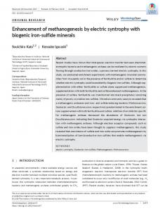
Figure 1. Fluorogram o f 5-15% SDSPAGE o f [35Slmethionine-labeled epithelial proteins from unwounded corneas (C) maintained 18 h in culture and from corneas 18 h postabrasion wound (W). [35S]Methionine ( 5 0 / t C i / m l ) was present for the entire culture time. Arrow indicates ll0-115-kD protein. Both C and W were loaded with 75,000 disintegrations per minute/lane; exposure time, 72 h. Molecular masses (in kilodaltons) determined from standard proteins are noted.
Purification of Chicken Gizzard Vinculin Vinculin was purified from 50 g of frozen chicken gizzards as described by Feramisco and Burridge (7), except that we used a DEAE 5 PW (Waters Associates, Milford, MA) column for the last purification step. Vinculin appeared as a single band in SDS-PAGE migrating slightly ahead of purified /~-galactosidase (116 kD). This molecular mass is in agreement with the report of Burridge et al. (5).
Quantification of Vinculin in Epithelial Protein Samples Rats were anesthetized with an intraperitoneal injection of chloralhydrate (35 rag/100 g of body weight) (Squibb, Princeton, NJ) followed by topical application of proparacaine HCI (Akorn, Metairie, LA), 3-mm scrape wounds were made in the central cornea and allowed to heal in vivo for 1, 3, 6, 18, 48, and 72 h and 7 d. The rats were killed with an intraperitoneal injection of sodium pentobarbital, and the eyes were stained and epithelium harvested as described above. Epithelium from unwounded corneas served as control. The epithelium was immediately frozen in liquid nitrogen and proteins solubilized in 2% SDS, 2 M urea, 0.1 mM phenyl methyl sulfonyl fluoride in 0.064 M Tris-HCI, pH 8.9. Protein amounts were quantitated by Pierce Chemical Co. (Rockford, IL) micro BCA assay using bovine albumin as standard. Duplicate samples of I and 5 p,g of total epithelial protein were then applied to nitrocellulose paper using a Minifold II slot blot apparatus (Schleicher and Schuell, Keene, NH). The blot was blocked with 5 % BSA in PBS for 1 h and then incubated overnight in 1% BSA containing antivinculin at a 1:50 dilution. After 3 x lO-min washes with PBS, the blot was incubated for 2 h in 1% BSA containing goat anti-mouse IgG [nsI] (ICN Radiochemicals, Irvine, CA) at a concentration of 1.2/tCi/rnl. The blot was washed 4 x 20-rain with PBS, dried, and exposed to x-ray film (X-Omat; Eastman Kodak Co., Rochester, NY) for 6 h at -80°C. Autoradiograms were then scanned using a soft laser scanning densitometer (Zeineh LKB Instruments, Galthersburg, MD) and the area under the peaks was determined with a vidcoplan II (Carl Zeiss, Inc., Thornwood, NY). Amount of vinculin in the epithelial protein samples was calculated based on comparison with a standard curve made using purified chicken gizzard vinculin in the above procedure. The standard curve was linear over the range of 2.5-250 ng of purified vinculin with a correlation factor of 0.99. No reaction was observed when the primary antibody was omitted or preabsorbed with purified chicken gizzard vinculin.
peritoneal injection of sodium pentobarbital and the corneas excised. The corneas were fixed for 5 min in methanol (-25°C), and the membranes were permeabilized for 5 min with 0.1% NP-40 in PBS. The whole corneas were incubated with antivinculin (1:40) for 30 min at room temperature, followed by washing 3 x 5-min in PBS and incubation for 30 min at room temperature with fluorescein-conjugated goat anti-mouse IgG (Organon Teknika-Cappel). The corneas were washed, prepared as flat mounts, and viewed with a photomicroscope III (Carl Zeiss, Inc.) equipped for epifluorescence. S~ondary antibody controls were performed by omitting the primary antibody. Specificity of the primary antibody was tested by preabsorbing the antivinculin with purified chicken gizzard vinculin (100 /tg/ml) overnight at 4°C. For localization in cross sections of migrating epithelium, rat corneas were frozen in Tissue Tek II OCT compound (Lab Tek Products, Naperville, IL). 6-/tin cryostat sections were placed on gelatin-coated slides and air-dried overnight at 370C followed by rehydration in PBS and washing in 1% BSA for 10 min. Alternatively, sections wore fixed in 3% paraformaldehyde in PBS and used immediately. Monoclonal antivinculin was applied and incubated 1 h in a moist chamber. The sections were washed with PBS, incubated for 1 h in FITC-conjugated goat anti-mouse IgG (Organon Teknika-Cappel), washed, and mounted with a medium consisting of PBS, glycerol, and paraphenylene diamine. Then they were viewed and photographed using a photomicroscope Ill equipped for epi-illumination (Carl Zeiss, Inc.). To test for nonspecific binding, sections wore viewed when either the primary or secondary antibodies were omitted or when the antivinculin was preabsorbed with purified chicken vinculin.
Results
3-mm scrape wounds were made in the central cornea as described above and allowed to heal in vivo for 20 h. The rats were killed with an intra-
Rat corneal epithelium consists of 5-7 layers of cells: a basal cell layer, 2-3 layers of wing cells, and 2-3 layers of flattened superficial cells. After an abrasion wound in which basement membrane is left intact, epithelium migrates into the wound area as a unified sheet with the leading edge of the sheet tapering down to a single cell layer. Migration does not require mitosis; cell elongation and flattening provide coverage of the wound. After epithelial wound closure, the rate of mitosis increases, and the epithelium regains its normal stratification (18).
The Journal of Cell Biology, Volume 109, 1989
572
lmmunolocalization of Vinculin in Flat Mounts and Cryostat Sections of Rat Corneas after Scrape Wounds
Figure 2. 5-15% SDS-PAGE of corneal
epithelium harvested 18 h postabrasion wound. Epithelium was extracted in 0.1 M ammonium acetate in a glass-glass homogenizer. Homogenate was centrifuged at 12,000 g for 10 min. Supernatant (S), 20 tzg/lane, and pellet (P), 40 ~tg/lane, were applied to SDS-PAGE. Arrow indicates ll0--ll5-kD protein. Silver stain. Molecular masses (in kilodaltons) determined from standard proteins are noted.
A ll0-115-kD protein is present in greater amounts in epithelium migrating over denuded basement membrane than in stationary epithelium (Fig. 1). Fig. 1 shows the incorporation of [s~S]methionine into proteins by the epithelium during organ culture; the ll0-115-kD protein is radiolabeled in migrating epithelium to a far greater extent than in stationary, unwounded corneal epithelium. This protein could be extracted in low-ionic-strength buffers such as 0.1 M ammonium acetate (Fig. 2). It could also be extracted in 0.1 M phosphate buffer, pH 7.0, or 10 mM Tris-HC1, pH 7.4. To determine whether the protein was vinculin, protein samples from migratory epithelium inside the 3-mm wound area (W) t, migratory epithelium from outside the original 3-mm wound periphery (WP), and unwounded control epithelium (C) were reacted with a monoclonal antibody against chicken vinculin. After staining of the blot for total protein, the ll0-115-kD protein could be detected in greater amounts in migratory epithelium (W + WP) than in C epithelium (Fig. 3 a). In an identical blot, antibody against vinculin reacted with bands corresponding to the llO-115-kD protein in W and WP with no detectable reaction in control epithelium (Fig. 3 b). To ensure specificity of the antibody, it was preabsorbed with purified chicken gizzard vinculin and then reacted with proteins from migratory epithelium in Western blots. The reaction of antivinculin and the ll0--ll5-kD protein was completely blocked (Fig. 3 c). No reaction was seen when the primary or secondary antibody was omitted (data not shown). To quantify alterations of vinculin levels during epithelial migration, rat epithelium was assayed with immunoslot blots (Fig. 4). In both W and WP epithelium migrating over a scrape wound, vinculin was present at maximal levels 6 h after wounding representing 2.72 + 0.14/zg and 1.60 + 0.001 #g/100/zg of total epithelial protein, an increase of 27- and 16-fold over control levels, respectively. The increase in vinI. Abbreviations used in this paper: C, controlepithelium;W, wound;WP,
wound periphery.
Zieskeet al. Vinculin Synthesis during Epithelial Migration
culin levels in WP was seen as early as 1 h after wounding; the level was 11-fold higher than control. Vinculin levels in W could not be measured at 1 and 3 h since only very small amounts of epithelium have migrated into the original 3-mm wound area at these time points. By 48 h after wounding, 24 h after wound closure, levels in W and WP dropped to 0.84 + 0.11 and 0.90 5:0.11 #g of vinculin/100 #g of total epithelial protein, and, by 72 h, the level of vinculin returned to control levels. The amount ofvinculin remained at control levels 7 d after wounding. In whole amounts of rat corneas, 20 h postscrape wound in vivo, vinculin was detected as punctate spots near the leading edge of migrating epithelium around the entire wound margin (Fig. 5). Behind the leading edge where the epithelium is more stratified, fewer punctate spots could be detected. Punctate spots were not detected when the primary antibody was omitted or when the antibody was preabsorbed with vinculin. Vinculin was also immunolocalized in cross sections of cryostat sections (Fig. 6). Intensity of binding was far greater in migrating than in stationary epithelium, corroborating the immunoslot blot data (Fig. 4). In corneas 6 and 18 h postabrasion wound, vinculin was localized, at the leading edge of migrating epithelium, along the basal cell membranes adjacent to the basement membrane and in patches
Figure 3. (a) Blot of 5-15% SDS-PAGE: W, 20 tzg of protein from
epithelium harvested 18 h postabrasion wound that has migrated into original 3-mm abrasion wound area as described (16); WP, 20 #g of protein from epithelium harvested 18 h postabrasion wound outside original 3-mm abrasion wound periphery; C, 20/~g of protein from control, unwounded epithelium. Blot was stained with 0.1% naphthol blue-black in 45% methanol and 10% acetic acid to reveal total proteins present. Arrow indicates ll0-115-kD protein. Molecular masses (in kilodaltons) determined from standard proteins are noted. (b) Immunoblot of 5-15% SDS-PAGE identical to blot on left. Blot was reacted with antivinculin as described in Materials and Methods. Arrow indicates immunoreaction with protein comigrating with ll0-115-kD protein. (c) Immunoblot of 5-15% SDS-PAGEidentical to Win a and b reacted with antivinculin preabsorbed overnight with purified chicken vinculin (100 /~g/ml). Note lack of reaction to ll0-115-kD protein.
573
between cells as well as diffusely throughout the cell (Fig. 6 b). Behind the leading edge at 18 h, vinculin was localized as punctate spots along the basal cell membranes adjacent to the basement membranes (Fig. 6 c). In unwounded corneas, vinculin was localized in far lesser amounts with only diffuse binding and along cell membranes of superficial cells. Vinculin was not localized in corneas reacted with antivinculin preabsorbed with purified chicken vinculin. Identical localizations of vinculin were seen when the tissue was cryostat sectioned, fixed in 3 % paraformaldehyde, and used immediately for immunolocalization.
z n.; I0 re'
2
a.
0 0
z -J
I
z
Discussion =I.
0 lh
3h
6h
18h
48h
72h
7d control
TIME AFTER WOUNDING
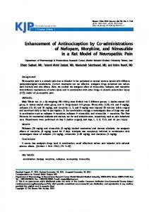
Figure 4. Vinculin concentrations in W (black bars) and WP (crosshatched bars) epithelium quantitated as described in Materials and Methods. Wound, epithelium that has migrated into original 3-mm wound area; WP, epithelium harvested from outside original WP; control, epithelium removed from unwounded corneas. Error bars are 5- SEM. n = 8. Note: SEM for 6 h WP is 5- 0.001 and is contained in border of bar. Vinculin concentrations in 1- and 3-h W could not be determined since only very small amounts of epithelium have migrated into the original 3-mm wound area at these time points.
Corneal epithelial migration in response to a wound involves major changes in epithelial architecture and protein synthesis. The hemidesmosomes, the normal cell-substrate adherens junctions, disappear (25), the number of cell layers decreases, the cells flatten and elongate, and mitosis diminishes (18). In addition, the rates of protein and glycoprotein synthesis increase dramatically during epithelial migration (16, 44). The enhanced synthesis of vinculin suggests that a new method of ceU-substrate adhesion is activated during epithelial migration. Although our data do not demonstrate that vinculin is localized in focal contacts in our system, the localization of vinculin along the basement membrane zone is consistent with reported localization of focal contacts in cultured cells.
Figure 5. Micrographs showing immunolocalization of vinculin in flat mounts of corneas allowed to heal 20 h in vivo after 3-mm-diam abrasion. Arrows indicate leading edge of migrating epithelial sheet. (a) Phase-contrast of area shown in b. Bar, 50 ~m. (b) Same area as a showing punctate binding of vinculin antibody at leading edge. Bar, 50 ~,m. (c) Lower magnification showing punctate localization around wound edge. Bar, 125 ~m. (d) Preabsorption control showing lack of punctate staining. Bar, 50 ~m. Defect area = area of wound remaining to be covered by migrating epithelium.
The Journal of Cell Biology, Volume 109, 1989
574
Figure 6. Micrographs showing immunolocalization of vinculin in cryostat sections. (a) Control, unwounded cornea. Large arrows indicate punctate binding along membranes of superficial cells. Bar, 50/~m. Note: a is printed at 6x the intensity of b, c, and d to allow detail to be seen. (b) Leading edge of migrating sheet of epithelium 6 h postabrasion wound. Large arrows indicate binding along basement membrane; small arrows indicate punctate binding between cells. Note overall increase in binding compared to controls. Similar localization was seen 18 h postabrasion wound. Bar, 50/~m. (c) Migrating epithelium behind leading edge, 18 h postabrasion wound. Arrows indicate punctate binding along basement membrane. Bar, 50/zm. (d) Preabsorption control showing reaction of antivinculin preabsorbed with chicken vinculin in leading edge of migrating epithelium. Bar, 50/~m. In b and d, note that tip of leading edge of migrating epithelium has been artefactually separated from basement membrane during sectioning.
Since the first report by Geiger (8) that vinculin is localized in focal contacts in cultured chick fibroblast cells, vinculin has been used as a marker of focal contacts in epithelial and nonepithelial tissues. The increased presence of vinculin and its localization along the basement membrane zone in migratory corneal epithelium suggest that focal contacts might be involved in the linkage between the epithelial sheets and their substrate during migration. The present report of increased synthesis of vinculin and its localization at the leading edge is, to the best of our knowledge, the first in an in vivo system showing migration of an intact epithelial sheet. Focal contacts and vinculin have been demonstrated in various in vitro systems involving cell spreading and growth. Soong (37) localized vinculin in rat corneal epithelial cells grown on collagen-coated glass slides. He found that vinculin was localized at the termini of actin bundles in the most motile cells at the edge of the cell colony. Vinculin and focal contacts have also been demonstrated in cell culture in the motile cells of neurites (17), human erythroleukemia cells (22), platelets (2), retinal pigmented epithelial cells (29), and chick fibroblasts (11). In all of these systems, vinculin has been hypothesized to be involved in linking the cell cytoskeleton to its substrate during cell movement. During migration, vinculin is localized along the basement membrane between cells and also diffusely throughout
Zieske et al.
Vinculin Synthesis during Epithelial Migration
the cells (Figs. 5 and 6). The diffuse localization may represent the cellular pool of vinculin required for the formation of cell-cell and cell-substrate junctions. In flat mounts, the localization of vinculin is maximal at the rear of the first cell, perhaps providing an anchor for the cell to push against. This localization differs from that of Soong (37) who found in cultured cells that the localization was at the front of the cell. The localization of vinculin in cell-cell junctions in the suprabasal cells of the migrating sheet correlates with the report of Gipson and Anderson (14) who found actin bundles in the same area in migrating epithelium. These data suggest that the upper layers of cells are actively involved in the migration process forming junctions and that they are not merely passively carried along by actively migrating basal cells. Perhaps our most important finding is the increase up to 27-fold in the amount of vinculin present in migratory compared with stationary epithelium. This type of increase was also observed in cultured fibroblasts (40) where fibroblasts grown on a substrate promoting focal contacts expressed up to 20-fold more vinculin than fibroblasts grown on a poorly adherent substrate. Geiger et al. (12) postulate that vinculin may control its own synthesis and that increased synthesis follows sequestering of the vinculin into junctions as would occur during the onset of migration. Another possible expla-
575
1. Antler, A. M., M. E. Greenberg, G. M. Edelman, and H. Hanafusa. 1985. Increased phosphorylation of tyrosine in vinculin does not occur upon transformation by some avian sarcoma viruses. Mol. Cell Biol. 5:263267. 2. Asyee, G. M., A. Sturk, and L. Muszbek. 1987. Association of vinculin to the platelet cytoskeleton during thrombin-induced aggregation. Exp. Cell Res. 168:358-364. 3. Burridge, K., and J. R. Feramisco. 1980. Microinjection and localization ofa 130K protein in living fibroblasts: a relationship to actin and fibronectin. Cell. 19:587-595. 4. Burridge, K., and P. Mangeat. 1984. An interaction between vinculin and talin. Nature (Lond.). 308:744-746. 5. Burridge, K., K. Fath, T. Kelly, G. Nuckolls, and C. Turner. 1988. Focal adhesions: transmembrane junctions between the extracellular matrix and the cytoskeleton. Annu. Rev. Cell Biol. 4:487-525. 6. Evans, R. R., R. M. Robson, and M. H. Stromer: 1984. Properties of smooth muscle vinculin. J. Biol. Chem. 259:3916-3924. 7. Feramisco, J. R., and K. Burridge. 1980. A rapid purification of ~-actinin, filamin, and a 130,000-dalton protein from smooth muscle. J. Biol. Chem. 255:1194-1199. 8. Geiger, B. 1979. A 130K protein from chicken gizzard: its localization at the termini of microfilament bundles in cultured chicken cells. Cell. 18:193-205. 9. Geiger, B., K. T. Tokuyasu, A. H. Dutton, and S. J. Singer. 1980. Vinculin, an intracellular protein localized at specialized sites where microfilamerit bundles terminate at cell membranes. Proc. Natl. Acad. Sei. USA. 77:4127-4131. 10. Geiger, B., A. H. Dutton, K. T. Tokuyasu, and S. L Singer. 1981. Immunoelectron microscope studies of membrane-microfilament interactions: distributions of ~-actinin, tropomyosin, and vinculin in intestinal epithelial brush border and chicken gizzard smooth muscle cells. J. Cell. Biol. 91:614-628. 11. Geiger, B., Z. Avnur, G. Rinnerthaler, H. Hinssen, and V. J. Small. 1984. Microfilament-organizing centers in areas of cell contact: cytoskeletal interactions during cell attachment and locomotion. J. Cell Biol. 99(Suppl.) 83s-91 s. 12. Geiger, B., T. Volk, T. Volberg, and R. Bendori. 1987. Molecular interactions in adherens-type contacts. J. Cell. Sci. Suppl. 8:251-272. 13. Gibbins, J. R. 1973. Epithelial migration in organ culture: role of protein synthesis as determined by metabolic inhibitors. Exp. Cell Res. 80:281-290. 14. Gipson, I. K., and R. A. Anderson. 1977. Actin filaments in normal and migrating corneal epithelial cells. Invest. OphthalmoL & Visual Sci. 16:161-166. 15. Gipson, I. K., and R. A. Anderson. 1980. Effect of lectins on migration of the corneal epithelium. Invest. Ophthalmol. & VisualSci. 19:341-349. 16. Gipson, I. K., and T. C. Kiorpes. 1982. Epithelial sheet movement: protein and glycoprotein synthesis. Dev. Biol. 92:259-262. 17. Halegoua, S. 1987. Changes in the phosphorylation and distribution of vinculin during nerve growth factor induced neurite outgrowth. Dev. Biol.
121:97-104. 18. Hanna, C. 1966. Proliferation and migration of epithelial cells during corneal wound repair in the rabbit and the rat. Am. J. Ophthalmol. 61:55-63. 19. Herman, B., M. A. Harrington, N. E. Olashaw, and W. J. Pledger. 1986. Identification of the cellular mechanisms responsible for platelet-derived growth factor induced alterations in cytoplasmic vinculin distribution. J. Cell. Physiol. 126:115-125. 20. Honvitz, A., K. Duggan, C. Buck, M. C. Beckerle, andK. Burridge. 1986. Interaction of plasma membrane fibronectin receptor with talin: a transmembrane linkage. Nature (Lond.). 320:531-533. 21. Iwashita, S., N. Kitamura, and M. Yoshida. 1983. Molecular events leading to fusiform morphological transformation by partial src deletion mutant of Rous sarcoma virus. Virology. 125:419--431. 22. Jarvinen, M., J. Ylanne, T. Vartio, and I. Virtanen. 1987. Tumor promoter and fibronectin induce actin stress fibers and focal adhesion sites in spreading human erythroleukemia (HEL) cells. Eur. J. Cell Biol. 44:238-246. 23. Jockusch, B. M., and G. Isenberg. 1981. InTeractionofot-actinin and vinculin with actin: opposite effects on filament network formation. Proc. Natl. Acad. Sci. USA. 78:3005-3009. 24. Kellie, S., B. Patel, A., Mitchell, D. R. ChriU:hley, N. M. Wigglesworth, and J. A. Wyke, 1986. Comparison of the relative importance of tyrosinespecific vinculin pbosphorylation and the loss of surface-associated fibronectin in the morphology of cells transformed by Rous sarcoma virus. J. Cell Sci. 82:129-142. 25. Khodadoust, A. A., A. M. Silverstein, K. R. Kenyon, and J. E. Dowling. 1968. Adhesion of regenerating corneal epithelium: the role of basement membrane. Am. J. Ophthalmol. 65:339-348. 26. Koteliansky, V. E., and G. N. Gneushev. 1983. Vinculin localization in cardiac muscle FEBS (Fed. Fur. Biochem. Soc.) Left. 159:158-160. 27. Miles Laboratories. 1980. Canalco SAGE Kit. Miles Laboratories, Inc., Elkhart, IN. i-16. 28. Nigg, E. A., B. M. Sefton, S. J. Singer, and P. K. Vogt. 1986. Cytoskeletal organization, vinculin-phosphorylation, and fibronectin expression in transformed fibroblasts with different cell morphologies. Virology. 151:50-65. 29. Opas, M., K. Turksen, and V. I. Kalnins. 1985. Adhesiveness and distribution of vinculin and spectrin in retinal pigmented epithelial cells during growth and differentiation in vitro. Dev. Biol. 107:269-280. 30. Otto, J. J. 1983. Detection of vinculin-binding proteins with an ~2~lvinculin gel overlay technique. J. Cell Biol. 97:1283-1287. 31. Pardo, J. V., J. D. Siliciano, and S. W. Craig. 1983. Vinculin is a component of an extensive network of myofibral-sarcolemma attachment regions in cardiac muscle fibers. J. Cell Biol. 97:1081-1088. 32. Richardson, K. C., L. Jarett, and E. H. Finke. 1960. Embedding in epoxy resins for ultrathin sectioning in electron microscopy. Stain Technol. 35:313-323. 33. Rohrschneider, L., and M. J. Rosok. 1983. Transformation parameters and pp60src localization in cells infected with partial transformation mutants of Rous sarcoma virus. Mol. Cell Biol. 3:731-746. 34. Sehroer, E., and A. Wegner. 1985. Purification and characterization of a protein from chicken gizzard, which inhibits actin polymerization. Fur. J. Biochem. 153:515-520. 35. Sefton, B. M., T. Hunter, E. H. Ball, and S. J. Singer. 1981. Vinculin: a cytoskeletal target of the transforming protein of Rous sarcoma virus. Cell. 24:165-174. 36. Singer, I. 1., and P. R. Paradiso. 1981. A transmembrane relationship between fibronectin and vinculin (130 kd protein): serum modulation in norreal and transformed hamster fibroblasts. Cell. 24:481-492. 37. Soong, H. K. 1987. Vinculin in focal cell-to-substrate attachments of spreading corneal epithelial cells. Arch. Ophthalmol. 105:1129-1132. 38. Spurr-Michard, S. J., M. Barza, and I. K. Gipson. 1988. An organ culture system for study of adherence of Pseudomonas aeruginosa to normal and wounded corneas. Invest. Ophthalmol. & Visual Sci. 29:379-386. 39. Towbin, H., T. Staehelin, and J. Gordon. 1979. Electrophoretic transfer of proteins from polyacrylamide gels to nitrocellulose sheets: procedure and some applications. Proc. Natl. Acad. Sci. USA. 76:4350-4354. 40. Ungar, F., B. Geiger, and A. Ben Ze'ev. 1986. Cell contact- and shapedependent regulation of vinculin synthesis in cultured fibroblasts. Nature (Lond.). 319:787-791. 41. Wilkins, J. A., and S. Lin. 1982. High-affinity interaction of vinculin with actin filaments in vitro. Cell. 28:83-90. 42. Wilkins, J. A., and S. Lin. 1986. A reexamination of the interaction of vinculin with actin. J. Cell Biol. 102:1085-1092. 43. Wray, W., T. Boulikas, V. P. Wray, and R. Hancock. 1981. Silver staining of proteins in polyacrylamide gels. Anal. Biochem. 118:197-203. 44. Zieske, J. D., and I. K. Gipson. 1986. Protein synthesis during corneal epithelial wound healing. Invest. Ophthalmol. & Visual Sci. 27:1-7. 45. Zieske, J. D., S. C. Higashijima, S. J. Spurr-Michaud, and I. K. Gipson. 1987. Biosynthetic responses of the rabbit cornea to a keratectomy wound. Invest. Ophthalmol. Vis. Sci. 28:1668-1677.
The Journal of Cell Biology, Volume 109, 1989
576
nation for the increase in vinculin synthesis is suggested by the report of Herman et al. (19) who found that, following disruption of focal contacts, vinculin was not recycled, and that the return of vinculin to adhesion plaques required de novo mRNA transcription and protein synthesis. If vinculin could not be reused during a period of rapid turnover of focal contacts such as would be expected in migration, large amounts of vinculin would have to be synthesized. Finally, it is of interest that using SDS-PAGE we detected a large increase in the amount of a protein corresponding to vinculin. No increase in any other focal contact component (e.g., integrin, talin, ~ actinin, or actin) was detected (44). Although this may reflect only the limitation of our assay, it is intriguing to speculate that vinculin may be the central point of focal adhesion formation. This work has been supported by grants EY05665 (to J. D. Zieske) and EY03306 (to I. K. Gipson) from the National Institutes of Health. Received for publication 12 September 1988 and in revised form 27 March 1989.
References