Atypical Erythema Multiforme
T AD
CASE REPORT
Erythema Multiforme Simulating Fixed Drug Eruption Fiks İlaç Erüpsiyonunu Taklit Eden Eritema Multiforme Seval Doğruk Kaçar1 , Pınar Özuğuz1 , Serap Polat1, Ciğdem Tokyol2 1
Afyon Kocatepe University School of Medicine, Department of Dermatology, Afyonkarahisar 2 Afyon Kocatepe University School of Medicine Department of Pathology, Afyonkarahisar
Abstract Erythema multiforme is an acute, sometimes recurrent skin condition with characteristic clinical presentation. Herpes simplex virus infections are the most common trigger. Fixed drug eruption is a form of drug eruption which may simulate erythema multiforme both clinically and histopathologically. We herein present a young girl with a sole, non-targetoid, purpuric lesion recurrent in nature, with herpes labialis history and absence of a preceeding drug history. Although both diseases can not be differentiated from the histopathological point of view, the lesion is accepted as an atypical variant of erythema multiforme with the history and clinical appearence. Key Words: Herpes associated erythema multiforme, differential diagnosis, fixed drug eruption Özet Eritema multiforme tipik klinik görünümüyle akut ve rekürren seyredebilen bir deri hastalığıdır. Herpes simpleks virüs enfeksiyonları en sık tetikleyicidir. Fiks ilaç erüpsiyonu eritema multiformeyi klinik ve histopatolojik olarak taklit edebilen bir ilaç döküntüsüdür. Burada lezyonu ortaya çıkmadan önce ilaç anamnezi vermeyen ancak herpes labialis öyküsü bulunan, ağız kenarında rekürren karakterli, non targetoid, purpurik tek bir lezyonu olan kız çocuğu sunulmuştur. Her iki hastalık histopatolojik olarak ayırt edilemese de lezyon öykü ve klinik görünüm ile atipik bir eritema multiforme varyantı olarak kabul edilmiştir. Anahtar Kelimeler: Herpes ilişkili eritema multiforme, ayırıcı tanı, fiks ilaç erüpsiyonu
E
rythema multiforme (EM) is usually characterized by target lesions on acral areas with or without mucosal lesions. Although various etiologies exist, herpes associated erythema multiforme (HAEM) is the most common entity, constituting approximately 70% of the cases. HAEM usually presents with recurrent episodes (1). On the other hand, fixed drug eruption (FDE) usually presents with sharply marginated, round to oval macules that may sometimes reveal central vesiculation or bullae formation. It characteristically recurs at the same site or sites each time the drug is administered. Acute lesions
Corresponding Author: Seval Doğruk Kaçar, MD, Ass.Prof. Afyon Kocatepe University School of Medicine Department of Dermatology 03100 Afyonkarahisar, Turkey E-mail:
[email protected]
Tıp Araştırmaları Dergisi; 2015: 13(3):131-133
usually develop 30 minutes to 8 hours after drug administration. Antibiotics and nonsteroidal antiinflammatory drugs are the most commonly implicated agents (2). The two diseases may simulate each other both clinically and histopathologically.
Case Report An 11-year-old girl presented to our dermatology outpatient clinic with redness on the right side of her chin which had started three days ago. The lesion was preceeded by flu-like symptoms, fever of 38˚C, nausea and vomiting that continued for a few days. She had also ongoing labial herpes for 12 days and a recent history of labial herpes 2 weeks ago. The patient had no drug and sensitizer history, not even an antipyretic drug. She had similar lesions that regressed spontaneously in 4-5 days, at similar locations, 4 times in the previous three years, with herpes labialis preceeding the lesions
131
Kaçar et al.
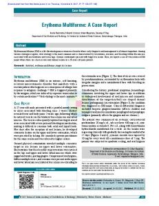
each time. But this time the lesion had augmented color. On dermatological examination, there was an oval, sharply bordered, slightly desquamated purpuric patch, inferomedial to the right labial commissure with a few petechiae at the bottom of this lesion and herpetic vesicules at the mid lower lip. There was a slightly hypopigmented macule on the left lower lip (Figure 1). No mucosal involvement was detected. She had no drug history. A punch biopsy was performed with the diagnosis of fixed drug eruption, erythema multiforme and pressure induced purpura and histopathologically; focal parakeratosis, spongiosis, lymphocyte exocytosis and a few necrotic keratinocytes in epidermis, subepidermal edema and vesiculation, dermal lymphocyte infiltration and erythrocyte
extravasation were observed (Figure 2a, b). The lesion was diagnosed as EM with history, clinical and histopathological findings. The lesion resolved spontaneously in a week,although only a topical antibiotic cream was applied after biopsy.
Discussion The exact role of Herpes Simplex Virus (HSV) in the etiology and pathogenesis of EM has been proven in the last decade in many studies when the viral genomes were detected in lesional skin (3,4). In a study by Ng and colleagues, HSV DNA has been detected in 60% of patients clinically diagnosed with recurrent HAEM and in 50% of patients with recurrent idiopathic EM, using polymerase chain reaction (PCR) of skin biopsy
Figure 1. Oval, sharply bordered, slightly desquamated purpuric patch, inferomedial to the right labial commissure, with a few petechiae at the bottom; and herpetic vesicule in lower lip.
Figure 2a. Focal parakeratosis, spongiosis, lymphocyte exocytosis, a few necrotic keratinocytes in the epidermis (HEx100).
Tıp Araştırmaları Dergisi; 2015: 13(3):131-133
Figure 2b. In a higher magnification necrotic keratinocytes (arrow) (HEx200).
132
Atypical Erythema Multiforme
CASE REPORT specimens (4). Delayed hypersensitivity reaction to herpes DNA fragments is thought to play a role in the pathogenesis of HAEM (3). On the other hand, drugs such as non-steroid antiinflammatory drugs, antibiotics and anticonvulsants are found to be responsible for about 10% of the EM cases. Food additives and some topical medications can also play a role in the etiology of EM which are interrogated in the present case (1). The clinical presentation is about 7-21 days after herpes infection, with mild prodromal symptoms. There is usually a predilection for acral surfaces and mucosal lesions are rare in HAEM. The characteristic target lesion of EM, also named ‘bull’s eye’, has three concentric rings; an innermost bullae or a crust due to epidermal necrosis, and two additional rings surrounding it (5). There are also some atypical presentations which are usually related to drug induced EM, such as two concentric rings, polycyclic and arcuate lesions with pronounced blisters (4). Mucosal lesions may exist in the form of erythema, erosions and ulcers which are more prominent in drug induced EM (1,4). The current case did not correlate with the characteristic clinical presentation of HAEM as the sole lesion located on the side of mouth, was neither symmetric nor targetoid in appearence. But there was an obvious relation with herpes infection in the history, and clinically both preceeding and coexisting herpes labialis was present. Histopathologically in the early lesions of EM; a vacuolar interface dermatitis showing lymphocyte infiltration along the dermoepidermal junction, hydropic changes and dyskeratosis indicating apoptosis in keratinocytes in epidermis are usually observed (6). The findings in our case was compatible except for erythrocyte extravasation which is not a common feature in EM. At this time, the history gave a clue: nausea and vomiting preceeded the lesions. The increased intraarterial pressure with emesis was thought to contribute to the pathogenesis of this microscopic sign as well as the purpuric view macroscopically. Fixed drug eruption (FDE) has similar clinical and pathological findings with EM. FDE presents with fewer lesions, usually single as in our case.
Tıp Araştırmaları Dergisi; 2015: 13(3):131-133
The lesions recur at the same site and may be reproduced by continuing administration of the responsible drug. Characteristically, sharply marginated, round to oval itchy plaques of erythema and edema that become dusky violaceous or brown, and sometimes with central vesiculation, bullae or necrosis are observed (2). Histopathologically, vacuolar interface dermatitis is also a characteristic feature in FDE, but a deeper extension of the infiltrate and melanin incontinence is usually observed (6). Necrotic keratinocytes can also be a prominent feature. Although our case mimics FDE both clinically and histopathologically, the significant relation to herpes infection, absent drug history and recurrences in similar but not exactly the same location, all make us consider the final diagnosis of EM. In conclusion, EM should be considered in acute, self-limited lesions with a history of accompanying herpes infection, even when the characteristic target lesions are missing. History taking is still a very important part of dermatological diagnosis.
References 1.
2.
3.
4.
5. 6.
Sokumbi O, Wetter DA. Clinical features, diagnosis, and treatment of erythema multiforme: a review for the practicing dermatologist. Int J Dermatol 2012; 51:889-902. Breathnach SM. Drug reactions. In: Burns T, Breathnach S, Cox N, Griffiths C (eds). Rook's Textbook of Dermatology. Oxford: Blackwell Publishing, 2010; 75.28. Aurelian L, Ono F, Burnett J. Herpes simplex virus (HSV)-associated erythema multiforme (HAEM): A viral disease with an autoimmune component. Dermatol Online J 2003;9:1. Ng PP, Sun YJ, Tan HH, Tan SH. Detection of herpes simplex virus genomic DNA in various subsets of erythema multiforme by polymerase chain reaction. Dermatology 2003;207:349–53. Huff JC. Erythema multiforme. Dermatol Clin 1985; 3:141–152. Weedon D. Weedon’s Skin Pathology, 3rd ed. Edinburgh: Churchill Livingstone/Elsevier, 2010; 202–207, 536–8.
133