Cancer Biol Med 2016. doi: 10.28092/j.issn.2095-3941.2015.0094
ORIGINAL ARTICLE
Establishment and application of a multiplex genetic mutation-detection method of lung cancer based on MassARRAY platform Hong-Xia Tian, Xu-Chao Zhang, Zhen Wang, Jian-Guang Chen, Shi-Liang Chen, Wei-Bang Guo, Yi-Long Wu Medical Research Center, Guangdong Lung Cancer Institute, Guangdong General Hospital, Guangdong Academy of Medical Sciences, Guangzhou 510080, China
ABSTRACT
KEYWORDS
Objective: This study aims to establish a method for highly parallel multiplexed detection of genetic mutations in Chinese lung cancer samples through Agena iPLEX chemistry and matrix-assisted laser desorption ionization time-of-flight analysis on MassARRAY mass spectrometry platform. Methods: We reviewed the related literature and data on lung cancer treatments. We also identified 99 mutation hot spots in 13 target genes closely related to the pathogenesis, drug resistance, and metastasis of lung cancer. A total of 297 primers, composed of 99 paired forward and reverse amplification primers and 99 matched extension primers, were designed using Assay Design software. The detection method was established by analyzing eight cell lines and six lung cancer specimens. The proposed method was then validated through comparisons by using a LungCartaTM kit. The sensitivity and specificity of the proposed method were evaluated by directly sequencing EGFR and KRAS genes in 100 lung cancer cases. Results: The proposed method was able to detect multiplex genetic mutations in lung cancer cell lines. This finding was consistent with the observations on previously reported mutations. The proposed method can also detect such mutations in clinical lung cancer specimens. This result was consistent with the observations with LungCartaTM kit. However, an FGFR2 mutation was detected only through the proposed method. The measured sensitivity and specificity were 100% and 96.3%, respectively. Conclusions: The proposed MassARRAY technology-based multiplex method can detect genetic mutations in Chinese lung cancer patients. Therefore, the proposed method can be applied to detect mutations in other cancer tissues. Lung neoplasms; driver genes; mutation; multigene testing; MassARRAY
Introduction Lung cancer is the leading cause of cancer-related deaths worldwide, and its rate of occurrence has been increasing. Precision medicine or personalized medicine based on genomic changes is a new breakthrough that allows more targeted therapy than traditional radiation and chemotherapy. Some of these mutations have been identified as predictive of clinical responses to targeted therapeutic agents in non-small cell lung cancer (NSCLC), such as the improved response to gefitinib or erlotinib in patients with certain epidermal growth factor receptor (EGFR) mutations and the response of crizotinib in patients with echinoderm Correspondence to: Yi-Long Wu E-mail:
[email protected] Received November 3, 2015; accepted January 5, 2016. Available at www.cancerbiomed.org Copyright © 2016 by Cancer Biology & Medicine
microtubule-associated protein-like 4-anaplastic lymphoma kinase (EML4-ALK) fusion mutation1,2. Given that many clinically relevant biomarkers continue to be identified, and targeted drugs are developed for personalized treatment, multigene mutation screening will be a requirement in routine clinical practice. Genetic mutations can be detected by employing iPLEX mass-modified single-base extension technology and matrixassisted laser desorption ionization time-of-flight (MALDITOF) mass spectrometry on MassARRAY platform. The target sequences are amplified with amplification primers and extension primers, which are located adjacent to the interrogated loci, and they are used for single-base extension reactions to hybridize and elongate the extension primers at the nucleotide position of interest. Mutations are distinguished via detection and resolution of the massmodified extension bases through MALDI-TOF. MassARRAY platform is an ideal platform for the mutation screening of multiple genes. LungCarta TM Panel 3,4 can
Cancer Biol Med Vol 13, No 1 March 2016
analyze 214 mutations in 26 oncogenes. However, this method presents several limitations. For instance, the panel is expensive and does not include the recently identified molecular targets of interest. Therefore, a multigene detection method that is suitable for Chinese lung cancer patients should be established. Briefly, the motivations of developing this new method are as follows: (1) MALDI-TOF method can simultaneously interrogate multiple gene mutation loci, which are screened for in-routine clinical practice. (2) If all the genes are analyzed individually, most analyses are certainly not possible because of the complete depletion of tissues and nucleic acid. However, multiplexing, which is employed in this method, reduces the amount of the clinical sample used. (3) Some novel molecular targets can be enrolled in the new method. (4) The custom panel in the new method is more costeffective than the commercial panel because the former is established by ourselves.
Materials and methods Patient specimens and cell lines We obtained a cohort of 106 lung cancer specimens from the Guangdong Lung Cancer Institute of Guangdong General Hospital between January 2014 and December 2014. An informed consent was obtained from each patient, which was approved by the ethics committee of Guangdong General Hospital. All samples, which were stored at –80 °C after being frozen in liquid nitrogen, were assessed by two pathologists to ensure that >50% of each sample consisted of tumor tissue. We used eight NSCLC cell lines (i.e. H460, PC9, H1650, H1975, A549, GLC82, L78, and HCC827), which were purchased from the cell bank of the Chinese Academy of Sciences in Shanghai.
Reagents and instruments The following materials were used in this study: QIAsymphony DNA Mini Kit (Qiagen, Valencia, Germany); LungCarta kit, PCR Accessory Set, iPLEX Pro Reagent Kit, and SpectroCHIP® (Agena Bioscience, San Diego, USA); H 2 O (Sigma-Aldrich, USA); QIAsymphony SP (Qiagen, Valencia, Germany); Thermo NanoDrop 1000 (Thermo, MA, USA); MassARRAY® Nanodispenser and MassARRAY® Analyzer (Agena Bioscience, San Diego, USA); ABI 3730xl Sequencing Machine (Life Technologies, New York, USA); and polymerase chain reaction (PCR) machine (Life Technologies, New York, USA).
69
Preparation of polygenic primer panel Determination of related driver genes of lung cancer We reviewed the related literature, data on lung cancer treatments, and gene spectrum characteristics of the Chinese lung cancer population. A total of 13 target oncogenes (i.e. EGFR, KRAS, ALK, FGFR1, FGFR2, FGFR3, PIK3CA, BRAF, PTEN, MET, ERBB2, AKT1, and STK11), which were closely related to the pathogenesis, drug resistance, and metastasis of lung cancer and associated with relevant transduction pathways, were enrolled in the polygenic primer panel5–12.
Selected mutation hot spots of driver genes We determined the Catalogue of Somatic Mutation in Cancer (COSMIC) identifier numbers of the following 13 genes by browsing the COSMIC database: EGFR(ENST00000275493), KRAS(ENST00000256078), ALK(ENST00000389048), FGFR1(ENST00000447712), FGFR2(ENST00000358487), FGFR3(ENST00000440486), PIK3CA(ENST00000263967), BRAF(ENST00000288602), PTEN(ENST00000371953), MET(ENST00000318493), ERBB2(ENST00000269571), AKT1(ENST00000349310), and STK11(ENST00000326873). In accordance with the mutation frequencies of 13 oncogenes in lung cancer, 99 mutation hot spots associated with lung cancer were added to the polygenic primer panel (Table 1).
Designed polygenic primer panel The genome sequence numbers of the following 13 target genes were identified in GenBank: EGFR(NG_007726.3), KRAS(NG_007524.2), ALK(NG_009445.1), FGFR1(NG_007729), FGFR2(NG_012449.1), FGFR3(NG_012632.1), PIK3CA(NG_012113.2), BRAF(NG_007873.3), PTEN(NG_007466.2), MET(NG_008996.1), ERBB2(NG_007503.1), AKT1(NG_012188.1), and STK11(NG_007460.2). We marked mutant loci in genomic DNA (gDNA) sequences in accordance with the mutation label and format requirements of the MassARRAY platform. The relevant parameters of Assay Design Software (ADS) were adjusted to include all 99 loci in the polygenic primer panel. The highest number of loci that can be detected simultaneously was set to 10 mutant loci. A total of 99 loci were randomly distributed in 12 wells through ADS on the basis of primer design (avoidance of the formation of dimers/mismatches). A total of 297 primers, including 99 paired forward and reverse amplification primers and 99 matched single-base extension primers, were designed. The target sequences were amplified by
70
Tian et al. Method for multiplex detection of genetic mutations of lung cancer
Table 1 The invented kit contains protein mutation locus of 13 genes Gene
Protein mutation locus
EGFR
p.G719SCDA p.E746_A750delELREA p.E746_T751>A p.E746_S752>V p.L747_T751delLREAT p.L747_P753>S p.L747_S752delLREATS p.L747_E749delLRE p.L747_A750>P p.T790M p.L858R p.L861Q
KRAS
p.G12CRSDVA p.G13CRSDAV p.Q61H
ALK
p.C1156Y p.F1174ILVSC p.L1196M p.F1245IVCL p.G1269A p.R1275QL
FGFR1
p.S125PL p.T141A p.K656E
FGFR2
p.S252W p.P253RL p.Y375CH p.C382RY p.N549HSK p.M640VTI p.I642VTM p.A648TD p.K659E
FGFR3
p.R248C p.S249C p.G370C p.S371C p.Y373C p.G380R p.A391EV p.K650EQMT p.G697C
PIK3CA
p.E542KQVAG p.E545KQAGVD p.H1047RLY
BRAF
p.G469AVER p.D594NHGVA p.L597RQPSV p.V600MLEAGKRD
PTEN
p.R130GQPL p.R173CH
MET
p.N375S p.R988CS p.T1010I p.Y1253D
AKT1
p.E17K
ERBB2
p.S310FY p.R678Q p.L755S p.D769YHN p.A775_G776insYVMA p.V777LM p.V842I
STK11
p.D194YN p.P281L p.F354L
amplification primers, and the extension primers were located on the base before the mutation sites, which were complementary to the amplification products and performed single-base extension reactions.
Configuration of polygenic primer panel The primers were synthesized by Shanghai Sangon Biological Engineering Technology and Service Co., Ltd. The polygenic primer panel was configured as follows: (1) The amplification primers were first diluted to 10 μM, and the working liquid was a mixture of all primers in each well, including 0.5 μM of each primer. In accordance with the parameters of ADS, the forward and reverse amplification primers were distributed into 12 pipes. The forward primer P1-F and reverse amplification primer P1-R comprised group 1, the forward primer P2-F and reverse amplification primer P2-R comprised group 2, and so on. (2) The extension primers were first diluted to 500 μM and then mixed on the basis of individual molecular weights. The extension primers of E1 comprised group 1, the extension primers of E2 comprised group 2, and so on (E1–E12). A total of 12 amplification primers corresponded to 12 extension primers (e.g. P1 corresponded to E1, etc.).
Establishment of detection method The detection method was verified by analyzing 8 cell lines (i.e. H460, PC9, H1650, H1975, A549, GLC82, HCC827, and H1299) and 6 lung cancer specimens. The proposed method
was then validated through comparisons by using a LungCartaTM kit or previously reported results. The gDNA of a healthy person (from foreskin tissue; with informed consent) was used as a negative control, and H2O was used as a blank control. Each sample required 120 ng gDNA for 12 wells (10 ng/well). The procedure was conducted in a MassARRAY system platform. The experiments using the LungCartaTM kit were performed in accordance with the manufacturer’s protocol. The polygenic primer kit was used in the detection method as follows.
PCR gDNA was extracted from cell lines and patient specimens in accordance with the manufacturer’s protocol. Afterward, gDNA was quantified using a NanoDrop ND-1000 spectrophotometer. gDNA was then amplified using a PCR Accessory Set. The thermocycling cocktail comprised 0.5 μL of PCR buffer (10×), 0.4 μL of MgCl2 (25 mM), 0.1 μL of dNTPs (25 mM), 0.2 μL of PCR enzyme (5 U/μL), 1 μL of amplification primer mix (P1-P12), 1 μL of gDNA (10 ng/μL), and H 2 O to a total volume of 5 μL. The thermocycling conditions were 94 °C for 2 min; followed by 45 cycles of 94 °C for 30 s, 56 °C for 30 s, and 72 °C for 1 min; and a final extension step of 72 °C for 5 min.
Shrimp alkaline phosphatase (SAP) reaction dNTPs in the PCR products were removed via SAP. For this reaction, 0.3 μL of SAP (1.7 U/μL) and 0.17 μL of SAP buffer
Cancer Biol Med Vol 13, No 1 March 2016
(10×) were added to step 1 PCR products, and H 2 O was added to a total volume of 7 μL. The reaction conditions were 37 °C for 40 min and 85 °C for 5 min.
Extension reaction A single-base extension reaction was performed with iPLEX Pro Reagent Kit to hybridize and elongate the extension primers at the nucleotide position of interest. For the singlebase extension, 0.2 μL of TypePlex buffer (10×), 0.2 μL of TypePlex termination mix (10×), 0.041 μL of TypePlex thermosequenase enzyme (33 U/μL), and 0.804 μL of extension primers (E1–E12) were mixed with step 2 products, and H2O was added to a total volume of 9 μL. The reaction conditions were 94 °C for 30 s; followed by 5 cycles of 94 °C for 5 s, 52 °C for 5 s, and 80 °C for 5 s, with this step repeated 35 times; and a final extension at 72 °C for 3 min.
71
cases. EGFR and KRAS mutations were detected through Sanger sequencing in accordance with a previously published protocol 13 . The sequencing data were analyzed using Sequencing Analysis Software v5.2 (Applied Biosystems).
Results Polygenic primer panel containing mutation sites We tested 99 mutation hot spots in 13 target genes (i.e. EGFR, KRAS, ALK, FGFR1, FGFR2, FGFR3, PIK3CA, BRAF, PTEN, MET, ERBB2, AKT1, and STK11), which were closely related to the pathogenesis, drug resistance, and metastasis of lung cancer and associated with relevant transduction pathways. The detailed protein mutation sites of genes are listed in Table 1.
Desalination For desalination, 41 μL of H2O and 15 mg of Clean Resin (96-well microplates) were added to step 3 extension products, and the plate was rotated for 30–60 minutes. The plate was then centrifuged at 3,200 g for 5 min.
Spotter and analysis The supernatant from the step 4 extension products was spotted onto matrix-precoated SpectroCHIP® through MassARRAY® Nanodispenser and scanned using MassARRAY® Analyzer. The results were analyzed using MassArray® Workstation Software (v.4.0). The mutation was distinguished with TOF mass spectrometry on the basis of different molecular weights. The peaks in the mass spectrum were identified as mutations.
Analysis of sensitivity and specificity The newly established methods were evaluated by Sanger sequencing of EGFR and KRAS genes in 100 lung cancer
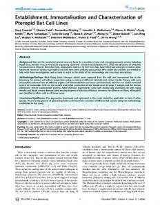
Establishing a detection method in cell lines The detection method used with the polygenic primer kit was established by analyzing 8 cell lines of lung cancer. The results from the newly established method were consistent with the previously reported mutations in cell lines. All the cell lines were confirmed by Sanger sequencing, and the sequencing results from A549 are shown in Figure 1. The detailed mutation sites within genes and proteins are listed in Table 2. The negative control and blank control did not detect mutations.
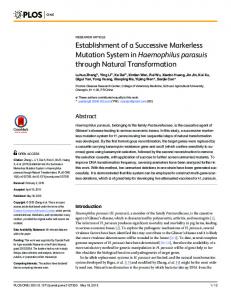
Established detection method in lung cancer specimens The detection method used with the polygenic primer kit was established by analyzing 6 lung cancer tissue specimens, and this method was validated through comparison by using a LungCartaTM kit (Table 3). An FGFR2 gene mutation was
Figure 1 KRAS_G12S mutation was detected in A549 cell lines using Sanger sequencing.
72
Tian et al. Method for multiplex detection of genetic mutations of lung cancer
Table 2 The result of established method was validated in cell lines Cell line
ATCC previously reported
Result of established method
H460
KRAS mutation PIK3CA mutation
KRAS_Q61H (c.183A>T) PIK3CA_E545K (c.1633G>A )
PC9
EGFR_ Exon19 deletion
p.E746_A750delELREA (c.2235-2249del15)
H1650
EGFR_ Exon19 deletion
p.E746_A750delELREA (c.2235-2249del15)
H1975
EGFR_L858R EGFR_T790M
EGFR_L858R (c.2573T>G) EGFR_T790M (c.2369C>T)
A549
KRAS mutation
KRAS_G12S (c.34G>A)
GLC82
EGFR_L858R
EGFR_L858R (c.2573_2574TG>GT)
HCC827
EGFR Exon19 deletion
p.E746_A750delELREA (c.2236_2250del15)
H1299
EGFR/ALK/KRAS negative
No mutation
Table 3 The result of established method was validated by comparing with LungCartaTM in lung cancer tissue samples Sample number
Result of LungCartaTM
Result of established method
K1736T
EGFR_L858R MET_N375S
EGFR_L858R (c.2573T>G) MET_N375S (c.1124A>G)
K1744T
EGFR_DEL
EGFR_p.E746_A750delELREA (c.2235_2249del15)
K1748T
STK11_F354L
STK11_F354L (c.1062C>G)
K1745T
No mutation
No mutation
K1746T
No mutation
No mutation
K1747T
No mutation
FGFR2_N549H (c.1645A>C)
Figure 2 FGFR2 gene mutation was detected by the newly developed method in K1747T tissue samples of lung cancer. C: mutant allele; A: wild-type allele.
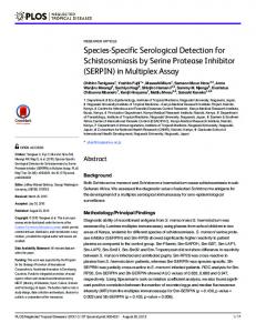
found in a sample of lung cancer tissue (K1747T) through the newly established method. However, the LungCartaTM kit cannot detect the mutation (Figure 2). Unfortunately, other methods cannot perform validation because of the depletion of the material. With respect to other clinical lung cancer specimens, the findings were consistent with the observations with LungCartaTM kit (Figure 3).
Sensitivity and specificity of the established method For 100 samples, 99 mutation hot spots in 13 targeted genes were analyzed using the established method. For comparison, EGFR and KRAS mutations were detected via Sanger sequencing. With Sanger sequencing as the gold standard, the sensitivity of the established method was 100%, and its specificity was 96.3% (positive predictive value, 95.8%;
Cancer Biol Med Vol 13, No 1 March 2016
73
Figure 3 Coexistence of _L858R and _N375S was detected by the newly developed method and LungCartaTM in K1736T tissue sample of lung cancer. (A) EGFR_L858R was detected by LungCartaTM. (B) MET_N375S was detected by LungCartaTM. (C) EGFR_L858R was detected by the newly developed method. (D) MET_N375S was detected by the newly developed method.
74
Tian et al. Method for multiplex detection of genetic mutations of lung cancer
negative predictive value, 100%). A detailed comparison of the results is shown in Table 4. Table 4 The result of the newly developed method compared with Sanger sequencing in 100 cases of lung cancer samples Result of new method
Result of Sanger sequencing Total Mutation
Wild type
Mutation
46
2
48
Wild type
0
52
52
46
54
10 0
Total
Discussion Quantitative PCR, Sanger sequencing, fluorescence in situ hybridization, and immunohistochemistry are the main single-gene detection methods for the molecular classification of lung cancer. These methods present low throughput, and they are also time consuming and expensive. Moreover, these methods significantly restrict the screening of ROS1, RET, MET, HER2, NRTK, and NRG in clinical practice with the exception of EGFR, ALK, and KRAS oncogenes14. Patients with newly diagnosed lung cancer who are undergoing surgery (major surgery or minor surgical biopsy) exhibit considerable cancer tissues, but only a small sample can be usually acquired from advanced patients through percutaneous biopsy, bronchoscopy biopsy, and endobronchial ultrasound biopsy. As such, any tissue barely remains for characterization of other important molecules and exploratory research on drug resistance mechanisms after diagnosis and molecular detection of individual genes. Considering the complexity of tumor driver genes and the limitations of single-gene detection techniques, we need to establish a multiplex genetic mutation-detection method for lung cancer in clinical practice and translational medicine. Next-generation sequencing (NGS) is currently one of the most important approaches to precise treatment employed by studies on the domain of life sciences, but the complexities of NGS technologies and data analysis are slowing their wide spread availability in the diagnostics laboratories. So a robust genotyping platform which can carry-on daily routine testing is need. Other multiplex genetic mutation-detection methods have been reported, such as Cancer Personalized Profiling by deep sequencing and SNaPShot7,15. Molecular characterization via MALDI-TOF on MassARRAY platform, which is characterized by high throughput, high sensitivity, and a simple operation, can be implemented by combining
single-base extension and mass spectrometry technology. This approach also minimizes the weaknesses of traditional single-gene tests, such as high cost and their time-consuming and tedious procedures8–10. Given the short segments of the PCR amplification products, not only fresh tissue specimens but also formalin-fixed paraffin-embedded (FFPE) specimens, pleural effusion, and biopsy samples can be used for effective detection3,4,8,9. LungCartaTM Panel, which is commercially available, can facilitate comprehensive screening of relevant biomarkers of lung cancer 4,10,11 . However, this method presents several limitations; for example, the panel is confidential. The LungCartaTM Panel is also expensive, and it does not include recently identified molecular targets of interest. According to the review of the related literature and data on lung cancer treatments, 99 mutation hot spots in 13 oncogenes of lung cancer were enrolled into the polygenic primer panel, such as MET mutations, which are associated with targeting the therapy resistance of EGFR tyrosine kinase inhibitors (TKIs)16,17. The mutations in multiple loci of the ALK gene are also associated with the resistance of crizotinib; for instance, the recently reported C1156Y, L1196M, F1174L, and G1269S mutations in ALK gene are identified as resistance mutations following crizotinib treatment in lung cancer. Some mutations are highly sensitive to the structurally unrelated ALK inhibitor TAE68412,18. In recent years, FGFR signaling pathways have been shown to demonstrate abnormal activation in various tumors19,20, and one FGFR2 gene mutation was detected in one clinical specimen through the newly established method. However, this mutation cannot be detected using the LungCartaTM kit. Unfortunately, the mutation cannot be confirmed by another method because of the depletion of the material. This observation demonstrates that the multiplex genetic mutation-detection method may provide accurate variant information for patients. MALDI-TOF mass spectrometry also presents advantages because of its high accuracy and sensitivity compared with the direct sequencing method21. At present, Sanger sequencing is generally regarded as the gold standard for detecting somatic mutations in tumor specimens. However, Sanger sequencing suffers from limited sensitivity for low-level mutant alleles22, particularly in FFPE specimens; this sequencing also takes a slow turnaround time 2 3 . Given that MALDI-TOF mass spectrometry established detection limits of 1%–5% mutant alleles24, the specificity of the in-house panel compared with that of Sanger sequencing is 96.3%, which may be associated with the high sensitivity of mass spectrometry and may compensate for the low sensitivity of direct sequencing. The proposed MALDI-TOF multiplex mutation-detection method demonstrates the following advantages: (1) The novel method examines oncogenes and multiple drugresistant loci in parallel, which are associated with lung-
Cancer Biol Med Vol 13, No 1 March 2016
cancer-targeted therapy. The screening results optimize the molecular characterization of the disease and provide information regarding targeted therapy. (2) The polygenic primer panel can reduce the patient’s molecular testing costs. Following clinical and translational applications, the proposed method provides direct economic benefits and optimizes clinical resources. (3) Moreover, the proposed method shows high throughput, and it can simultaneously detect 99 loci of 13 oncogenes in 12 wells. Thus, a sensitive highly parallel approach is provided, and the patient diagnostic samples are used efficiently for an accurate molecular classification. Furthermore, we can freely enroll updated mutations to the in-house panel based on the flexibility of the MassARRAY platform. Thus, the newly developed method is extensible. However, the newly developed method also presents limitations. For instance, this method is based on a panel of preselected sites. Therefore, the mutational patterns in tumor suppressors, such as P53 gene 25 , which is highly random and usually disperse across the gene, cannot be comprehensively detected with the newly developed method. For these mutation patterns, Sanger sequencing or NGS may represent a good alternative. In conclusion, successful establishment of the novel detection method, which demonstrates a great potential for lung cancer treatment, can significantly reduce the costs of biomarker tests.
Acknowledgements This work was supported by the Special Fund for Research in the Public Interest from the National Health and Family Planning Commission of PRC (Grant No. 201402031), the Key Lab System Project of the Guangdong Science and Technology Department (Grant No. 2012A061400006), and the Special Fund for Research in the Public Interest and Capacity Building from the Guangdong Science and Technology Department (Grant No. 2014A020212225). This article was published orginally in Chinese Journal of Clinical Oncology 2015; 42(17): 856-61 (in Chinese).
Conflict of interest statement No potential conflicts of interest are disclosed.
References 1.
Mok TS, Wu YL, Thongprasert S, Yang CH, Chu DT, Saijo N, et al. Gefitinib or carboplatin-paclitaxel in pulmonary adenocarcinoma. N Engl J Med. 2009; 361: 947-57.
2.
Soda M, Choi YL, Enomoto M, Takada S, Yamashita Y, Ishikawa S, et al. Identification of the transforming EML4-ALK fusion gene in
75
non-small-cell lung cancer. Nature. 2007; 448: 561-6.
3.
Okamoto I, Sakai K, Morita S, Yoshioka H, Kaneda H, Takeda K, et al. Multiplex genomic profiling of non-small cell lung cancers from the LETS phase III trial of first-line S-1/carboplatin versus paclitaxel/carboplatin: results of a West Japan Oncology Group study. Oncotarget. 2014; 5: 2293-304.
4.
Quinn AM, Hickson N, Adaway M, Priest L, Jaeger E, Udar N, et al. Diagnostic mutation profiling and validation of non small cell lung cancer small biopsy samples using a high throughput platform. J Thorac Oncol. 2015; 10: 784-92.
5.
An SJ, Chen ZH, Su J, Zhang XC, Zhong WZ, Yang JJ, et al. Identification of enriched driver gene alterations in subgroups of non-small cell lung cancer patients based on histology and smoking status. PLoS One. 2012; 7: e40109.
6.
Kris MG, Johnson BE, Berry LD, Kwiatkowski DJ, Iafrate AJ, Wistuba II, et al. Using multiplexed assays of oncogenic drivers in lung cancers to select targeted drugs. JAMA. 2014; 311: 1998-2006.
7.
Su J, Zhang XC, An SJ, Zhong WZ, Huang Y, Chen SL, et al. Detecting the spectrum of multigene mutations in non-small cell lung cancer by Snapshot assay. Chin J Cancer. 2014; 33: 346-50.
8.
Pearce M, Cullinan A, Hogg G, Hosseini D, Ehrich M. Mutation profiling in tumor samples using the Sequenom OncoCartaTM? Panel. Nature Methods. 2009; 6: vii-viii.
9.
Fumagalli D1, Gavin PG, Taniyama Y, Kim SI, Choi HJ, Paik S, et al. A rapid, sensitive, reproducible and cost-effective method for mutation profiling of colon cancer and metastatic lymph nodes.
BMC Cancer. 2010; 10: 101. 10. Sherwood JL, Müller S, Orr MC, Ratcliffe MJ, Walker J. Panel based MALDI-TOF tumour profiling is a sensitive method for detecting mutations in clinical non small cell lung cancer tumour. PLoS One. 2014; 9: e100566. 11. Wen YS, Cai L, Zhang XW, Zhu JF, Zhang ZC, Shao JY, et al. Concurrent oncogene mutation profile in Chinese patients with stage Ib lung adenocarcinoma. Medicine (Baltimore) 2014; 93: e296. 12. Heuckmann JM, Hölzel M, Sos ML, Heynck S, Balke-Want H, Koker M, et al. ALK mutations conferring differential resistance to structurally diverse ALK inhibitors. Clin Cancer Res. 2011; 17: 7394-401. 13. Zhou Q, Zhang XC, Chen ZH, Yin XL, Yang JJ, Xu CR, et al. Relative abundance of EGFR mutations predicts benefit from gefitinib treatment for advanced non-small-cell lung cancer. J Clin Oncol. 2011; 29: 3316-21. 14. Larsen JE1, Minna JD. Molecular Biology of Lung Cancer: Clinical Implications. Clin Chest Med. 2011; 32: 703-40. 15. Newman AM, Bratman SV, To J, Wynne JF, Eclov NC, Modlin LA, et al. An ultrasensitive method for quantitating circulating tumor DNA with broad patient coverage. Nat Med. 2014; 20: 548-54. 16. Krishnaswamy S, Kanteti R, Duke-Cohan JS, Loganathan S, Liu W, Ma PC, et al. Ethnic differences and functional analysis of MET mutations in lung cancer. Clin Cancer Res. 2009; 15: 5714-23. 17. Shieh JM, Tang YA, Yang TH, Chen CY, Hsu HS, Tan YH, et al. Lack of association of C-Met-N375S sequence variant with lung
76
Tian et al. Method for multiplex detection of genetic mutations of lung cancer
cancer susceptibility and prognosis. Int J Med Sci. 2013; 10: 988-94.
18. Choi YL, Soda M, Yamashita Y, Ueno T, Takashima J, Nakajima T. EML4-ALK mutations in lung cancer that confer resistance to ALK inhibitors. N Engl J Med. 2010; 363: 1734-9.
19. Tchaicha JH, Akbay EA, Altabef A, Mikse OR, Kikuchi E, Rhee K, et al. Kinase domain activation of FGFR2 yields high-grade lung adenocarcinoma sensitive to a Pan-FGFR inhibitor in a mouse model of NSCLC. Cancer Res. 2014; 74: 4676-84.
20. Liao RG, Jung J, Tchaicha J, Wilkerson MD, Sivachenko A, Beauchamp EM, et al. Inhibitor-sensitive FGFR2 and FGFR3 mutations in lung squamous cell carcinoma. Cancer Res. 2013; 73: 5195-205.
21. Ulivi P, Delmonte A, Chiadini E, Calistri D, Papi M, Mariotti M, et
StripAssay for detecting KRAS mutations in non small cell lung carcinomas. J Exp Clin Cancer Res. 2012; 31: 79. 23. Querings S, Altmüller J, Ansén S, Zander T, Seidel D, Gabler F, et al. Benchmarking of mutation diagnostics in clinical lung cancer specimens. PLoS One. 2011; 6: e19601. 24. Kriegsmann M, Arens N, Endris V, Weichert W, Kriegsmann J. Detection of KRAS, NRAS and BRAF by mass spectrometry-a sensitive, reliable, fast and cost-effective technique. Diagn Pathol. 2015; 10: 132. 25. Gao W, Mady HH, Melhem MF, Keohavong P. Analysis of p53 mutations in histologically normal lung tissues and lung tumors from non-small cell lung cancer patients. Mol Carcinog. 2009; 48: 633-41.
al. Gene mutation analysis in EGFR wild type NSCLC responsive to erlotinib: are there features to guide patient selection? Int J Mol Sci. 2014; 16: 747-57.
22. Jancik S, Drabek J, Berkovcova J, Xu YZ, Stankova M, Klein J, et al.
Cite this article as: Tian HX, Zhang XC, Wang Z, Chen JG, Chen SL, Guo WB, et al. Establishment and application of a multiplex genetic mutation-
A comparison of direct sequencing, pyrosequencing, high
detection method of lung cancer based on MassARRAY platform. Cancer Biol
resolution melting analysis, TheraScreen DxS, and the K-ras
Med. 2016; 13: 68-76. doi: 10.28092/j.issn.2095-3941.2015.0094