Microsc. Microanal., page 1 of 10 doi:10.1017/S1431927615014920
© MICROSCOPY SOCIETY OF AMERICA 2015
Evaluating the Use of Synthetic Replicas for SEM Identification of Bloodstains (with Emphasis on Archaeological and Ethnographic Artifacts) Policarp Hortolà1,2,* 1
Àrea de Prehistòria, Universitat Rovira i Virgili (URV), Avinguda de Catalunya 35, ES-43002 Tarragona, Catalonia, Spain Institut Català de Paleoecologia Humana i Evolució Social (IPHES), Carrer de Marcel·lí Domingo s/n, Edifici W3 Campus Sescelades, ES-43007 Tarragona, Catalonia, Spain
2
Abstract: Some archaeological or ethnographic specimens are unavailable for direct examination using a scanning electron microscope (SEM) due to methodological obstacles or legal issues. In order to assess the feasibility of using SEM synthetic replicas for the identification of bloodstains (BSs) via morphology of red blood cells (RBCs), three fragments of different natural raw material (inorganic, stone; plant, wood; animal, shell) were smeared with peripheral human blood. Afterwards, molds and casts of the bloodstained areas were made using vinyl polysiloxane (VPS) silicone impression and polyurethane (PU) resin casting material, respectively. Then, the original samples and the resulting casts were coated with gold and examined in secondary-electron mode using a high-vacuum SEM. Results suggest that PU resin casts obtained from VPS silicone molds can preserve RBC morphology in BSs, and consequently that synthetic replicas are feasible for SEM identification of BSs on cultural heritage specimens made of natural raw materials. Although the focus of this study was on BSs, the method reported in this paper may be applicable to organic residues other than blood, as well as to the surface of other specimens when, for any reason, the original is unavailable for an SEM. Key words: blood smears, erythrocytes, scanning electron microscopy, haemotaphonomy, specimen preparation
I NTRODUCTION The presence of blood residues on archaeological artifacts has been previously reported by using a variety of techniques, including microscopy (e.g., Loy, 1983; Gramly, 1991; Loy & Dixon, 1998; Balme et al., 2001; Robertson et al., 2009; Jones, 2009; Fraser et al., 2013). In addition, several results on ethnographic artifacts housed in museums have revealed the preservation of adhering blood residues capable of being identified via microscopy (e.g., Torrence, 1993; Mazel et al., 2006; Robertson, 2011). This technique has been recommended as a screening method to determine which artifacts are most likely to retain blood residues (Malainey, 2011: 219–220). The presence of erythrocytes or red blood cells (RBCs) in a smear is evidence of blood (Fiori, 1962). The microscopy identification of bloodstains (BSs) needs the presence of any morphologically recognizable corpuscle characteristic of blood. Erythrocytes are only present in blood and are the most abundant formed element in blood. Also, they are the corpuscle that occupies more blood volume. In mammals, the proportion of blood volume occupied by RBCs exclusively (hematocrit value) is around 40% (mean ± standard deviation = 40.3 ± 2.5; estimated from data provided by Johnn et al., 1992). This means that nearly a half of mammalian blood volume corresponds to RBCs. Received March 31, 2015; accepted July 13, 2015 *Corresponding author.
[email protected]
Previous results on both experimental and archaeological stone tools suggest that several kinds of organic residues—including blood—can be identified using an incident-light (metallographic) microscope (e.g., Wadley et al., 2004; Lombard, 2005, 2008; Lombard & Wadley, 2007; Wadley & Lombard, 2007). Nevertheless, scanning electron microscopy is the most appropriate microscopy technique for examining BSs in situ (i.e., without removing them from their substrate) because scanning electron microscopes (SEMs) provide the best visualization of in-smear RBCs. This visualization is achieved by some critical SEM features: large depth of field, shadow-relief effect, and high resolution when examining bulk objects (Goldstein et al., 2003: 1–2). On the other hand, some archaeological or ethnographic specimens are unavailable for direct SEM examination due to methodological obstacles or legal issues. Such obstacles are that the specimen cannot be accommodated into the SEM chamber, or that the specimen is an immovable structure such as a ritual pole or a ceremonial altar. Legal issues include acquiring permission from a museum curator or private collector for study of the specimen. The increased size of specimen chambers in variablepressure and extended low-vacuum (“environmental”) SEMs has resulted in a concomitant increase in the size of samples that can be accommodated in an SEM. Currently, the technical problem of the sample dimension has been mostly overcome with the advent of the Large Chamber SEM (LC-SEM) (Klein & Brandt, 2006; Huq et al., 2008). Thus, the MIRA-X LC-SEM developed by VisiTec
2
Policarp Hortolà
Microtechnik—which at present is the largest series model SEM in the world—has a chamber inner volume of 3.5 m3, and can accommodate specimens up to 1.5 m in diameter. The main concern with LC-SEMs is their cost and, as a result, limited availability in electron microscopy facilities offered by most universities and research centers. A customary solution to the SEM chamber constraint and legal issues has been to make synthetic replicas capable of capturing details at the submicrometer level and, consequently, suitable for SEM use. The suitability of highresolution synthetic replicas for SEM use does not preclude that loss of some detail may occur (e.g., Rose, 1983, Fig. 4). The general principles of SEM replication can be found in the scientific literature (e.g., Barnes, 1978; Rose, 1983; Bromage, 1985). This technique involves a two-step procedure. First, a mold (the “impression” or “negative”) of the original surface is made from a dental elastomer (siliconebased rubber-like impression material). Subsequently, this mold is used to create a matching cast (the “replica” or “positive”) by pouring a (usually epoxy or polyurethane) synthetic resin into the mold. Direct SEM examination of molds, avoiding casting, has also been carried out (e.g., Greenfield, 1999; Li et al., 2012). Rose (1983, and references therein) enumerated six advantages of using synthetic replicas instead of original specimens. In brief: (1) ability of studying successive stages of change using a single specimen, (2) capability of being made in the field or in other outsidelab settings, (3) easy transport, (4) possibility of examining parts of large specimens in small SEM chambers without damage to the original, (5) inspection of porous bones or teeth that cannot be adequately evacuated in a conventional (high-vacuum) SEM, and (6) versatility for studying a wide spectrum of specimen types. A remarkable advantage of SEM replication is that it is a non-destructive technique. The ethical implications of the use of destructive techniques at the interface between archaeology and archaeometry has been examined previously (Galanidou, 2006, and references therein). The feasibility of using synthetic replicas does not depend only on their fidelity, but also on the absence of damage produced to the original specimen by the replication process. On the other hand, because not all sites and artifacts are viable for studying preserved micro-residues, criteria for site and sample selection—as those suggested by Langejans & Lombard (2015)—must be taken into account. SEM replication has been applied to a wide variety of specimens, from dental ceramic crowns to fossil plant impressions (e.g., Shipman, 1986; D’Errico & Villa, 1997; Haverkort & Lubell, 1999; Pickering et al., 2000; Galbany et al., 2004, and references therein; Ungar et al., 2006; Fiorenza et al., 2009; Frayer et al., 2010; Rhyu et al., 2010; Moisan, 2012; Trifkovic et al., 2012). In forensic science, SEM replicas have been recently used for ballistics investigations (Cominato et al., 2015). Galbany et al. (2006, and references therein) have highlighted high-quality replication as a way to allow museum curators to loan tooth casts to researchers for analysis without any risk of further damage to
the original specimens. For our purposes, this assertion can be adapted by saying that high-quality synthetic replication allows SEM examination of BSs from museum specimens without risk of damage. In research on prehistoric technology, high-resolution synthetic replicas have been employed for SEM examining artifact use-wear (e.g., D’Errico et al., 2003; Ollé & Vergès, 2008), but applying such SEM replicas to organic residues on implements has not received the same interest by researchers. Previously, I reported studies dealing with SEM examination and imaging of erythrocytes in BSs (e.g., Hortolà, 1992, 2002, 2013). In those studies, only original specimens were used. In this paper, I report an assessment of the feasibility of using SEM replicas for the identification of BSs via RBC morphology.
MATERIALS
AND
METHODS
Original Samples Three fragments of different natural materials (inorganic, plant, animal) were smeared with a nonhomogeneous, thinto-thick film of peripheral human blood from an adult male (Table 1). Previous to blood smearing, potential dust on the BS substrates was removed by applying several consecutive air streams using a bulb syringe. Blood smearing was carried out by sliding the bleeding epidermis on the raw material fragments, after pricking the author’s left-hand middle finger with a sterile clinical lancet. Based on the mechanism that created them, the BSs fell into the category “transfer stains”, subcategory “swipe pattern”, according to the terminology recommended by the Scientific Working Group on Bloodstain Pattern Analysis (SWGSTAIN, 2009). The BSs were air-dried indoors at a room temperature of 22°C and stored until the next day (ca. 24 h) to carry out the following steps.
Molding The molding method was modified from Ollé (2003). The molding materials and devices used in this work were manufactured by Heraeus Kulzer GmbH (Hanau, Germany). Of each original sample, a mold was made using a vinyl polysiloxane (VPS) silicone impression material (Provil novo Light C.D. 2 regular set; dispensed in two components, a green base and a matching grayish catalyst). Previous to molding, potential dust on the original samples was removed by applying several consecutive air streams with a bulb syringe. Then, the impression material was put down on the BSs and their surrounding areas using an ad-hoc gun for dispensing the two impression components and a cartridgecoupled tip for mixing them homogenously at a dosing ratio of 1:1 in volume and for directing the mixture to the area under analysis (Mixpac Cartridge Delivery System 2). The molds were cured at room temperature until completely solidified, which occurred after ca. 15 min and was verified by puncturing gently with a scalpel blade. Then, without
SEM of Bloodstain Replicas Table 1.
Characteristics of the Fragments of Different Natural Materials used as BS Substrate.*
Material Origin Stone Wood Shell
3
Basalt (a fine-grained igneous extrusive volcanic rock) Mastic tree (Pistacia lentiscus Linnaeus, 1753; Magnoliopsida: Anacardiaceae) Mediterranean mussel (Mytilus galloprovincialis Lamarck, 1819; Bivalvia: Mytilidae)
Bloodstain Substrate
Substrate Preparation
Block-split angular pebble, flat surface/section Branch lengthwise section, flat surface/section Valve outside, convex surface/section
Sanded down with a very-coarse grit flint paper (CAMI grade #16) Split in half, and then smoothed down with a fine grit flint paper (CAMI grade #100) Cleansed with and rinsed in tap water, and then air dried
*The volcanic rock was selected due to its use in the manufacture of Melanesian Club Heads (Hitchcock, 2004). The bivalve species was selected due to the resemblance of its shell with that of the giant mussel “choro zapato” (Choromytilus chorus Molina, 1782; Bivalvia: Mytilidae), which was employed in Tierra del Fuego for multifunctional shell knives (Gusinde, 1987 [1937]: 475, cited in Lucero, 2004: 18–19). The plant species was not selected ex profeso, but used according to availability. The color of the fragments was typical of each corresponding raw material in the procurement area (black in both basalt stone and Mediterranean mussel shell, dirty-white in mastic tree wood). CAMI, Coated Abrasives Manufacturers’ Institute (U.S.).
detaching the original samples from the molds, they were strengthened by covering with a silicone putty (Provil novo Putty regular set; dispensed in two components, a green base and a matching grayish catalyst), acting as a “back mold”. The two putty components were mixed by hand at an approximate ratio of 1:1 in volume until a homogenous greenish mass. After ca. 5 min hardening at room temperature, the strengthened molds were detached from the original samples, which in total were in contact with the impression material ca. 20 min. Then, in order to obtain a receptacle-like morphology, a small wall was build around each strengthened mold using the same silicone putty. To avoid potential dust on the molds, they were preserved in self-sealed plastic bags until proceeding to the following step.
Casting The casting method was modified from Ollé (2003). The casting materials used in this work were manufactured by Synthesia Española S.A. (La Llagosta, Spain). Just after making each mold, a matching cast was obtained using a polyurethane (PU) resin (Feropur 55; dispensed in two components, a colorless base and a yellowish hardener). First, the two casting components were mixed at a dosing ratio of 1:1 in weight in a plastic urinalysis container, and then the mixture was poured into the molds. Then, the casts were left to harden at room temperature until completely solidified, which occurred after 1 h and was verified by observing the changes in color (from yellowish to whitish), opacity (from translucent to opaque), and state of matter (from liquid to solid; confirmed by engrasping the plastic urinalysis receptacle containing the mixture surplus). Finally, each cast was preserved in a self-sealed plastic bag. In order to avoid potential dust deposit on and/or damage to their target surface, the casts were left without detaching them from their molds until proceeding to the following step.
Scanning Electron Microscopy After casting, the original samples and their resulting casts were coated with gold by a cathodic sputter coater (SCD 004,
BAL-TEC AG, Balzers, Liechtenstein) for 3 min at 30 mA (layer thickness ≈20 nm). Previous to coating, potential dust on the original specimens was removed by applying several consecutive air streams with a bulb syringe. In the case of the casts, since they had been left without detaching from their molds, such a cleaning was considered unnecessary and consequently not done. After coating, landmark numbers to locate potential areas of interest were written on the specimens, and subsequently they were examined via secondary electrons with a high-vacuum SEM (JSM-6400, JEOL Ltd., Tokyo, Japan). Microscope conditions were 15 kV accelerating voltage, 6 × 10⁻¹¹ A probe current, and 11–20 mm working distance (13–18 mm for the stone specimen, 12–18 mm for the wood specimen, and 11–20 mm for the shell specimen). Digital SEM micrographs were obtained at a magnification of 20× , 500× , 1,000× , and 2,000× using an SEM-coupled imaging system (INCAEnergy, Oxford Instruments Analytical Ltd, Bucks, UK), working at an imagerecording resolution of 1024 × 832 pixels, medium recording speed, 8-bit data, and synchronized mains. Previous to SEM examination, potential dust on originals and replicas was removed by applying several consecutive air streams with a bulb syringe.
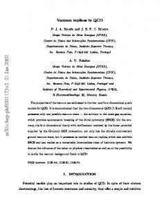
RESULTS In all the specimens, replicas demonstrated adequate capability for reproduction of the RBC morphology in BSs. On the other hand, both with the naked eye and under a lowpower microscope no BS remains were found on the molds when detached from the originals. SEM results are displayed in Figures 1–5. Figures 1–2 correspond to single views of the originals and replicas micrographed at 500× , St being for stone, W for wood, and Sh for shell. Figure 1 displays the originals. St and W are micrographs of the smear-substrate boundary, where the substrate can be seen on the left side. Sh is a micrograph of an ultrathin bloodstained area in the smear’s periphery; some loss of the shell periostracum can be observed at the upper part of the image. Figure 2 shows the replicas. In St, a thin bloodstained area contiguous to the smear-substrate
4
Policarp Hortolà
Figure 1. Single views of the originals, micrographed at 500× . St and W are micrographs of the smear-substrate boundary, where the substrate can be seen at the left side. Sh is a micrograph of an ultrathin bloodstained area in the smear’s periphery; some loss of shell periostracum can be observed at the upper part of the image. Working distances: 16 mm (St), 12 mm (W), and 18 mm (Sh). St, stone; W, wood; Sh, shell.
Figure 2. Single views of the replicas, micrographed at 500× . In St, a thin bloodstained area contiguous to the smearsubstrate boundary can be seen. A comparison original versus replica of this area is exhibited below (Fig. 3). In W, an area in the smear’s periphery is shown. In Sh, an ultrathin bloodstained area where RBCs are nearly arranged in monolayer, as in a clinical smear test, is displayed. Working distances: 13 mm (St), 16 mm (W), and 11 mm (Sh). St, stone; W, wood; Sh, shell.
boundary can be seen. A comparison of the original versus replica of this area is exhibited below (Fig. 3). In W, an area in the smear’s periphery is shown. In Sh, an ultrathin bloodstained area where RBCs are nearly arranged in monolayer, as in a clinical smear test, is displayed. Figures 3−5 correspond to related views of the originals and matching replicas, o being for original, r for replica, and the subscript indicating the original magnification. Figure 3 displays comparative views of the stone specimen. Micrographs o20× and r20× show a partial view of the BS at very low magnification, in the smear’s periphery. Macrocracking corresponds to the thickest areas of the BS. The silhouette of the pale, thinner smeared area—outlining the thicker one—suggests that it has been caused by blood trickling on the (impermeable) basaltic substrate. Micrographs o1,000× and r1,000 × display RBCs in the thinner area of the two bloodstained ones shown in o20× and r20× . The imaged area is the same as in Figure 2 St, at twofold magnification. Micrographs o2,000× and r2,000× depict detail of the same area as in o1,000× and r1,000× , at twofold magnification. A rouleau composed of three RBCs is shown at the top of
these micrographs. Figure 4 shows comparative views of the wood specimen. Micrographs o20× and r20× show a partial view of the smear at very low magnification, in the smear’s periphery. The bright bodies seen in the original are probably residues of conducting silver; in any case, they are of postmolding origin, given that they do not appear in the replica. Micrographs o1,000× and r1,000× display RBCs in the bloodstained area shown in o20× and r20× . Micrographs o2,000× and r2,000× depict detail of the same area as in o1,000× and r1,000× , at twofold magnification. A slight difference in RBC aspect between the original (flatter and more “outlined”) and the replica (bulkier and less “outlined”) can be noted. As can be perceived, this difference does not affect display of RBCs in both specimens. Figure 5 exhibits comparative views of the shell specimen. Micrographs o20× and r20× show a partial view of the smear center at very low magnification. External growth rings of the shell are observable. Macrocracking corresponds to the thickest areas of the BS. Areas where the periostracum is eroded can be seen in the form of pale scratches. These (periostracum-lost) pale scratches were used as an examining guide to find matching areas in
SEM of Bloodstain Replicas
Figure 3. Comparative views (original versus replica) of the stone specimen. Micrographs o20× and r20× show a partial view of the BS at very low magnification, in the smear’s periphery. Macrocracking corresponds to the thickest areas of the BS. The silhouette of the pale, thinner smeared area—outlining the thicker one—suggests that it has been caused by blood trickling on the (impermeable) basaltic substrate. Micrographs o1,000× and r1,000× display RBCs in the thinner area of the two bloodstained ones shown in o20× and r20× . The imaged area is the same as in Figure 2 St, at twofold magnification. Micrographs o2,000× and r2,000× depict detail of the same area as in o1,000× and r1,000× , at twofold magnification. A rouleau composed of three RBCs is shown at the top of these micrographs. Working distances: 17 mm (o) and 13 mm (r). O, original; r, replica; subscript = original magnification.
5
6
Policarp Hortolà
Figure 4. Comparative views (original versus replica) of the wood specimen. Micrographs o20× and r20× show a partial view of the smear at very low magnification, in the smear’s periphery. The bright bodies seen in the original are probably residues of conducting silver; in any case, they are of post-molding origin, given that they do not appear in the replica. Micrographs o1,000× and r1,000× display RBCs in the bloodstained area shown in o20× and r20× . Micrographs o2,000× and r2,000× depict detail of the same area as in o1,000× and r1,000× , at twofold magnification. A slight difference in RBC aspect between the original (flatter and more “outlined”) and the replica (bulkier and less “outlined”) can be noted. As can be perceived, this difference does not affect display of RBCs in both specimens. Working distances: 14 mm (o) and 16 mm (r). O, original; r, replica; subscript = original magnification.
SEM of Bloodstain Replicas
Figure 5. Comparative views (original versus replica) of the shell specimen. Micrographs o20× and r20× show a partial view of the smear center at very low magnification. The shell external growth rings are observable. Macrocracking corresponds to the thickest areas of the BS. Areas where the periostracum is eroded can be seen in the form of pale scratches. These (periostracum-lost) pale scratches were used as an examining guide to find matching areas in the replicas. Micrographs o1,000× and r1,000× display RBCs in the bloodstained area shown in o20× and r20× . Micrographs o2,000× and r2,000× depict detail of the same area as in o1,000× and r1,000× , at twofold magnification. Working distances: 20 mm (o) and 13 mm (r). O, original; r, replica; subscript = original magnification.
7
8
Policarp Hortolà
the replicas. Micrographs o1,000× and r1,000× display RBCs in the bloodstained area shown in o20× and r20× . Micrographs o2,000× and r2,000× depict detail of the same area as in o1,000× and r1,000× , at twofold magnification. A technical artifact found in the original shell specimen after gold coating was that the molded area was brighter than the non-molded one when seen under tilted incident light, both to the naked eye and with a low power microscope. This difference in brightness had its equivalent in texture under the SEM, where the molded area appeared smoother than the non-molded one. This extra brightness phenomenon was mimicked by the corresponding replica.
D ISCUSSION Different BS substrates were used with the aim of encompassing a range of natural raw materials used in the manufacture of archaeological and ethnographic artifacts, and that could potentially be bloodstained after use. Examples of such artifacts are stone clubs (e.g., Haddon, 1900; Taylor, 2001: 13–16; McNiven & von Gnielinski, 2004), wooden arrows (e.g., Lyons, 1922; Bush, 1985; Waguespack et al., 2009), and shell knives (e.g., Gusinde, 1924; Landtman, 1927: 28; Holdgate, 1961). A sole bloodstained fragment per raw material was examined because the RBC count in whole blood is very high in all vertebrate classes, especially in mammals (several authors, in Ragan, 2003, Tables B.1. and B.2.). This RBC count is related to the mean corpuscular volume, each tending to vary inversely (Ragan, 2003). In adult humans, a RBC count reference range (in million RBCs per microliter) is 4.5–5.9 in males and 4.5–5.1 in females (several authors, in Perkins, 2003, Table A.1.). According to this range, a single human blood drop of 60 μL can contain 270–354 million RBCs in males, and 270– 306 million RBCs in females. Therefore, any small human BS provides an extremely large quantity of RBCs. The fact that no BS remains were found to be transferred to the molds when detaching them from the originals would indicate that the BS-substrate adhesion forces are higher than the BS-mold ones. It is also worth mentioning that the original specimens were not examined until the molds were made because, on the one hand, the best imaging quality is achieved by coating the specimens—which applies to both highvacuum and variable-pressure SEMs—and, on the other, impressions should logically be made on the naked original surface and not on the already coated one. Regarding such specimen coating, it is well-known that the best SEM image resolution is obtained by using a sample metal coating (gold, gold:palladium 4:1 alloy, platinum, etc.) instead of a non-metal (carbon) coating. Consequently, a customary metal coating (gold) was used on the examined samples. Because the goal of this work was to gain insight into the identification of bloodstains rather than into the ultrastructure of RBCs, no SEM magnification above 2,000× was used because this value is sufficient to examine RBCs. In fact, a magnification of 850× is enough to clearly see RBCs in human BSs, as discussed elsewhere (Hortolà, 2013). It must also be noted that, despite using landmarks to locate potential
areas of interest, finding the same area in both the original and the replica was difficult, especially in the wood specimen. The slight difference in RBC aspect between the original wood specimen and its replica could be due to any artifact produced during the replication process. In the replica, the bulky shape of typical RBCs (discocytes) is displayed better than in the (post-molded) original. This suggests that this artifact is not due to the chemical action of the molding compounds, but rather to a physical action on the original, perhaps excessive mechanical pressure at the time of making the “back mold”. The effect of gold coating on the molded area of the shell specimen is probably due to an effect of the molding compounds on the naked periostracum of the shell. This phenomenon should not be critical because the interest of this study is to avoid using original specimens. It is not expected that an (archaeological, ethnographic) piece would be subjected to treatments such as a gold coating. Methodologically, limitations of the technique derive from the necessity that the surface of the replica to be examined be more or less flat, as occurs in the case of BSs. Replicas of specimens such as pollen grains, fungal spores, hair, feathers, etc. should be suitable only on condition that a partial view of them be sufficient for identification purposes. Finally, it deserves mention that, in this paper, care was taken to use accurate terminology (“impression” instead of “negative replica”, “replica” instead of “positive replica”), avoiding the usual conceptual inaccuracies already noted by Barnes (1978). Also, the terms “specimen” and “sample” have been used with different meaning. “Specimen” denoted a solid object physically separated (i.e., each individual fragment smeared with blood), while “sample” signified the sole part of the solid object that was the study target (i.e., each individual BS).
CONCLUSIONS The obtained results suggest that PU resin casts obtained from VPS silicone molds can preserve RBC morphology in BSs, and consequently that synthetic replicas are feasible for the SEM identification of BSs on cultural heritage specimens made of natural raw materials. Once the molds have been made in situ, casts from them can be made either at the same place or somewhere else—the previous molds or the finished casts being taken in person or sent by post or courier service—and finally SEM examined anywhere in the world. Although the focus of this study was on BSs, the method reported in this paper may be applied to organic residues other than blood (muscle, skin, tendon, etc.), as well as to the naked surface of other specimens (small engravings made by aboriginal or prehistoric peoples, trial pieces of evidence related to crime investigation, etc.) when, for any reason, the original is unavailable for an SEM.
ACKNOWLEDGMENTS A. Solé (colleague at IPHES) instructed me in synthetic replication. The Service of Scientific and Technical Resources
SEM of Bloodstain Replicas
(URV) provided me the use of its electron microscopy facility. This work was supported by research grants MINECO CGL2012-38434-C03-03 and MINECO CGL2010-15326 (Government of Spain), and GENCAT 2014 SGR 901 (Government of Catalonia).
REFERENCES BALME, J., GARBIN, G. & GOULD, R.A. (2001). Residue analysis and palaeodiet in arid Australia. Aust Archaeol 53, 1–6. BARNES, I.E. (1978). Replication techniques for the scanning electron microscope 1. History, materials and techniques. J Dent 6, 327–341. BROMAGE, T.G. (1985). Systematic inquiry in tests of negative/ positive replica combinations for SEM. J Microsc 137, 209–216. BUSH, T. (1985). Form and decoration of arrows from the highlands of Papua New Guinea. Rec Aust Mus 37, 255–293. COMINATO, L., VALLE, F., PIERINI, G., BONINI, P., BISCARINI, F. & D’ELIA, M. (2015). Flattening mountains: Micro-fabrication of planar replicas for bullet lateral striae analysis. Forensic Sci Int 247, 97–104. D’ERRICO, F., JULIEN, M., LIOLIOS, D., VANHAEREN, M. & BAFFIER, D. (2003). Many awls in our argument. Bone tool manufacture and use in the Châtelperronian and Aurignacian levels of the Grotte du Renne at Arcy-sur-Cure. In The Chronology of the Aurignacian and of the Transitional Technocomplexes. Dating, Stratigraphies, Cultural Implications. Proceedings of Symposium 6.1 of the XIVth Congress of the UISPP, University of Liège, Belgium, September 2–8, 2001, Zilhão, J. & D’Errico, F. (Eds.), pp. 247–270. Lisboa: Instituto Português de Arqueologia. D’ERRICO, F. & VILLA, P. (1997). Holes and grooves: The contribution of microscopy and taphonomy to the problem of art origins. J Hum Evol 33, 1–31. FIORENZA, L., BENAZZI, S. & KULLMER, O. (2009). Morphology, wear and 3D digital surface models: Materials and techniques to create high-resolution replicas of teeth. J Anthropol Sci 87, 211–218. FIORI, A. (1962). Detection and identification of bloodstains. In Methods of Forensic Science, vol. 1, Lundquist, F. (Ed.), pp. 243–290. New York: John Wiley & Sons. FRASER, D., DEROO, C.S., CODY, R.B. & ARMITAGE, R.A. (2013). Characterization of blood in an encrustation on an African mask: Spectroscopic and direct analysis in real time mass spectrometric identification of haem. Analyst 138, 4470–4474. FRAYER, D.W., FIORE, I., LALUEZA-FOX, C., RADOVČIĆ, J. & BONDIOLI, L. (2010). Right handed Neandertals: Vindija and beyond. J Anthropol Sci 88, 113–127. GALANIDOU, N. (2006). Analytical and ethical issues concerning organic residues on Paleolithic chipped stone tools from NW Greece. J Field Archaeol 31, 351–362. GALBANY, J., ESTEBARANZ, F., MARTÍNEZ, L.M., ROMERO, A., DE JUAN, J., TURBÓN, D. & PÉREZ-PÉREZ, A. (2006). Comparative analysis of dental enamel polyvinylsiloxane impression and polyurethane casting methods for SEM research. Microsc Res Tech 69, 246–252. GALBANY, J., MARTÍNEZ, L.M. & PÉREZ-PÉREZ, A. (2004). Tooth replication techniques, SEM imaging and microwear analysis in primates: Methodological obstacles. Anthropologie (Brno) 42, 5–12.
9
GOLDSTEIN, J.I., NEWBURY, D.E., ECHLIN, P., JOY, D.C., LYMAN, C.E., LIFSHIN, E., SAWYER, L. & MICHAEL, J.R. (2003). Scanning Electron Microscopy and X-Ray Microanalysis. 3rd ed., vol. 1. New York, NY: Springer. GRAMLY, R.M. (1991). Blood residues upon tools from the East Wenatchee Clovis site, Douglas County, Washington. Ohio Archaeol 41, 4–9. GREENFIELD, H.J. (1999). The origins of metallurgy: Distinguishing stone from metal cut-marks on bones from archaeological sites. J Archaeol Sci 26, 797–808. GUSINDE, M. (1924). Cuarta expedición a la Tierra del Fuego. Informe del Jefe de Sección [Fourth expedition to Tierra del Fuego. Report of the Section Head]. Publ Mus Etnol Antropol Chile 4, 7–67. HADDON, A.C. (1900). A classification of the stone clubs of British New Guinea. J Anthropol Inst Great Brit Ireland 30, 221–250 + Plates XIX–XXIII. HAVERKORT, C.M. & LUBELL, D. (1999). Cutmarks on Capsian human remains: Implications for Maghreb Holocene social organization and palaeoeconomy. Int J Osteoarchaeol 9, 147–169. HITCHCOCK, G. (2004). Torres Strait origin of some stone-headed clubs from the Torassi or Bensbach River area, southwest Papua New Guinea. Mem Queensl Mus Cult Herit Ser 3, 305–313 + Appendix [by Jamieson, D.N., Szymanski, R. & Rout, B.]. HOLDGATE, M.W. (1961). Man and environment in the south Chilean islands. Geogr J 127, 401–414. HORTOLÀ, P. (1992). SEM analysis of red blood cells in aged human bloodstains. Forensic Sci Int 55, 139–159. HORTOLÀ, P. (2002). Red blood cell haemotaphonomy of experimental human bloodstains on techno-prehistoric lithic raw materials. J Archaeol Sci 29, 733–739. HORTOLÀ, P. (2013). Human bloodstains on biological materials: High-vacuum scanning electron microscope examination using specimens without previous preparation. Microsc Microanal 19, 415–419. HUQ, S., ABIDI, B., PAGE, D., FRAFJORD, J., DEKANICH, S. & ABIDI, M. (2008). 3D modeling from Large Chamber SEM images for micro-scale material characterization. In 2nd International Joint Topical Meeting on Emergency Preparedness and Response and Robotic and Remote Systems [CD-ROM], American Nuclear Society (Ed.), pp. 53–60. La Grange Park, IL: American Nuclear Society. JOHNN, H., PHIPPS, C., GASCOYNE, S., HAWKEY, C. & RAMPLING, M.W. (1992). A comparison of the viscometric properties of the blood from a wide range of mammals. Clin Hemorheol 12, 639–647. JONES, P.J. (2009). A microstratigraphic investigation into the longevity of archaeological residues, Sterkfontein, South Africa. In Archaeological Science Under a Microscope. Studies in Residue and Ancient DNA Analysis in Honour of Thomas H. Loy, Haslam, M., Robertson, G., Crowther, A., Nugent, S. & Kirkwood, L. (Eds.), pp. 29–46. Canberra: ANU E Press. KLEIN, M. & BRANDT, T. (2006). Development of a high precision positioning system (PS) for a large chamber scanning electron microscope (SEM). Proc ASPE Spring Top Meet 38, 9–11. LANDTMAN, G. (1927). The Kiwai Papuans of British New Guinea. A Nature-Born Instance of Rosseau’s Ideal Community. London: Macmillan. LANGEJANS, G.H.J. & LOMBARD, M. (2015). About small things and bigger pictures: An introduction to the morphological identification of micro-residues on stone tools. In Use-Wear and Residue Analysis in Archaeology, Marreiros, J.M., Gibaja Bao, J.F. & Ferreira Bicho, N. (Eds.), pp. 199–219. Cham (CH): Springer.
10
Policarp Hortolà
LI, X.J., MARTINÓN-TORRES, M., MEEKS, N. & XIA, Y. (2012). Scanning electron microscopy imaging of tool marks on Qin bronze weapons using silicone rubber impressions. In Historical Technology, Materials and Conservation. SEM and Microanalysis, Meeks, N., Cartwright, C., Meek, A. & Mongiatti, A. (Eds.), pp. 62–68. London: Archetype Publications. LOMBARD, M. (2005). Evidence of hunting and hafting during the Middle Stone Age at Sibidu Cave, KwaZulu-Natal, South Africa: A multianalytical approach. J Hum Evol 48, 279–300. LOMBARD, M. (2008). Finding resolution for the Howiesons Poort through the microscope: Micro-residue analysis of segments from Sibudu Cave, South Africa. J Archaeol Sci 35, 26–41. LOMBARD, M. & WADLEY, L. (2007). The morphological identification of micro-residues on stone tools using light microscopy: Progress and difficulties based on blind tests. J Archaeol Sci 34, 155–165. LOY, T.H. (1983). Prehistoric blood residues: Detection on tool surfaces and identification of species of origin. Science 220, 1269–1271. LOY, T.H. & DIXON, E.J. (1998). Blood residues on fluted points from eastern Beringia. Am Antiq 63, 21–46. LUCERO, M. (2004). Evaluación del Uso de Artefactos de Concha en el Poblamiento Inicial del Semiárido de Chile [Evaluating the use of shell artifacts in the early peopling of semiarid Chile]. BA Thesis. Santiago de Chile: Departamento de Antropología, Facultad de Ciencias Sociales, Universidad de Chile. http://tesis.uchile.cl/ handle/2250/106400 (retrieved October 3, 2006). LYONS, A.P. (1922). The arrows of the Upper Morehead River (Papua) bush tribes. Man 22, 145–147 + Plate K. MALAINEY, M.E. (2011). A Consumer’s Guide to Archaeological Science. Analytical Techniques. New York: Springer. MAZEL, V., RICHARDIN, P. & CHARLIER, P. (2006). Restes biologiques dans les patines rituelles de la statuaire Dogon (Mali) [Biological remains in ritual patina of Dogon statuary (Mali)]. In 1er Colloque International de Pathographie. Loches, Avril 2005 [Conference Proceedings], Charlier, P. (Ed.), pp. 131–144. Paris: De Boccard. MCNIVEN, I.J. & VON GNIELINSKI, F. (2004). Stone club head manufacture on Dauan Island, Torres Strait. Mem Queensl Mus Cult Herit Ser 3, 291–304. MOISAN, P. (2012). The study of cuticular and epidermal features in fossil plant impressions using silicone replicas for scanning electron microscopy. Palaeontol Electron 15(2), 23A (9 pp). http://palaeo-electronica.org/content/2012-issue-2-articles/279replicas-for-sem-examination (retrieved February 28, 2014). OLLÉ, A. (2003). Variabilitat i Patrons Funcionals en els Sistemes Tècnics de Mode 2. Anàlisi de les deformacions d'ús en els conjunts lítics del Riparo Esterno de Grotta Paglicci (Rigano Garganico, Foggia), Aridos (Arganda, Madrid) i Galeria-TN (Atapuerca, Burgos) [Variability and functional patterns in Mode 2 technical systems. Use-wear analysis in lithic assemblages of Grotta Paglicci’s Riparo Esterno (Rignano Garganico, Foggia), Áridos (Arganda, Madrid) and Galería-TN (Sierra de Atapuerca, Burgos)]. PhD Thesis. Tarragona: Departament d’Història i Geografia, Facultat de Lletres, Universitat Rovira i Virgili. http://www.tdx.cat/handle/10803/ 8603 (retrieved September 18, 2007).
OLLÉ, A. & VERGÈS, J.M. (2008). SEM functional analysis and the mechanism of microwear formation. BAR Int Series 1783, 39–49. PERKINS, S.L. (2003). Normal blood and bone marrow values in humans [Appendix A]. In Wintrobe’s Clinical Hematology, 11th ed., vol. 2, Greer, J.P., Foerster, J., Lukens, J.N., Rodgers, G.M., Paraskevas, F. & Glader, B. (Eds.), pp. 2697–2706. Philadelphia, PA: Lippincott Williams & Wilkins. PICKERING, T.R., WHITE, T.D. & TOTH, N. (2000). Cutmarks on a Plio-Pleistocene hominid from Sterkfontein, South Africa. Am J Phys Anthropol 111, 579–584. RAGAN, H.A. (2003). Comparative hematology [Appendix B]. In Wintrobe’s Clinical Hematology, 11th ed., vol. 2 Greer, J.P., Foerster, J., Lukens, J.N., Rodgers, G.M., Paraskevas, F. & Glader, B. (Eds.), pp. 2707–2719. Philadelphia, PA: Lippincott Williams & Wilkins. RHYU, Y.S., CHUNG, Y.J. & UHM, C.S. (2010). Scanning electron microscopic observation of human skin replica. Korean J Microsc 40, 267–270. ROBERTSON, G. (2011). Changing perspectives in Australian archaeology, part VII. Aboriginal use of backed artefacts at Lapstone Creek rockshelter, New South Wales: An integrated residue and use-wear analysis. Tech Rep Aust Mus, Online 23, 83–101. ROBERTSON, G., ATTENBROW, V. & HISCOCK, P. (2009). Multiple uses for Australian backed artefacts. Antiquity 83, 296–308. ROSE, J.J. (1983). A replication technique for scanning electron microscopy: Applications for anthropologists. Am J Phys Anthropol 62, 255–261. SHIPMAN, P. (1986). Studies of hominid-faunal interactions at Olduvai Gorge. J Hum Evol 15, 691–706. SWGSTAIN (2009). Scientific Working Group on Bloodstain Pattern Analysis: Recommended terminology. Forensic Sci Commun 11 (2). http://www.fbi.gov/hq/lab/fsc/backissu/ april2009/standards/2009_04_standards01.htm (retrieved May 8, 2009). TAYLOR, C.F. (2001). Native American Weapons. London: Salamander Books. TORRENCE, R. (1993). Ethnoarchaeology, museum collections and prehistoric exchange: Obsidian-tipped artifacts from the Admiralty Islands. World Archaeol 24, 467–481. TRIFKOVIC, B., BUDAK, I., TODOROVIC, A., HODOLIC, J., PUSKAR, T., JEVREMOVIC, D. & VUKELIC, D. (2012). Application of replica technique and SEM in accuracy measurement of ceramic crowns. Meas Sci Rev 12, 90–97. UNGAR, P.S., GRINE, F.E., TEAFORD, M.F. & EL ZAATARI, S. (2006). Dental microwear and diets of African early Homo. J Hum Evol 50, 78–95. WADLEY, L. & LOMBARD, M. (2007). Small things in perspective: The contribution of our blind tests to micro-residue studies on archaeological stone tools. J Archaeol Sci 34, 1001–1010. WADLEY, L., LOMBARD, M. & WILLIAMSON, B. (2004). The first residue analysis blind tests: Results and lessons learnt. J Archaeol Sci 31, 1491–1501. WAGUESPACK, N.M., SUROVELL, T.A., DENOYER, A., DALLOW, A., SAVAGE, A., HYNEMAN, J. & TAPSTER, D. (2009). Making a point: Wood- versus stone-tipped projectiles. Antiquity 83, 786–800.