with allows to display a virtual bronchoscopy in real time and we demonstrate an ... In the medical environment, navigation systems have started to call attention.
Evaluation and Extension of a Navigation System for Bronchoscopy inside Human Lungs Ingmar Wegnera , Juergen Biedererb , Ralf Tetzlaffb , Ivo Wolfa , Hans-Peter Meinzera German Cancer Research Center, of Medical and Biological Informatics, b Division of Radiology, Im Neuenheimer Feld 280, 69120 Heidelberg, Germany a Division
ABSTRACT For exact orientation inside the tracheobronchial tree, clinicians are in urgent need of a navigation system for bronchoscopy. Such an image guided system has the ability to show the current position of a bronchoscope (instrument to inspect the inside of the lung) within the tracheobronchial tree. Thus orientation inside the complex tree structure is improved. Our approach of navigated bronchoscopy considers the problem of using a static image to navigate inside a constantly moving soft tissue. It offers a direct guidance to a preinterventionally defined target inside the bronchial tree to save intervention time spent on searching the right path and to minimize the duration of anesthesia. It is designed to adapt to the breathing cycle of the patient, so no further intervention to minimize the movement of the lung has to stress the patient. We present a newly developed navigation sensor with allows to display a virtual bronchoscopy in real time and we demonstrate an evaluation on the accuracy within a non moving ex vivo lung phantom. Keywords: Electromagnetic Tracking, Enhanced Reality, Image-Guided Therapy, Minimally Invasive Surgery, Navigated Bronchoscopy
1. INTRODUCTION AND MEDICAL BACKGROUND Specialized navigation systems have become omnispresent in daily life. The most common navigation system is the global positioning system (GPS) built into almost every new car model. By using signals from at least three satellites the current position on earth is determined. Special navigation softwares display the current position onto a map of roads or offer a guidance to a specified destination by displaying the directions at the right moment. In the medical environment, navigation systems have started to call attention. Such systems assist for example a neurosurgeon to approach a certain spot inside the brain without harming vulnerable regions. But since the techniques of different medical procedures vary considerably, navigation systems have to be specialized in each procedure. Several research groups are working on the development of a navigation system for the lung. Here the complex structure can cause problems to directly approach a defined target. With the use of a video bronchoscope (flexible endoscope) video images from the inside of the bronchial tree can be displayed. But these images are depending on rotation and thus not very intuitive to orient oneself. Furthermore the amount of branching points aggravates the problem. Therefore additional information about the global position and orientation inside the bronchial tree would assist the intervention. In the past the incidence of peripheral lesions has increased.1 But the diameter of the bronchoscope limits the depth of insertion. To overcome this problem, smaller catheters are used, which Copyright 2007 Society of Optical Engineering. This paper was published in Proc. SPIE Vol.6509, article number 65091H, Medical Imaging 2007: Visualization and Image-Guided Procedures; Kevin R. Cleary, Michael I. Miga (Eds) and is made available as an electronic reprint with permission of SPIE. One print or electronic copy may be made for personal use only. Systematic or multiple reproduction, distribution to multiple locations via electronic or other means, duplication of any material in this paper for a fee or for commercial purposes, or modification of the content of the paper are prohibited.
can not provide an image. In most cases fluoroscopy or computer tomography (CT) scanning is used to monitor the current position of the catheter and to control the position before a therapeutic procedure (e.g. endoluminal brachytherapy) is done. Here a navigation system could assist as well, if the tracking device is small enough to fit into the catheter. Its position could be superimposed on a preinterventionally acquired CT-scan. But this also implies, that the navigation system is accurate in real time. In order to perform an image guided navigation, some kind of patient specific data containing the anatomy of the patient is mandatory. So far CT scans provide the best resolution in space. With an appropriate sequence even the structure of thin bronchial walls can be captured and stored as a digital dataset. Because patients who have a suspicious lung lesion usually undergo a CT-scan, this dataset is generally available and can be used for virtual bronchoscopy. Here the voxels of the dataset are divided into air and tissue voxels by means of gray value thresholding or other more distinguished segmentation algorithms. In a virtual 3D scene only tissue voxels are displayed so that the tracheobronchial tree of the specific patient can be examined in virtual space without the typical risk of pneumothorax or bleeding.2 Virtual bronchoscopy may be a promising attempt for identifying obstructions and endoluminal lesions and might enable the examination of the tracheobronchial tree beyond stenosis.3 But for a more thorough inspection of a suspicious lesion or possibly a biopsy or therapeutic intervention a lung specialist has to carefully steer a catheter or a bronchoscope through the patients tracheobronchial tree. This can be greatly assisted by an image guiding navigation system on the basis of the existing CT dataset. Similar to a GPS navigation system it can show the current global position of the instrument drawn onto the orthogonal slices of the CT dataset. In order to transfer the tracked position (real) into the CT dataset (virtual) a registration of real and virtual data has to be accomplished. By localizing three or more corresponding landmarks in real and in virtual space, a rigid registration can be abtained. But in contrast to the analogy of a city map the soft tissue of the lung moves over the time (breathing motion) causing a rigidly registered navigation system to become imprecise. Thus a soft tissue approach, which has the ability to successively register the virtual data with the real tracheobronchial tree, has to be implemented.4
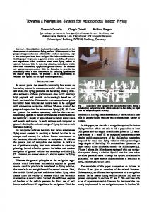
Figure 1. Screenshot of a navigated bronchoscopy showing a 3-D visualization of the surface of a bronchial tree. The current matched position of the tracked instrument is represented by a cone. A red path leads from the root to the defined target to show the direction.
Currently developed navigation systems for bronchoscopy can be sub-divided by the technique used to determine the position of the instrument. Merritt et al. are using image to image registration where the real image of a video bronchoscope is registered to a predicted virtual image.5 This eliminates the movement of the lung because the registration is done locally and performed over and over again. But this technique also is limited by the high computation time.6 Furthermore poor image quality (e.g. due to bronchial secretion) or a sudden strong deformation of the bronchi (e.g. cough) causes a loss of position information. Mori et al. are using a hybrid tracking system.7 A five degrees of freedom (DOF) electromagnetic tracking (EMT) sensor provides a three dimensional position and a two dimensional orientation without direct view to it. The missing dimension of orientation is the rotation (roll angle) which is needed to suggest a direction in virtual bronchoscopy. In order
to supplement the necessary information of rotation, Mori et al. perform an image to image registration at the tracked position. The performance is improved compared to the approach of Merritt et al. because the number of registered images is decreased. But other problems caused by the use of image registration still remain. Hautmann et al. are neglecting the missing rotational information of the used 5DOF sensor and rather focus on increasing the accuracy of the position tracking.8 They described the usefulness of a navigation system for bronchoscopy in a prospective study using an EMT sensor placed inside the working channel of a bronchoscope. From the variety of commercially available tracking systems they have chosen a system distributed by Northern Digitally Inc. (NDI; Waterloo, Ontario, Canada) called AURORA. The system can track up to eight 5DOF sensors and has a well supported platform for development of custom made specialized tracking sensors. During the past two years an other attempt to develop a prototype of an image guided navigation system for bronchoscopy was made at the division of medical and biological informatics at the German Cancer Research Center Heidelberg, Germany.9 This approach uses an electromagnetic tracking system (AURORA II, NDI) is used to monitor three 5DOF reference sensors and one instrument sensor. The reference sensors are fixed onto the patient’s thorax to track the overall movement of the patient or a possible movement of the examination table. Depending on the desired visualization a navigation sensor has to be chosen. In case of a virtual 3-D visualization of the instrument relative to the surface of the tracheobronchial tree a 5DOF sensor is suitable (see fig. 1). The missing information of rotation can be neglected. The available two degrees of orientation can be used to improve accuracy.10 If the assistance of a virtual bronchoscopy is desired, six degrees of freedom are needed to rotate the virtual scene according to the instruments rotation. A 6DOF sensor is available from NDI, but yet too big to be pushed through the working channel or a small catheter or to be fixed onto the tip of the bronchoscope. Thus a special 6DOF sensor was developed (see 2.2). In order to compute the transformation from real space to virtual space, three or more anatomical landmarks are interactively defined in virtual space and encountered inside the real bronchial tree under vision of a bronchoscope. Bifurcation can provide obvious landmarks. As known, the area spread up by the selected landmarks directly affects the quality of the transformation. Thus the placement of one landmark at the end of the trachea (bifurcatio tracheae) and two landmarks at easily discernible peripheral bifurcations enhance the quality. But as already mentioned this is limited by the diameter of the bronchoscope. So it will not be possible to enclose the entire navigation area with the landmarks. The software component of the navigation system is developed using the open source toolkit MITK.11 With it the bronchial tree can be segmented and converted into a tree based representation, storing the center line and the diameter of each representative position. Furthermore other regions of interest such as lesions and nodules can be segmented and stored as surfaces to be displayed during the intervention. A preinterventional planning module enables the interactive definition of the navigation target along with the virtual landmarks. A simulation of the guidance showing the same interface as during the intervention is offered. This way a virtual bronchoscopy can be done as well. Different visualizations can be used to provide additional informations during guidance. Several reconstructed 2-D slices of the CT-scan, a virtual 3-D scene showing the desired elements as partly transparent surfaces, a virtual bronchoscopy from the view of the current position of the bronchoscope or a video image of the bronchoscope extended by means of augmented reality can be used to maintain orientation. During the intervention the navigation system is calibrated by locating the real landmarks. This only represents a very rough registration of the static virtual data and the moving soft tissue. Thus a special constraint is used to successively match the tracked position with the moving tissue (see section 2.1). All necessary steps for planning, computation, and execution can be performed. To show its usability, several tests were made.
2. METHODS 2.1. Compensation of Lung Movement A soft tissue navigation system has to successively adapt to the moving investigation area. In the field of bronchoscopy, a strong constraint can isolate the navigation volume. The assumption, that the bronchoscope always stays inside the bronchial tree and doesn’t penetrate through the bronchial wall is fair for bronchoscopy because the end of the bronchoscope is blunt and cannot disrupt the connective tissue of the bronchial wall. Together with the center line representation of the bronchial tree, this can be combined to a movement compensating
Figure 2. Sketch of the technique used to compensate lung movement. The tracked position is matched onto the center line of the bronchial tree description by its perpendicular (left). Discrepancy in case of a lung movement along the bronchi (right).
technique, where the tracked position of the bronchoscope is matched onto the center line of the tree by dropping a perpendicular (see left fig. 2). The moving tracked position of the steady held bronchoscope is then transfered into an almost steady position inside the static virtual bronchial tree. It can not be fully stabilized because the motion along the bronchi caused by the breathing can not be isolated from the motion caused by a real movement of the sensor (see right fig. 2). But this assumption can also decrease the accuracy by matching the real position onto the center line in case the diameter of the bronchi is larger than of the sensor. But since the video image of the bronchoscope is available as long as the latter can be pushed forward, this limitation can be dealt with. Furthermore the compensation technique can be adapted by additional informations such as the description of a regular respiratory motion or by other constraints. EU patent PCT/EP2005/002244 and US patent 10,590,195 describing the method are pending.
2.2. Development of a 6DOF Sensor for Bronchoscopy
Figure 3. Developed 6DOF bronchoscope tracking sensor to be used with Aurora tracking system by NDI. The two sensor coils are placed on each side of the bronchoscope heading in different directions (non-parallel) (left). A shrink hose is used to stabilize and isolate the coils (right). The size of the sensor is to be optimized.
Metallic (especially ferromagnetic) materials can disturb the quality of an electromagnetic tracking system.12 Thus several tests with different sensors (5DOF, 6DOF) were made to ensure the performance of an electro-
magnetic tracking sensor inside or adjacent to a bronchoscope. Measurements at different distances between the sensor and the bronchoscopes tip were made to detect an optimal position of the sensor. The distances went from 100 mm to 10 mm in steps of 10 mm and from 10 mm to 0 mm (inside bronchoscope) in steps of 1mm. A digital caliper (resolution 0.01mm; precision +/- 0.02 mm) was used to adjust the position. The EMT field generator was positioned underneath the sensor inside a non-metallic, wooden cupboard with a distance of 136.80 mm +/-0.02 mm between generator and sensor. Measured values were collected over 1000 iterations for each measurement, giving information about the variance of position and orientation. The interpretation of the collected data was then performed in Matlab. Furthermore tests analyzing the quality of the electromagnetic tracking system together with a carbon sliding bed of an examination table were made to ensure unrestricted functionality. Since no severe disturbance was observed the next tests are to be made with a complete operation table also containing metal units.
Figure 4. Screenshot of the software NDI 6D Architect showing the definition of the developed 6DOF bronchoscope sensor (2D and 3D views). Here the non parallel orientation of the two 5DOF sensor coils can be seen.
Early stages of the prototypic navigation system were using a 5DOF sensor placed inside the working channel of the bronchoscope. This way the system could be easily converted into a navigation system for catheters and also be used with any brand of bronchoscope. A good experience due to the construction of the tip was made with a fiber bronchoscope distributed by Karl Storz GmbH (Tuttlingen, Germany) as well as with a CCD bronchoscope distributed by Richard Wolf GmbH (Knittlingen, Germany). To be able to insert additional instruments through the working channel into the lung, e.g. to extract a tissue sample or to place an irradiation catheter at the right position, the location of the tracking sensor had to be changed. Having evaluated the functional capability of an EMT sensor next to a bronchoscope, we were able to build a 6DOF sensor consisting of two 5DOF NDI sensor coils (ø 0.8mm) located on each side of the tip of the bronchoscope (see Fig. 4). The sensors are non parallel to each other and connected to one port of the AUTORA system control unit. To permanently fasten the sensor coils onto the tip of the bronchoscope a shrink hose (wall thickness shrunken 0.8mm) was used. The overall diameter of the trackable tip of the bronchoscope now reaches from 6.6mm to 8.2mm. As a definition file was necessary to merge the two 5DOF sensors into one 6DOF information, the NDI 6D Architect tool was used. Nine collections of sensor data were gained by differently positioning the sensor inside the measurement volume of the tracking system. For this special sensor the calculated parameters were the following: Sensor 1 2
X -1.24 1.24
Y 3.07 -3.07
Z 0.06 -0.06
OX 0.1427 -0.2326
OY 0.0236 -0.0659
OZ -0.9895 -0.9703
This definition can either be loaded into the tracking module of our navigation system, or it can be written
onto a SROM device placed inside the connector of the sensor. The two 5DOF sensor coils now act as one sensor and provide 6DOF information. Thus, also rotation along the axis of the sensor (roll angle) can be observed in real time and with high performance (40fps).
2.3. Evaluation of the Navigation System using an ex vivo lung phantom
Figure 5. An ex vivo lung phantom was used to evaluate the ability to monitor the right position of the sensor. Here the lung is inflated to test its quality after deep freeze. The heart is explanted as well to keep the lung in the right shape.
The division of radiology at the German Cancer Center is examining the motion of lung carcinoma using an ex vivo lung phantom (see fig. 5). Together with the heart a porcine lung explant is embedded into a plastic container representing a thorax. The trachea is attached to a tube leading out of the container and the lid is closed to construct a vacuum. At the end of the shape a balloon, representing the diaphragm, can be filled with water. The amount of water can be periodically changed over time so that respiratory motion can be reproduced. The phantom doesn’t contain any metallic objects so that electromagnetic interference is avoided making it perfectly suited for testing a navigation system. An evaluation design was developed to measure the accuracy of the prototype using the phantom, so far without soft tissue movement. The investigation area was focused onto the peripheral lung. Thus a 5DOF flex cord sensor equivalent to a catheter (distributed by NDI) is used to reach into the deep periphery of the lung (ø 1.8 mm). At the beginning of the evaluation the lung phantom was scanned by a Toshiba CT-scanner (Aquilion S16; 16x0.5 mm collimation, pitch 0.625, FOV 400 mm, 120 mAs, matrix 512x512, reconstructed 0.78 mm in-plane, 0.4 mm (z-plane) 0.5 mm slice thickness). The dataset then was transfered from the scanner to a PACS workstation (CHILI GmbH, Heidelberg, Germany) in order to process the volume inside a MITK plugin. The tracheobronchial tree was segmented by using a regular region growing approach (upper threshold = -1005 HU). Then the segmented voxels were transformed into a tree based representation. Furthermore the segmentation was filtered by marching cubes to extract its surface. The generated data was transfered to a standard workstation (3GHz Pentium IV, 1GB RAM, ATI Radeon X800Pro) build up next to the CT-scanner. Then the lung phantom was carefully positioned on a wooden cupboard and the NDI field generator was placed underneath the phantom. Three 5DOF reference sensors were fixed onto the surface of the phantom and a forth 5DOF was pushed through the working channel of a bronchoscope (Richard Wolf GmbH; flexible bronchoscope DAFE; ø 6 mm). The video signal of the bronchoscope was processed by a video controller (Richard Wolf) and streamed into the workstation to be displayed into the software of the navigation system. A technician was operating the
software and a physician (radiologist) was performing the intervention while observing the displayed visualization. The intervention started with the planning procedure where the two previously generated datasets were loaded. Then the navigation target was defined by selecting the position on the virtual tree. The path from the root to the target was extracted and visualized in 3-D. Then three landmarks were defined on the surface of the virtual bronchial tree. The physician then located the same landmarks in real using the video image to steer the sensor to touch the landmark. During the performed evaluation the same bifurcations were used as landmarks. Finally he pulled the sensor out of the bronchoscope, the navigation system was enabled, and he pushed the sensor forward to the defined position and placed the sensor as precisely as possible. Each duration of the workflow was recorded. Then the sensor was fixed onto the tube (trachea), the virtual tracked and matched positions were stored, and the cable of the sensor was unplugged. In order to measure the distance between the virtual positions and the real position the phantom was carefully carried to the ct-scanner and a CT-dataset was acquired. With this workflow five CT-scans could be acquired measuring the performance of five independent navigation interventions.
Figure 6. Analyzing the acquired CT data: The distance between the matched position and the real tip of the sensor is measured by using MITK.
To calculate the accuracy the acquired CT datasets were analyzed. In order to measure the distance between the matched position and the real tip of the sensor, the dataset, which was used during planning, had to be registered to the dataset acquired after navigation. This was done using a point based registration technique implemented with MITK and ITK. Then the distance between the tip and the stored matched position could be calculated (see fig. 6) by MITK.
3. RESULT The analysis of the CT-scans taken after each navigation demonstrate an accuracy better than of 8.2 mm (average = 5.03 mm; best = 2.17 mm; see fig. 7). To discover a sideeffect of the constraint used to compensate respiratory motion, the accuracy relative to the diameter of the bronchi was calculated (see fig. 8) but no significance could be found.
Figure 7. Distance between the real and matched position at the end of each navigation compared to the diameter of the bronchi.
Figure 8. Distance between real and tracked position relative to the diameter of the bronchi.
Furthermore to detect an inaccuracy because of a bad calibration, the accuracy relative to the distance between sensor and closest landmark was calculated (see fig. 9). Here the dependency between the quality of the calibration and the accuracy of the system is slightly indicated.
Figure 9. Distance between real and tracked position relative to the distance between the real position and the closest landmark.
Figure 10. Duration of different steps in workflow of each navigation as well as the median and root mean square (RMS).
During the five interventions the prototype of the navigation system, showed, that an inexperienced user can orient himself inside the tracheobronchial tree soley based on its virtual representation. The duration of the interventions were promising (most time consuming navigation: No. 2 = 10.68 minutes; average = 7.9 minutes; see fig. 10).
Figure 11. Distance between the tracked and the matched position.
To analyze the effect of the compensation technique onto the navigation system, the distance between the position, which is delivered by the tracking system, and the position, to which the compensation algorithm matches the tracked position onto, is visualized (see fig. 11). Overall the prototype showed a promising performance. Limitations discovered were the straight tip of the 5DOF catheter which at a bifurcation hardly allows to deviate from its path of predilection to alternative bronchi that would be accessible with a curved tip through simple rotation of the catheter. Preliminary tests on the lung phantom indicated good results of the developed 6DOF bronchoscope tracking sensor. With this sensor the working channel of the bronchoscope remains free whereas the tracking system still receives information in 6DOF in real time. The rotation (roll angle) was successfully tracked so that the virtual bronchoscopy showed almost the same picture than the video image. The optics of a bronchoscope is distorted though in order to capture the lateral bronchial walls. Plans for the future contain amongst others the placement of the two 5DOF coils underneath the isolation tube of the bronchoscope. At present all four ports of the Aurora tracking system are used but due to our positive experience made with a dual sensor on one port, two reference sensors can be merged to one port and the freed port can be used to track an instrument inserted through the working channel and pushed forward into the periphery of the tracheobronchial tree while still tracking the bronchoscope. The evaluation of a disturbance of a tracking sensor caused by the tip of a bronchoscope showed that there is a disturbance at certain distances between sensor and tip of the bronchoscope. Not inside, but shortly in front of the working channel, the tracked position gets shifted in position and orientation (see fig. 12). A general behavior of the distortion can not be recognized. The result depends on the used bronchoscope as well as the sensor. But the intensity of the distortion was observed not to be too strong to not use it for soft tissue navigation in case a succsessive registration approach is used to correct the information. So the result were promising if the already integrated compensation technique is used ( also see fig. 11). Thus the movement of the lung is compensated as well as the minimal shift of the position caused by the electromagnetic distortion.
4. DISCUSSION Several factors, that influence the accuracy of the evaluation, were discovered and could not be eliminated. First of all, the quality of calibration is known to be directly dependable. As figure 9 compared to figure 7 shows, the distance between the landmarks and the target affects the accuracy. Thus the landmarks should be defined
0.4 0.2
Error [mm]
0 −0.2 −0.4 −0.6 x
−0.8
y z
−1 −1.2
1
2
3
4
5
6
7
8
9
10
11
12
8
9
10
11
12
Experiment #
−4
1.4
x 10
x y z
1.2
Variance [mm]
1 0.8 0.6 0.4 0.2 0
1
2
3
4
5
6
7
Experiment #
Figure 12. Result of a series of measurements (here 5DOF): position error (top) and variance (bottom) at different distances between sensor and tip of bronchoscope. Experiment 1 was done without bronchoscope, experiment 2 to 12 were done with a distance of 100mm to 0mm between sensor and bronchoscope in steps of 10mm. A shift of aproximately 1.2 mm appears in z axis if the sensor is inserted into the working channel.
at the most peripheral bifurcation, which is possible to reach with the bronchoscope. Furthermore the limited resolution of the CT dataset might impair visibility of objects smaller than the voxel size. This might cause a displacement of the real sensor in data. Furthermore the image to image registration, that had to be used to compare the positions, is an other influencing factor. The fixation of the sensor together with the positioning of the phantom onto the CT-scanners slide are other influencing factors. The initial evaluation of the accuracy is satisfactory for assisting the orientation inside the tracheobronchial tree. Thus the duration of the intervention can be lowered by a direct guidance. The system is not yet precise enough to guide a needle in order to extract a tissue sample. In this case an endobronchial ultrasound (EBUS) transbronchial needle aspiration (TBNA) can be used to visualize of the current surrounding tissue. The presented evaluation was performed in stable mode. The next evaluation will be analyzing the performance in dynamic mode (simulated respiratory motion) where we expect the system to increase its performance. The mentioned discrepancy of the constraint could be by an indication factor showing the current precision of the system according to the diameter of the current bronchi. A similar indicator, displaying the distance between tracked and matched position has already been integrated but not yet tested. Several advancements in motion compensation were developed over time. One early approach was to store the tracked position of the sensor over the time to build up a second tree based representation. This tree then was registered with the preinterventional computed bronchial tree. Tests on a self constructed lung phantom9 showed that this approach was not leading to better results in motion compensation. As long as the sensor doesn’t change its position relative to the bronchoscope, the observed distortion of the sensor might be eliminated by the calibration as well as by the compensation algorithm. Further tests on designing a prototype of a tracked bronchoscope which is usable in clinical environment, have to be done. The evaluation of the sensor distortion was difficult to set up. The movement of the sensor over all measurements could not be precisely avoided because the bronchoscope had to carry the whole sensor during the last
measurements. Especially the setup for the NDI 6DOF sensor was difficult because it could not be inserted into the working channel. The diameter was small enough to be carried by the working channel but the stiff tip was to long to get over a curve inside the handle of the bronchoscope. But the measurements could be performed by introducing the bronchoscope from the opposite side while the sensor was not carried by the working channel. Future work will include the improvement of the accuracy of 5DOF and 6DOF information. An algorithm, that uses the current orientation of the 5DOF or 6DOF sensor to match the position into the bronchial tree more precisely is about to be implemented.10 Furthermore an artificial horizon is to be integrated into the visualization interface to permanently present the current rotation tracked by the 6DOF sensor.
REFERENCES 1. K. Stanley, “Prognostic factors for survival in patients with inoperable lung cancer.,” J Natl Cancer Inst (65), pp. 25–32, 1980. 2. K. Yeow, L. See, and K. Lui, “Risk factors for pneumothorax and bleeding after ct-guided percutaneous coaxial cutting needle biopsy of lung lesions.,” J Vasc Interv Radiol 12, pp. 1305–1312, 2001. 3. W. D. Wever, V. Vandecaveye, S. Lanciotti, and J. Verschakelen, “Multidetector ct-generated virtual bronchoscopy: an illustrated review of the potential clinical indications.,” Eur Respir J 23, pp. 776–782, 2004. 4. D. Hawkes, P. Edwards, D. Barratt, J. Blackall, G. Penney, and C. Tanner, Surgery Simulation and Soft Tissue Modeling, vol. 2673, Springer Berlin / Heidelberg, 2003. 5. S. Merritt, L. Rai, and W. E. Higgins, “Real-time ct-video registration for continuous endoscopic guidance,” SPIE Medical Imaging: Physiology, Function, and Structure from Medical Images (6143), pp. 370–384, 2006. 6. W. E. Higgins, J. P. Helferty, and D. R. Padfield, “Integrated bronchoscopic video tracking and 3d ct registration for virtual bronchoscopy,” SPIE Medical Imaging: Physiology and Function - Methods, Systems, and Applications (5031), pp. 80–89, 2003. 7. K. Mori, D. Deguchi, K. Akiyama, T. Kitasaka, C. R. Maurer, Y. Suenaga, H. Takabatake, M. Mori, and H. Natori, “Hybrid bronchoscope tracking using a magnetic tracking sensor and image registration,” in MICCAI (2), pp. 543–550, 2005. 8. H. Hautmann, A. Schneider, T. Pinkau, F. Peltz, and H. Feussner, “Electromagnetic catheter navigation during bronchoscopy: validation of a novel method by conventional fluoroscopy,” Chest (128(1)), pp. 382– 387, 2005. 9. I. Wegner, M. Vetter, M. Schoebinger, I. Wolf, and H.-P. Meinzer, “Development of a navigation system for endoluminal brachytherapy in human lungs,” SPIE Medical Imaging: Visualization, Image-Guided Procedures, and Display (6141), pp. 23–30, 2006. 10. B. Hummel and K. Tischler, “Robust, gps-only map matching: Exploiting vehicle position history, driving restriction information and road network topology in a statistical framework.,” in GIS Research UK Conference (GISRUK), pp. 68–77, 2005. 11. I. Wolf, M. Vetter, I. Wegner, M. Nolden, T. Boettger, M. Hastenteufel, M. Schobinger, T. Kunert, and H. Meinzer, “The medical imaging interaction toolkit (mitk),” SPIE (5367), pp. 16–27, 2004. 12. S. R. Kirsch, C. Schilling, and G. Brunner, “Assesment of metallic distortions of an electromagnetic tracking system,” SPIE Medical Imaging: Visualization, Image-Guided Procedures, and Display (6141), pp. 143–151, 2006.