1
Identification of a Potent Activator of Akt Phosphorylation from
2
a Novel Series of Phenolic, Picolinic, Pyridino and Hydroxamic
3
Zinc(II) Complexes
4
5
Savvas N. Georgiades,1 Lok Hang Mak,2 Inmaculada Angurell,1 Evelyn Rosivatz,2 M. Firouz
6
Mohd Mustapa,1 Christoulla Polychroni,1 Rudiger Woscholski2 and Ramon Vilar1
7 8
1
9
2
10
Department of Chemistry, Imperial College London, Exhibition Road, London SW7 2AZ, UK Division of Cell and Molecular Biology, Imperial College London, Exhibition Road,
London SW7 2AZ, UK
11 12
Corresponding author: Ramon Vilar
13
Email:
[email protected]
14
Fax number: +44 207594 1139
15 16 17 18 19
Keywords: Zinc, Hydroxamic Acid, Phosphorylation, Akt, PI3K
20 21 22 1
23
Abstract
24 25
Discovery of small molecule modulators of signalling pathways is currently a particularly
26
active area of research. We aimed at developing unprecedented metal-based activators of Akt
27
signalling, which can potentially find applications as tools for regulating glucose metabolism
28
downstream of Akt or serve as lead structures for developing anti-diabetic drugs. In this
29
context, a highly diverse library of eleven new zinc(II) complexes with phenolic, picolinic,
30
pyridino and hydroxamic ligands, all containing features beneficial for medicinal purposes,
31
was prepared and screened in an assay that detected levels of phospho-Akt in lysates from
32
NIH3T3 cells after treatment with the compounds. The complexes featuring hydroxamic-
33
based ligands were found to be the most prominent activators of Akt among the prepared
34
molecules, with the most efficient compound (11) acting at sub-micromolar concentrations.
35
Further characterisation revealed that this compound induces phosphorylation of the Akt
36
downstream effector glycogen synthase kinase 3β (GSK3β), but does not act as an inhibitor
37
of tyrosine phosphatases or PTEN.
38
2
39
Introduction
40
Small molecules with the ability to modulate cellular signalling have the potential to act as
41
spatial and temporal perturbagens of biological processes, and thus constitute a reversible,
42
practical, easily tuned and readily accessible alternative to genetic mutations in our study of
43
biological systems.[1] Using chemical tools to interfere with processes that involve protein
44
phosphorylation, for example, has a high likelihood of leading to significant biological
45
consequences (both structural and functional),[2] since it is now well established that in all
46
living systems phosphorylation serves as a universal signal that marks the activation or
47
deactivation of enzymes and signalling cascades.[3]
48 49
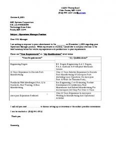
A particularly appealing target for modulation is the serine/threonine kinase Akt. The
50
activation of Akt (Figure 1) is due to the phosphorylation of two sites (Thr-308 and Ser-473)
51
by protein kinases that are controlled by phosphoinositide-3-kinase (PI3K).[4] PI3K is
52
activated by upstream signals such as insulin binding to its receptor. Numerous reports have
53
linked Akt phosphorylation to critical biological events such as cell growth, proliferation and
54
survival,[5] thus rendering it medicinally significant for cancer[6] and neurological diseases[7]
55
among others. Akt activation has also been implicated in cellular glucose uptake and
56
utilization, consequently Akt is also a highly relevant target in the development of anti-
57
diabetic drugs.[8]
58 59
Identification of new small molecules that make it possible to regulate at will the
60
phosphorylation levels of Akt might yield control over some of these downstream processes.
61
With a focus on the diabetes implications of Akt activation, we have previously shown that a
62
series of vanadium(IV) and vanadium(V) complexes with picolinic ligands were able to
63
induce Akt phosphorylation, acting by inhibiting PTEN phosphatase, an enzyme upstream of 3
64
Akt that directly counteracts the function of PI3K.[8a] In parallel, other laboratories have
65
suggested that protein tyrosine phosphatases (PTP) are possible targets for complexes of
66
vanadium.[9] Recent studies by Sakurai describing metal-based small molecules with the
67
ability to enhance Akt phosphorylation levels have involved zinc(II) as the metal center.[10]
68
Most of these reports have suggested that zinc(II) complexes carry out their effects in a PI3K-
69
dependent mode,[11] and that relatively high concentrations of complexes are needed in order
70
to achieve significant Akt phosphorylation enhancements.
71
72 73 74
Figure 1. Simplified schematic representation of the biochemical pathways that connect the insulin receptor (IR), PI3K and PTEN with Akt and its downstream effector GSK3β.
75 76
Encouraged by the above findings and given the relatively limited number of zinc(II)
77
compound classes evaluated as Akt activators so far, we investigated a broader range of
78
zinc(II) compound families, with the aim of identifying novel and more potent small
79
molecules that can find applications as chemical perturbagens of Akt signalling and glucose
80
metabolism. Zinc(II) favorably exhibits low toxicity (especially in comparison to vanadium)
81
as well as the ability to afford complexes with a variety of coordination geometries, spanning 4
82
from tetra- to hexa-coordination. These properties were taken into account in our compounds
83
design, along with the inclusion of new phenolic, picolinic, pyridino and hydroxamic ligands,
84
that we considered likely to contribute favorable properties in terms of functional and
85
structural diversity, H-bonding ability, aqueous solubility, membrane transport and
86
pharmacokinetics. The synthesis of eleven new zinc(II) complexes (Figure 2) and their
87
biological evaluation leading to identification of a potent Akt activator is described herein.
88
89
Figure. 2 Proposed structures of all zinc(II) coordination compounds prepared in this study.
5
90
Results and Discussion
91
Synthesis of Ligands and Complexes
92
As part of our effort to discover new and effective metal-based activators of Akt signalling,
93
we prepared an extended series of zinc(II) complexes, which would be inclusive of a diverse
94
set of ligands. The compound design had to fit certain requirements: the complexes should
95
exhibit distinct coordination geometries and 3D-shapes; exhibit various patterns of hydrogen
96
bond donors and acceptors; be inclusive of pharmaceutically-beneficial functionalities; have
97
good aqueous solubility and membrane permeability; and be readily synthesized. We chose to
98
investigate four major classes of ligands, namely phenolic, picolinic (similar to the ones we
99
previously evaluated with vanadium), pyridino and hydroxamic. The functional groups
100
present in these ligands were deemed compatible with all of the above criteria, based on their
101
performance in a multitude of previous pharmaceutical applications. Eleven complexes were
102
synthesized in total, all of which are novel compounds.
6
103 104
Scheme 1. Synthesis of phenolic zinc(II) complexes 1-4 (yields: 1, 99%; 2, 54%; 3, X%; 4, 95%)
105
The first group of complexes (compounds 1-4, Scheme 1) includes polydentate Schiff base
106
ligands (or their reduced counterparts) derived from salicyl precursors, and were formed
107
using Zn(OAc)2 as the metal source. Compounds 1 and 2 exhibit an O-N-O coordination
108
pattern in their tridentate ligand. Compound 1 was obtained by condensation of glycine on
109
the 2-position of 2,4-di(formyl)-phenol in the presence of the metal, followed by a second
110
condensation of the remaining aldehyde moiety to an ethanolamine unit. Complex 2 was
111
obtained in one step by reacting an α-aminoacid, homocysteic acid, with a similar salicyl
112
aldehyde that features a sulfonic acid substituent on the 4-position. The chosen polar
113
functionalities in these compounds confer desired hydrophilicity and H-bonding ability to the
114
complexes, both beneficial for their aqueous solubility and potential to interact with
115
biomolecules. As suggested by our study and in agreement with a reported crystal structure of
116
1a,[12] complexes 1 and 2 were isolated in a hydrated state and in aqueous solution can 7
117
potentially reach hexa-coordination through further hydration. The fact that zinc(II) has no
118
specific preference for a fixed geometry allows water to occupy free coordination sites, which
119
is likely the case for other complexes in this study as well. Ligands for the synthesis of 3 and
120
4 were prepared from the same aromatic precursor as 2. In the case of 3 (which has a ligand-
121
to-metal stoichiometry of 2:1) the organic precursor was modified via reductive amination
122
with n-propylamine to afford the N-O bidentate amine ligand 3a prior to complexation. The
123
ligand in 4 (which exhibits an O-N-N-O coordination pattern) links two precursor molecules
124
via double imine formation upon their condensation with ethylenediamine. The complex
125
likely has distorted pyramidal geometry, as has been demonstrated previously for other
126
zinc(II)-salen complexes.[13]
127 128
Scheme 2. Synthesis of picolinic zinc(II) complex 5 (yield: 34%)
129 130
Another bidentate N-O ligand was derived from 5-aminopicolinic acid (Scheme 2). Although
131
this specific complex has not been previously reported, it should be noted that zinc(II)
132
complexes with other derivatives of picolinic acid have been studied previously as insulin
133
mimetics.[14] The amine handle was initially modified to afford benzylamine 5a[15] in order to
134
explore the impact of this bulky moiety on the biological activity of the corresponding
135
zinc(II) complex. Complex 5 was prepared by reacting the ligand with zinc sulfate under
136
basic conditions.
8
137
A group of four ligands were derived from 2-formylpyridines (Scheme 3) via imine
138
formation. In the case of compounds 6b and 7b (ligands for 6 and 7 respectively) the amine
139
components reacting with 2-formylpyridine contained an ethyl urea moiety. In the synthesis
140
of 8, propylamine was reacted with 5-nitro-2-formylpyridine, and the nitro/imine
141
intermediate 8a was reduced to the amine/amine ligand 8b. Complexation to obtain
142
complexes 6-8 was achieved with ZnSO4. In the case of 9, the zwitterionic amine taurine was
143
reacted with 2-formylpyridine under basic conditions, followed by complexation with ZnCl2
144
in the same pot. While this sub-set of molecules also offers extensive H-bonding patterns,
145
they have larger steric requirements compared with some of the asymmetric members of the
146
series.
147 148
Scheme 3. Synthesis of pyridino zinc(II) complexes 6-9 (yields: 6, 83%; 7, 51 %; 8, 30%; 9, 20%)
149 150
9
151
Hydroxamic complexes 10 and 11 were generated as shown in Scheme 4. A benzoic acid
152
functionalized at the 4-position with an amine handle (either directly attached to the ring or as
153
an aminomethylene moiety respectively) was converted to its corresponding methyl ester
154
using known procedures.[16] A critical step in ligand preparation was the transformation of
155
these esters to hydroxamic acids using hydroxylamine sulfate under basic aqueous conditions.
156
Conversion of methyl ester 10a to hydroxamic acid 10b was performed in one step[17] leaving
157
the free amine intact, while 11a underwent modification with Tf2O to form sulphonamide
158
11b, prior to its conversion to the desired hydroxamic acid 11c. Ligands 10b and 11c
159
afforded (2:1 ligand-to-metal) complexes upon reaction with Zn(OAc)2. Hydroxamates are
160
considered excellent chelators for metal ion transport through cell membranes,[18] while both
161
the hydroxamate[18] and sulphonamide[19] functionalities are of high medicinal relevance and
162
present in many bioactive compounds.
163 164
Scheme 4. Synthesis of hydroxamic zinc(II) complexes 10-11 (yields: 10, 92%; 11, 84%)
165 10
166 167
Biological evaluation of the effect of compounds on the Insulin/PI3K/PTEN/Akt
168
pathway.
169
Zinc complexes induce Akt activation
170
The zinc(II) ion and some of its complexes have been reported in the past to elicit activation
171
of Akt in several cell types.[11a, 20] The activation of Akt in these cases occurs via the PI3K
172
route and can be monitored by detecting the level of phosphorylation of residue Thr-308 or
173
Ser-473 of Akt. To examine whether the new zinc complexes prepared in this study induce
174
Akt activation, NIH3T3 cells were treated with 10 µM of each compound, followed by light
175
stimulation with 0.04 µM insulin. Western blots were run from the cell lysates and phospho-
176
Akt was quantified using a phospho-Akt (Ser473) antibody. Total Akt was also quantified as
177
loading control. Figure 3 (A and B) summarizes these results. A positive control (stimulation
178
with high insulin, 2 µM) and a negative control (low insulin equal to the concentration used
179
to activate the insulin receptor, 0.04 µM) were included for comparison. All compounds, with
180
the exception of 1-3 whose effect is negligible, appear to activate Akt by elevating phospho-
181
Akt levels to different extents. While the majority of the compounds exhibit Akt activation to
182
around 50% of the positive control, the two compounds derived from hydroxamic acid
183
ligands (10 and 11) stand out, as they appear to perform equally effective or considerably
184
better than the high insulin activation control respectively.
11
A
2 µM insulin 0.04 µM insulin
-
+ -
+
+
+
+
+
+
High insulin
Low insulin
1
2
3
4
5
Phospho-Akt Ser473 Akt
185
B
2 µM insulin 0.04 µM insulin
-
+ -
+
+
+
+
+
+
+
6
7
8
9
10
11
Phospho-Akt Ser473 Akt
186 187 188 189 190 191 192 193 194 195 196
High Low insulin insulin
Figure 3. A (compounds 1-5) and B (compounds 6-11): NIH3T3 cells were starved overnight, incubated with 10 µM of compound (or vehicle) for 15 min, followed by insulin stimulation as indicated. Cells were collected and analyzed by western blotting using phospho-Akt (Serine-473) and Akt antibodies. The blots in Figures 6A and 6B were quantified using ImageJ. The percentage of activation of Akt (p-Akt phosphorylation normalized for loading using the Akt blotting intensities) is shown. Low insulin stimulation (0.04 µM) was set to 0% and high insulin stimulation (2 µM) was set to 100%, respectively. Results (bar chart) are presented as the mean ± standard deviation of three independent experiments.
197 198
Compound 11, as the most promising Akt-activator, was subjected to further investigation.
199
First, we determined the cytotoxicity of this compound with the MTT assay on NIH3T3
200
cells.[21] MTT is reduced to formazan by living cells which can be quantified using a
201
spectrophotometer at an absorbance of 590 nm. As can be seen in Figure 4, compound 11
202
shows cytotoxicity effects on NIH3T3 cells at concentrations equal or higher than 100 µM.
12
203
At the concentration of 10 µM, which were used in the initial screen of all 11 compounds
204
(Figure 3), compound 11 has no cytotoxic effect on the cells.
205
206 207
Figure 4 Cytotoxicity of compound 11. NIH3T3 cells were treated with up to 1 mM of
208
compound 11. The results are presented as mean ± standard deviations (n = 4).
209 210
To determine the minimum concentration of compound 11 required to induce Akt activation,
211
NIH3T3 cells were treated with various concentrations of the complex. As can be seen in
212
Figure 5A, Akt activation commenced at a concentration of 500 nM of compound 11.
213
Moreover, when probing the compound-treated cell lysates for glucogen synthase kinase 3β
214
(GSK-3β) phosphorylation at Ser-9, we found that 11 was also able to stimulate GSK-3β
215
phosphorylation with similar potency (Figure 5A). GSK-3β, a key enzyme in the regulation
216
of glucose metabolism, is phosphorylated at Ser-9 by Akt, suggesting that the activation of
217
Akt induced by the Zn complexes is propagated downstream of Akt to enhance glycogen
218
synthesis. For comparison, the effect of compound 11 on the MAP kinase pathway was
219
investigated by probing cellular extracts for phosphorylation of p42 and p 44 MAP kinases.
220
As shown in Figure 5B, compound 11 fails to activate MAPK p44/p42 at a concentration of 13
221
500 nM implying that compound 11 might be a useful chemical tool to probe glucose
222
metabolism and the signalling controlling it.
223 224 225 226 227 228 229 230 231
Figure 5. (A) Activation of Akt and downstream element GSK-3β (Serine-9) with compound 11 at various concentrations. Starved cells were incubated with 100 nM, 250 nM and 500 nM of compound 11 for 15 min, followed by stimulation of insulin at a concentration of 0.04 µM. (B) Compound 11 does not increase phosphorylation levels of p44/p42 MAPK. Same cell lysates (from figure 5A) were probed with a phospho-p44/42 MAPK (Thr202/Tyr204) antibody.
232 233
Zinc complex-induced Akt activation is PI-3kinase dependent
234
To investigate whether the observed Akt phosphorylation is PI3K-mediated, we studied the
235
effects of compound 11 on Akt phosphorylation levels in the presence of LY-294002, a
236
known PI3K inhibitor.[22] If the observed Akt activation is PI3K dependent, the presence of
237
LY-294002 would be expected to abolish the activation induced by the zinc compound.
238
Indeed, use of LY-294002 abrogated the compound 11-induced Akt phosphorylation as
239
shown in Figure 6. To further confirm the hypothesis that the zinc complex-mediated
240
activation occurs via PI3K, we examined whether any of the prepared complexes can induce
241
Akt activation without any insulin stimulation. Notably, when insulin is completely deprived,
14
242
the compounds are unable to induce Akt phosphorylation (Figure 7), which suggests they do
243
not act directly at the insulin receptor level, but rather between receptor and PI3K. Taking
244
these observations together, we suggest that the zinc compounds induce Akt activation in a
245
PI3K-dependent fashion.
246
247 248 249 250
Figure 6. Effects of compound 11 on Akt phosphorylation with and without the PI3K inhibitor LY294002.
251 252 253 254 255 256 257 258 259 260 261 262 263
Figure 7. Phospho-Akt (Serine-473) blots after cells were treated either with the zinc complexes without any insulin stimulation or with insulin (2 µM). Loading control is performed using the detection of total Akt.
264 265
15
266
Zinc complex-induced Akt activation is not via inhibition of PTPases or PTEN
267
The PI3K/Akt cascade is modulated by phosphatases of the PTPase family and PTEN.
268
PTPases oppose the signalling mediated by receptor tyrosine kinases by removing tyrosine
269
phosphorylation, whereas PTEN directly antagonises PI3K by dephosphorylating
270
phosphatidylinositol-3,4,5-trisphosphate (PIP3), the product of PI3K activity. As both
271
PTPases and PTEN are upstream of Akt, their phosphatase activity can influence the level of
272
Akt activation. Indeed, it has been found that inhibition of PTPases and PTEN will increase
273
the cellular activity of Akt.[23]
274 275
Zinc(II) has been reported to have the ability to inhibit phosphatases such as λ-
276
phosphoprotein phosphatase,[24] protein tyrosine phosphatase 1B,[25] as well as PTEN.[11b] In
277
order to investigate whether the observed Akt activation was due to inhibition of PTEN and
278
PTPases, the two most promising Akt-activating compounds (hydroxamic subset) 10 and 11
279
were evaluated as PTEN and PTP-β inhibitors in vitro. As shown in Figure 8 the inhibition of
280
PTP-β and PTEN by compound 10 and 11 revealed a significantly higher potency towards
281
the PTP-β, but failed to demonstrate the nanomolar potency achieved for Akt
282
phosphorylation in cellulo. A similar picture was observed for other representative members
283
of the series (results not shown).
16
284 285 286 287 288 289 290 291 292 293 294
Figure 8. Inhibition of PTEN and PTP-β with zinc(II) complexes 10 and 11 using OMFP and p-NPP as substrates. Recombinant PTEN and PTP-β were pre-incubated with either compound 10 or 11 at various concentrations at room temperature. Reactions were initialized by adding 200 µM of OMFP for PTEN and 1 mM of p-NPP for PTP- β. The IC50 in the presence of compound 10 was calculated to be 116 ± 25 µM for PTEN and 42 ± 7 µM for PTP- β. The IC50 of compound 11 was determined to be 77 ± 5 µM for PTEN and 19 ± 1 µM for PTP-β. The results are presented as the mean ± the standard deviation of three independent experiments.
295 296 297
Conclusion
298 299
In summary, a series of eleven novel zinc(II) complexes incorporating medicinally-relevant
300
functionalities were prepared based on phenolic, picolinic, pyridino and hydroxamic ligands.
301
Screening of the compounds for their potential to upregulate Akt phosphorylation in cellulo,
302
revealed a sulphonamide-containing hydroxamic complex as a potent activator of Akt at sub-
303
micromolar concentrations. This lead structure can be potentially used as a chemical tool in
304
diabetes research, due to the fact that apart from activating Akt signalling, its effect is
305
propagated downstream of Akt towards GSK-3β, an important regulator of glycogen
306
synthesis/glucose metabolism. Our findings suggest that the observed effects on Akt
307
phosphorylation by the hydroxamic zinc complexes, while PI3K-mediated, are not due to the 17
308
inhibition of the tumor-supressor phosphatase PTEN or PTPases. This signifies a departure
309
from other zinc and vanadium complexes that have been shown to cause Akt activation by
310
inhibiting these phosphatases.
311 312
Future research will be directed towards elucidating the exact mechanism of action of
313
hydroxamic zinc complexes on Akt activation, as well as the development of improved Akt
314
activators based on evolution of this novel template.
315
316
Experimental Section
317
General Procedures
318
The purity of the organic chemicals was verified by 1H-NMR spectroscopy. 1H-NMR and
319
13
320
spectrometer. Infra-red spectra were recorded on a Perkin-Elmer FTIR spectrometer.
321
Electrospray ionisation mass spectra were recorded on a Bruker Daltronics Esquire 3000
322
spectrometer. Elemental analyses were performed by either London Metropolitan University
323
or University of Cambridge. The following compounds were prepared following literature
324
procedures: 1a,[12] 5a,[15] 10a,[16a] 10b[17] and 11a.[16b]
C-NMR spectra were recorded on a Bruker Avance 400 MHz Ultrashield NMR
325 326
Synthesis
327 328
Synthesis of 1
329
Intermediate 1a (0.107 g, 0.35 mmol, 1 equiv) was suspended in 45 mL of methanol.
330
Ethanolamine (42 µL, 0.70 mmol, 2 equiv) was added and the mixture was stirred at r.t.
331
overnight. The resulting solution was then cooled at 0°C and 2 mL of a methanolic solution 18
332
of NaBH4 (0.026 g, 0.70 mmol, 2 equiv) was added dropwise. The mixture was stirred
333
initially at 0°C for 1 h, then allowed to warm up to r.t. It was then filtered and the collected
334
yellow precipitate was washed with methanol and dried to afford 43% of imine intermediate
335
(0.053 g, 0.15 mmol). This compound was then dissolved in 10 mL of water to afford a clear
336
solution. Aqueous HCl 0.6 M (0.25 mL, 0.15 mmol, 1 equiv) was added to the solution and
337
the mixture stirred at r.t. briefly, before evaporating the solvent to dryness. The obtained solid
338
residue was washed with diethyl ether to afford complex 1 in nearly quantitative yield (0.058
339
g, 0.15 mmol). 1H-NMR (D2O, 4.79 ppm)- (ppm): 3.07 (t, 2H, J=5.4 Hz), 3.75 (t, 2H, J=5.4
340
Hz), 4.06 (s, 2H), 4.08 (s, 2H), 6.69 (bd, 1H, J=8.6 Hz), 7.24 (m, 2H), 8.26 (s, 1H). 13C-NMR
341
(D2O)- (ppm): 48.1, 50.4, 56.7, 56.8, 119.5, 122.4, 126.0, 135.2, 137.6, 149.9, 168.9.
342
Elemental analysis: calculated for C12H19ClN2O6Zn: C 37.13, H 4.93, N 7.22; found: C 36.57,
343
H 4.12, N 6.96.
344 345
Synthesis of 2
346
Racemic homocysteic acid (0.150 g, 0.82 mmol, 1 equiv) was added to 5 mL of water and the
347
resulting mixture was heated at 70°C. A solution of 3-formyl-4-hydroxy-benzenesulfonic
348
acid disodium salt (0.201 g, 0.82 mmol, 1 equiv) in 5 mL of water was then added and the
349
mixture was stirred at 70 °C for 90 mins. A solution of Zn(OAc)2∙2H2O (0.223 g, 1.02 mmol,
350
1.24 equiv) in 10 mL of water was finally added. During this last addition the pH of the
351
solution was adjusted to about 7 with aqueous NaOH 0.5 M. The solution was stirred at 70°C
352
for two more hours. The solvent was then evaporated to dryness and a yellow-orange residue
353
was obtained. Successive recrystallizations in water/methanol yielded compound 2 as a white
354
solid in 54% yield (0.217 g, 0.44 mmol). 1H-NMR (D2O, 4.79 ppm)- (ppm): 2.25 (m, 2H),
355
2.86 (m, 2H), 4.03 (t, 1H, J=6.2 Hz), 6.72 (d, 1H, J1=9.0 Hz), 7.57 (dd, 1H, J1=9.0 Hz, J2=
356
2.5 Hz), 7.66 (d, 1H, J2=2.5 Hz), 8.38 (s, 1H). 19
C-NMR (D2O)- (ppm): 29.9, 46.8, 66.3,
13
357
118.4, 122.2, 128.5, 130.9, 133.9, 169.7, 171.1, 179.1. Elemental Analysis: calculated for
358
C11H13NNa2O11S2Zn∙2H2O: C 24.16, H 3.16, N 2.56; found C 24.42, H 3.33, N 2.54.
359 360
Synthesis of 3a
361
3-Formyl-4-hydroxy-benzenesulfonic acid disodium salt (0.300g, 1.22 mmol, 1 equiv) was
362
dissolved in 40 mL of methanol, and propylamine (0.121 mL, 1.47 mmol, 1.2 equiv) was
363
added. The solution was stirred at r.t. for 3 h, at which point an aliquot was obtained to verify
364
by NMR that the imine intermediate had formed. The solution was then cooled in an ice-bath
365
and NaBH4 (0.185 g, 4.88 mmol, 4 equiv) was added. The mixture was stirred at 0°C for 1 h
366
and at r.t. overnight. A few drops of water were added and then the solvent was evaporated to
367
dryness. The residue was re-dissolved in methanol and insoluble impurities were removed by
368
filtration. The filtrate was concentrated under vacuum and diethyl ether was added to
369
precipitate the product. Compound 3a was obtained as a pink solid in 72% yield (0.255 g,
370
0.88 mmol). 1H-NMR (D2O, 4.79 ppm)- (ppm): 0.85 (t, 3H, J=7.4 Hz), 1.58 (bs, 2H), 2.88
371
(t, 2H, J=7.4 Hz), 4.02 (s, 2H), 6.55 (d, 1H, J=8.6 Hz), 7.45 (m, 2H). This ligand was used
372
for the synthesis of 3 without any further purification.
373 374
Synthesis of 3
375
To a solution of 3a (0.100 g, 0.35 mmol, 1 equiv) in 10 mL of methanol was added a solution
376
of Zn(OAc)2·2H2O (0.038 g, 0.175 mmol, 0.5 equiv) in 5 mL of methanol and the resulting
377
mixture was stirred at 50 °C overnight. The solvent was evaporated to dryness and complex 3
378
was obtained as a yellow residue. It was further purified by recrystallization in
379
methanol/diethyl ether. 1H-NMR (D2O, 4.79 ppm)- (ppm): 0.86 (t, 6H, J=7.1 Hz), 1.59 (bs,
380
4H), 2.90 (bt, 4H, J=6.7 Hz), 4.06 (s, 4H), 6.63 (d, 2H, J=9.2 Hz), 7.59 (m, 4H). Also
20
381
present: 1 equiv. of NaOAc impurity: 1.82 (s, 3H). Elemental Analysis: calculated for
382
C20H26N2Na2O8S2Zn∙NaOAc: C 38.86, H 4.30, N 4.12; found: C 38.77, H 4.41, N 3.98.
383 384
Synthesis of 4
385
A solution of ethylenediamine (0.027 mL, 0.41 mmol, 0.5 equiv) in 2 mL of methanol was
386
added to a solution of 3-formyl-4-hydroxy-benzenesulfonic acid disodium salt (0.202 g, 0.82
387
mmol, 1 equiv) in 40 mL of methanol and the reaction mixture was stirred at r.t for 1 h.
388
Zn(OAc)2·2H2O (0.090 g, 0.41 mmol, 0.5 equiv) was then added and the mixture was heated
389
at 50°C overnight. It was subsequently filtered, and the collected white residue was washed
390
with cold methanol and dried under vacuum to afford 4 in 95% yield (0.216 g, 0.39 mmol).
391
1
H-NMR (D2O, 4.79 ppm)- (ppm): 3.79 (s, 4H), 6.80 (d, 2H, J1=8.9 Hz), 7.55 (dd, 2H,
392
J1=8.9 Hz, J2=2.5 Hz), 7.64 (d, 2H, J2=2.5 Hz), 8.43 (s, 2H). 13C-NMR (D2O)- (ppm): 55.8,
393
119.2, 122.6, 128.3, 130.0, 133.1, 168.1, 170.8. Elemental Analysis: calculated for
394
C16H14N2Na2O9S2Zn∙0.5H2O: C 34.15, H 2.69, N 4.98; found: C 34.11, H 2.72, N 4.89.
395 396
Synthesis of 5
397
A solution of ZnSO4·7H2O (0.050 g, 0.175 mmol, 0.5 equiv) in 1.5 mL of water was added to
398
a solution of 5a (0.080 g, 0.35 mmol, 1 equiv) in 3 mL of EtOH/DMF (1:1). Aqueous NaOH
399
0.5 M (0.7 mL, 0.35 mmol, 1 equiv) was added and the mixture was stirred at r.t. for 3 h. The
400
resulting precipitate was then filtered off, washed with water and dried under vacuum to
401
furnish 34% of complex 5 (0.033 g, 0.06 mmol). 1H-NMR (DMSO-d6, 2.50 ppm)- (ppm):
402
4.34 (d, 4H, J=5.2 Hz), 7.12 (d, 2H, J=7.2 Hz), 7.24 (m, 2H), 7.32 (m, 8H), 7.45 (bs, 2H),
403
7.85 (d, 2H, J=8.8 Hz), 7.94 (bs, 2H). Elemental Analysis: calculated for C26H26N4O6Zn: C
404
56.17, H 4.71, N 10.08; found: C 56.18, H 4.60, N 10.14.
405 21
406
Synthesis of 6a
407
Ethyl isocyanate (0.29 mL, 3.66 mmol, 1 equiv) was slowly added to a cooled solution (–10
408
°C) of 1,3-diaminopropane (0.305 mL, 3.66 mmol, 1 equiv) in 35 mL of CH2Cl2. The mixture
409
was then allowed to stir overnight at r.t., resulting in precipitation of the desired urea product
410
(6a). This solid was collected by filtration. Additionally, the filtrate was dried under vacuum
411
to yield an oily residue, which upon treatment with diethyl ether afforded more of the
412
product. The combined product was washed with diethyl ether and dried. Overall, 63% of 6a
413
was obtained (0.335 g, 2.31 mmol).
414
J=7.0 Hz), 1.40 (m, 2H), 2.50 (m, 2H), 3.05 (m, 4H), 5.80 (bs, 2H). LC-MS (ESI+):
415
calculated for C6H15N3O (M+H) 146.13, found 146.13. This compound was used in the next
416
step without any further purification.
1
H-NMR (DMSO-d6, 2.50 ppm)- (ppm): 0.95 (t, 3H,
417 418
Synthesis of 6b
419
Pyridine-2-carboxaldehyde (0.10 mL, 1.0 mmol, 1 equiv) was slowly added (over 10 min) to
420
a cold solution (–10 °C) of 6a (0.145 g, 1.0 mmol, 1 equiv) in 2 mL of methanol, and the
421
mixture was allowed to stir overnight at r.t. The solvent was then removed under vacuum to
422
yield a white residue, which was washed several times with diethyl ether and dried to furnish
423
93% of 6b (0.217 g, 0.93 mmol). 1H-NMR (DMSO-d6, 2.50 ppm)- (ppm): 0.97 (t, 3H, J=7.2
424
Hz), 1.73 (m, 2H), 2.98 (m, 2H), 3.05 (apparent q, 2H, J=6.4 Hz), 3.62 (t, 2H, J=6.8 Hz),
425
5.76 (t, 1H, J=5.6 Hz), 5.86 (t, 1H, J=5.6 Hz), 7.45 (m, 1H), 7.86 (td, 1H, J1=7.6 Hz, J2=1.2
426
Hz), 8.34 (s, 1H), 8.63 (d, 1H, J=4.8 Hz), 8.94 (d, 1H, J1=7.6 Hz).
427
39.52 ppm)- (ppm): 16.2, 31.6, 34.5, 37.7, 58.4, 120.9, 125.5, 137.3, 149.8, 154.6, 158.5,
428
162.4. Elemental Analysis: calculated for C12H18N4O: C 61.52, H 7.74, N 23.91; found: C
429
61.52, H 7.64, N 23.84.
430 22
13
C-NMR (DMSO-d6,
431
Synthesis of 6
432
A solution of ZnSO4∙7H2O (0.100 g, 0.35 mmol, 0.5 equiv) in 2 mL of water was added to a
433
solution of 6b (0.163 g, 0.70 mmol, 1 equiv) in 2 mL of methanol, and the resulting mixture
434
was stirred at r.t. for 2 h. The solvent was then removed under vacuum, yielding complex 6 as
435
a pale brown solid in 83% yield (0.193 g, 0.29 mmol). 1H-NMR (DMSO-d6, 2.50 ppm)-
436
(ppm): 0.88 (t, 6H, J=7.2 Hz), 1.74 (m, 4H), 2.86 (m, 4H), 3.01 (apparent q, 4H, J=5.6 Hz),
437
3.65 (bs, 4H), 5.85 (bs, 2H), 6.17 (bs, 2H), 7.66 (bs, 2H), 7.94 (d, 2H, J=7.6 Hz), 8.08 (bs,
438
2H), 8.52 (s, 2H), 8.78 (bs, 2H). Elemental Analysis: calculated for C24H40N8O8SZn·H2O: C
439
42.14, H 6.19, N 16.38; found: C 42.33, H 5.69, N 16.33.
440
Synthesis of 7a
441
This compound was prepared in 28% yield, following an analogous method to the one used
442
for the synthesis of 6a. 1H-NMR (DMSO-d6, 2.50 ppm)- (ppm): 0.95 (t, 3H, J=7.1 Hz), 1.40
443
(m, 4H), 2.50 (bt, 2H, overlapping with DMSO peak), 3.00 (m, 4H), 5.71 (bt, 1H), 5.80 (bt,
444
1H). LC-MS (ESI+): calculated for C7H17N3O (M+H) 160.15, found 160.15. This compound
445
was used in the next step without any further purification.
446 447
Synthesis of 7b
448
This compound was prepared in 99% yield, following an analogous method to the one used
449
for the synthesis of 6b. 1H-NMR (DMSO-d6, 2.50 ppm)- (ppm): 0.96 (t, 3H, J=7.2 Hz), 1.40
450
(m, 2H), 1.61 (m, 2H), 2.99 (m, 4H), 3.62 (t, 2H, J=7.2 Hz), 5.71 (t, 1H, J=5.6 Hz), 5.80 (t,
451
1H, J=5.2 Hz), 7.45 (m, 1H), 7.86 (td, 1H, J1=7.6 Hz, J2=1.2 Hz), 7.93 (d, 1H, J1=7.6 Hz),
452
8.33 (s, 1H), 8.63 (d, 1H, J=4.8 Hz). 13C-NMR (DMSO-d6, 39.52 ppm)- (ppm): 16.2, 28.2,
453
28.4, 34.5, 60.6, 120.8, 125.5, 137.3, 149.8, 154.6, 158.5, 162.1. Elemental Analysis:
454
calculated for C13H20N4O: C 62.88, H 8.12, N 22.56; found: C 62.96, H 8.09, N 22.48.
455 23
456
Synthesis of 7
457
This compound was prepared in 51% yield, following an analogous method to the one used
458
for the synthesis of 6. 1H-NMR (DMSO-d6, 2.50 ppm)- (ppm): 0.93 (t, 6H, J=7.2 Hz), 1.31
459
(m, 4H), 1.61 (m, 4H), 2.95 (m, 8H), 3.60 (bs, 4H), 5.93 (bs, 2H), 6.02 (bs, 2H), 7.67 (bs,
460
2H), 7.97 (d, 2H, J=7.6 Hz), 8.10 (bs, 2H), 8.53 (s, 2H), 8.82 (bs, 2H). Elemental Analysis:
461
calculated for C26H44N8O8SZn∙2.5H2O: C 42.25, H 6.68, N 15.16; found: C 42.13, H 5.89, N
462
15.16.
463 464 465
Synthesis of 8a
466
A solution of 5-nitropyridine-2-carboxaldehyde (0.386 g, 2.54 mmol, 1 equiv) in 20 mL of
467
dry methanol was added dropwise to a solution of propylamine (0.24 mL, 2.92 mmol, 1.15
468
equiv) in 8mL of methanol. The mixture was stirred for 5 h at r.t. and the solvent was then
469
removed under vacuum, to afford 8a quantitatively as a dark oil (0.49 g, 2.54 mmol). 1H-
470
NMR (CDCl3,7.26 ppm)- (ppm): 1.00 (t, 3H, J=7.4 Hz), 1.79 (apparent sextet, 2H, J=7.2
471
Hz), 3.73 (td, 2H, J1=6.9 Hz, J2=1.3 Hz), 8.23 (d, 1H, J3=8.7 Hz), 8.47 (bs, 1H), 8.53 (dd,
472
1H, J3=8.7 Hz, J4=2.4 Hz), 9.47 (d, 1H, J4=2.4 Hz). 13C-NMR (CDCl3, 77.00 ppm)- (ppm):
473
11.9, 23.8, 63.5, 121.21, 131.6, 144.4, 144.9, 159.2, 159.9. This compound was used for the
474
next step without any further purification.
475 476
Synthesis of 8b
477
A solution of 8a (0.293 g, 1.52 mmol, 1 equiv) in 25 mL of dry methanol was mixed with
478
0.09 g of 10% Pd-C and cooled at 5°C. NaBH4 (0.23 g, 6.1 mmol, 4 equiv) was added
479
portionwise to the mixture over 30 min. The resulting solution was stirred at the same
480
temperature for 1 h, then allowed to warm up to r.t. It was subsequently filtered through celite 24
481
and the solvent was evaporated to dryness. The remaining residue was re-suspended in ethyl
482
acetate and the organic phase was washed with water. The aqueous phase was back-extracted
483
with CHCl3. All organic fractions were dried over MgSO4 and the solvent was evaporated to
484
dryness to afford 8b as a yellow oil in 55% yield (0.139 g, 0.84 mmol). 1H-NMR (CDCl3,
485
7.26 ppm)- (ppm): 0.93 (m, 3H), 1.54 (m, 2H), 2.61 (m, 2H), 3.66 (bs, 2H), 3.79 (s, 2H),
486
6.96 (dd, 1H, J1=8.2 Hz, J2= 2.8 Hz), 7.09 (d, 1H, J1=8.2 Hz), 8.06 (d, 1H, J2=2.8 Hz). 1H-
487
NMR (DMSO-d6, 2.50 ppm)- (ppm): 0.85 (m, 3H), 1.41 (m, 2H), 2.46 (m, 2H), 3.60 (s, 2H),
488
5.13 (bs, 2H), 6.88 (dd, 1H, J1’=8.3 Hz, J2’=2.7 Hz), 7.03 (d, 1H, J1’=8.3 Hz), 7.85 (d, 1H,
489
J2’=2.7 Hz).
490
136.9, 141.0, 149.9. 13C-NMR (DMSO-d6, 39.52 ppm)- (ppm): 12.3, 22.9, 51.1, 54.4, 121.0,
491
122.4, 135.8, 143.7, 147.3. This compound was used for the next step without any further
492
purification.
13
C-NMR (CDCl3, 77.00 ppm)- (ppm): 11.8, 23.2, 51.5, 54.7, 122.3, 122.6,
493 494
Synthesis of 8
495
A solution of ZnSO4·7H2O (0.121g, 0.42 mmol, 0.5 equiv) in 10 mL of methanol was added
496
to a solution of 8b (0.139 g, 0.84 mmol, 1 equiv) in 10 mL of methanol, and the resulting
497
mixture was stirred at r.t. for 3 h. The solvent was then evaporated to dryness and the residue
498
washed with dichloromethane and ethyl ether. Recrystallization in methanol/diethyl ether
499
afforded complex 8 as a yellow solid in 30% yield (0.066 g, 0.125 mmol). 1H-NMR (DMSO-
500
d6, 2.50 ppm)- (ppm): 0.81 (t, 6H, J=7.3 Hz), 1.50 (m, 4H), 2.62 (bs, 4H), 3.80 (bs, 4H),
501
5.58 (bs, 2H), 5.71 (bs, 4H), 7.18 (bs, 4H), 8.03 (bs, 2H). 13C-NMR (DMSO-d6, 39.52 ppm)-
502
(ppm): 11.8, 20.9, 50.3, 50.4, 123.3, 123.7, 134.7, 142.4, 145.6. LC-MS (ESI+): calculated
503
for C18H28N6Zn (M+H) 393.18, found 393.18. IR (cm-1): 1118 (SO42-), 1506 (m), 1581 (m),
504
1630 (m), 1735 (br), 2875 (br), 2964 (br), 3229, 3349. Elemental Analysis: calculated for
505
C18H34N6O6SZn·2H2O: C 38.33, H 6.79, N 14.90; found: C 38.58, H 6.15, N 14.27. 25
506 507
Synthesis of 9
508
Pyridine-2-carboxaldehyde (0.19 mL, 2 mmol, 1 equiv) was dissolved in 50 mL DMF in a
509
round-bottom flask. Taurine (0.25 g, 2 mmol, 1 equiv) was added to this solution, followed
510
by anhydrous triethylamine (0.28 ml, 2 mmol, 1 equiv). After stirring at r.t. until all
511
components had gone into solution, ZnCl2 was added (0.14 g, 1 mmol, 0.5 equiv) and the
512
reaction mixture was heated at 110°C for 4h. The resulting precipitate was filtered and
513
washed to afford 9 as a pink powder in 20% yield (0.10 g, 0.2 mmol). 1H-NMR (DMSO-d6,
514
2.50 ppm)- (ppm): 3.03 (bs, 4H), 4.24 (bs, 4H), 7.67 (t, 2H, J=6.4 Hz), 8.03 (d, 2H, J=8.0
515
Hz), 8.17 (bs, 2H), 8.36 (bs, 2H), 8.88 (bs, 2H). 13C-NMR (DMSO-d6, 39.52 ppm)- (ppm):
516
49.86, 54.46, 128.94, 141.11, 148.63, 163.60. IR (cm-1): 738, 1228, 1530, 1594, 2943, 3235,
517
3331. Elemental analysis: calculated for C16H18N4O6S2Zn: C 39.07, H 3.69, N 11.39; found:
518
C 39.13, H 3.73, N 11.45.
519 520
Synthesis of 10
521
4-Amino-N-hydroxybenzamide (10b) (0.20 g, 1.32 mmol, 1 equiv) was transferred to a
522
round-bottom flask and dissolved in methanol (13.2 mL). Zn(OAc)2.2H2O (0.145 g, 0.66
523
mmol, 0.5 equiv) was added, and the reaction was refluxed at 65°C for 12 h, during which a
524
white precipitate formed. After cooling at r.t., the mixture was filtered and the solid washed
525
several times with chilled water and dried under vacuum. Overall, 92% (0.235 g, 0.61 mmol)
526
of complex 10 was obtained. 1H-NMR (DMSO-d6, 2.50 ppm)- (ppm): 5.50 (s, 4H), 6.53 (d,
527
4H, J=8.6 Hz), 7.48 (d, 4H, J=8.6 Hz), 11.50 (s, 2H). 1H-NMR (D2O, 4.79 ppm, with 1%
528
formic acid-d2)- (ppm): 7.26 (d, 4H, J=8.0 Hz), 7.71 (d, 4H, J=8.0 Hz). LC-MS (ESI+):
529
calculated for C14H14N4O4Zn (M+H) 367.04, found 367.04. IR (cm-1): 1504, 1563, 1598,
26
530
3018, 3170, 3431. Elemental analysis: calculated for C14H16N4O5Zn: C 43.59, H 4.18, N
531
14.53; found: C 44.02, H 3.71, N 14.35.
532 533
Synthesis of 11b
534
Methyl 4-(aminomethyl)benzoate (11a) (1.65 g, 10 mmol, 1 equiv) was transferred to a
535
round-bottom flask, which was then sealed and purged with nitrogen. Anhydrous CH2Cl2 (33
536
mL) was added and the mixture was cooled at -78°C. Triethylamine (2.8 mL, 20 mmol, 2
537
equiv) was added, followed by dropwise addition of trifluoromethanesulfonic anhydride (1.7
538
mL, 10 mmol, 1 equiv) at -78°C. The mixture was allowed to stir over 3-4 h, with the
539
temperature slowly rising to 0°C. The reaction mixture was then diluted with CH2Cl2, and
540
washed once with aqueous NH4Cl (sat), once with aqueous NaCl (sat) and once with H2O.
541
The organic phase was dried over Na2SO4, then filtered and concentrated under vacuum. The
542
sample was applied to a silica column and eluted with hexane-ethyl acetate (first 2:1, then
543
1:1), to afford 11b in 88% yield (2.61 g, 8.8 mmol) as a white solid. 1H-NMR (CDCl3, 7.26
544
ppm)- (ppm): 3.92 (s, 3H), 4.51 (s, 2H), 5.43 (bs, 1H), 7.39 (d, 2H, J=8.3 Hz), 8.01 (d, 2H,
545
J=8.3 Hz).
546
127.61, 130.03, 130.19, 140.49, 166.89. LC-MS (ESI+): calculated for C10H10F3NO4S (M+H)
547
298.04, found 298.04. IR (cm-1): 1001, 1018, 1081, 1119, 1150, 1181, 1233, 1289, 1306,
548
1386, 1445, 1615, 1695, 3229. Elemental analysis: calculated for C10H10F3NO4S: C 40.41, H
549
3.39, N 4.71; found C 40.51, H 3.27, N 4.64.
13
C-NMR (CDCl3, 77.00 ppm)- (ppm): 47.65, 52.37, 119.62 (CF3 quartet),
550 551
Synthesis of 11c
552
Hydroxylamine sulfate (0.82 g, 5 mmol, 1 equiv) was transferred to a round-bottom flask
553
containing 5 g of ice. A solution of NaOH (1.0 g, 25 mmol, 5 equiv) in 5 mL of water was
554
added to the flask and the mixture was stirred at r.t. for 5 min. A catalytic amount of Na2SO3 27
555
(0.095 g, 0.75 mmol, 0.15 equiv) and methyl 4-[(trifluoromethylsulfonamido)methyl]-
556
benzoate (11b) (1.49 g, 5 mmol, 1 equiv) were then added in solid form, and the mixture was
557
heated at 45°C for 16 h. After cooling at r.t., the pH was modified to about 5 by dropwise
558
addition of aqueous H2SO4 10% (w/w), and the solvent was removed under vacuum. Cold
559
methanol was added to the solid residue and the resulting suspension was vigorously stirred
560
for 15 mins and filtered. The hydroxamic acid, free of most inorganic impurities, was
561
recovered by drying the methanolic filtrate. This sample was re-dissolved in CH2Cl2-CH3OH
562
90:10 and applied to a silica column. Elution took place with the same solvent system (first
563
90:10, then 85:15) to afford 76% (1.13 g, 3.8 mmol) of hydroxamic acid 11c as a white solid.
564
1
565
J=8.1 Hz), 9.04 (s, 1H), 10.02 (s, 1H), 11.22 (s, 1H).
566
(ppm): 46.34, 119.69 (CF3 quartet), 127.21, 127.38, 132.20, 140.22, 163.92. LC-MS (ESI+):
567
calculated for C9H9F3N2O4S (M+H) 299.03, found 299.03. IR (cm-1): 1511, 1542, 1577,
568
1618, 3388. Elemental analysis: calculated for C9H9F3N2O4S: C 36.24, H 3.04, N 9.39; found
569
C 36.25, H 3.02, N 9.11.
H-NMR (DMSO-d6, 2.50 ppm)- (ppm): 4.42 (s, 2H), 7.40 (d, 2H, J=8.1 Hz), 7.75 (d, 2H, 13
C-NMR (DMSO-d6, 39.52 ppm)-
570 571
Synthesis of 11
572
N-hydroxy-4-[(trifluoromethylsulfonamido)methyl]benzamide (0.63 g, 2.11 mmol, 1 equiv)
573
was transferred to a round-bottom flask and dissolved in absolute ethanol (15 mL). A solution
574
of NaOH (0.084 g, 2.11 mmol, 1 equiv) in 5 mL of water was added, and the mixture was
575
stirred at r.t. for 5 min. Solid Zn(OAc)2∙2H2O (0.23 g, 1.055 mmol, 0.5 equiv) was then
576
added, and the reaction was refluxed at 80°C for 16 h. The solvent was then removed under
577
vacuum and the solid residue washed extensively with cold water and dried under vacuum to
578
afford complex 11 in 84% yield (0.60 g, 0.89 mmol), as a white solid. 1H-NMR (DMSO-d6,
579
2.50 ppm)- (ppm): 4.27 (s, 4H), 7.35 (d, 4H, J=7.7 Hz), 7.66 (d, 4H, J=7.7 Hz), 11.82 (s, 28
580
2H). LC-MS (ESI+): calculated for C18H16F6N4O8S2Zn (M+H) 658.97, found 658.97. IR (cm-
581
1
582
Elemental analysis: calculated for C18H18F6N4O9S2Zn.H2O: C 31.07, H 2.90, N 8.05; found C
583
31.00, H 2.40, N 7.63.
): 874, 918, 1022, 1056, 1149, 1190, 1231, 1372, 1441, 1503, 1565, 1606, 3350 (br).
584 585
Biological Materials and Methods
586 587
Cell culture and Western blot analysis
588
NIH 3T3 fibroblasts were purchased from ATCC. Cells were grown in Dulbecco’s modified
589
Eagle’s Medium (DMEM) (Sigma) supplemented with 10% (v/v) bovine calf serum (ATCC)
590
in an atmosphere of 5% CO2 at 37°C. For testing the zinc complexes, cells were starved in
591
DMEM (without serum) for 16 h at 37°C. Subsequent to serum deprivation, cells were
592
treated with zinc compounds for 15 min in DMEM at 37°C. Following compound treatment,
593
cells were stimulated either with 2 µM or 0.04 µM insulin for 15 min. A control was included
594
where cells were neither treated with zinc compounds nor stimulated with insulin. The cells
595
were washed afterwards three times with ice-cold PBS and then scrapped in lysis and SDS-
596
PAGE buffer, containing 150 mM Tris-HCl, pH 6.8, 25% glycerol, 5% SDS, 5% β-
597
mercaptoethanol and 0.01% bromophenol blue. Cell lysates were separated on a 10% SDS-
598
polyacrylamide gel and transferred onto a nitrocellulose membrane. Membranes were
599
blocked with 5% skimmed milk in TBS/T (50 mM Tris, 150 mM NaCl, 0.1% Tween 20, pH
600
7.4) for 1 h at room temperature. Phospho-Akt (Ser473) rabbit monoclonal antibody, mass-
601
Akt rabbit polyclonal antibody, GSK-3β (Ser9) rabbit polyclonal antibody and phospho-
602
p44/42 MAPK (Thr202/Tyr204) rabbit polyclonal antibody (Cell Signaling) were used as
603
primary antibodies for immunoblotting analysis. Primary antibody incubation was performed
604
in 5% bovine serum albumin in TBS/T for 16 h at 4°C. Blots were washed for 15 min with
29
605
TBS/T and then incubated with horseradish peroxidase-conjugated secondary antibody
606
(Biorad) in blocking buffer for 1h and then probed with western blotting detection reagent
607
(ECL, GE healthcare).
608 609
PTEN expression and purification
610
PTEN was expressed as a glutathione S-transferase fusion-protein and purified according to
611
methods previously described.[26] Protein expression was induced in the E. coli strain XL-1
612
blue for 24 h using 1 mM isopropyl β-D-1-thiogalactopyranoside at 23°C. Cells were
613
harvested and stored at -20°C.
614
containing 50 mM Tris (pH 7.4), 1% Triton X-100, 10 mM benzamidine hydrochloride, 100
615
µg/mL soybean trypsin inhibitor, 1 mM 4-(2-aminoethyl)benzenesulfonyl fluoride
616
hydrochloride and 2 mM dithiothreitol (DTT). Lysis was performed by adding lysozyme to
617
the cell suspension at a concentration of 2 mg/mL and sonication. Cell debris was removed
618
by centrifugation at 18000 g for 1 h at 4°C. The supernatant was loaded onto a glutathione
619
sepharose column, pre-equilibrated with 50 mM Tris (pH 7.4), 140 mM NaCl and 2.7 mM
620
KCl. After loading, the column was washed twice with 50 mM Tris (pH 7.4), 140 mM NaCl,
621
2.7 mM KCl and 2 mM DTT. Another two washes were performed using the same buffer
622
with 500 mM NaCl. The GST-tagged PTEN was eluted using 20 mM glutathione in 50 mM
623
Tris (pH 7.4), 250 mM NaCl, 20% glycerol and 2 mM DTT. Protein concentration was
624
determined using Bradford assay.
The harvested cells were re-suspended in lysis buffer
625 626
PTEN activity assay
627
PTEN activity in the presence of zinc compounds was assessed using the fluorescence
628
substrate 3-O-Methylfluorescein phosphate (OMFP).[27] OMFP was dissolved in DMSO to a
629
concentration of 20 mM and then further diluted with 1% DMSO to the tested concentrations.
30
630
Assays were performed in 100 mM Tris (pH 7.4) containing 2 mM DTT at room temperature
631
(20 °C). For inhibition studies PTEN was pre-incubated with zinc compounds at RT for 10
632
min. Reactions were then initialised by adding OMFP to a final concentration of 200 µM.
633
The hydrolysis of OMFP to OMF was monitored by measuring the change of fluorescence
634
units (FU) in a 96-well microtiter plate (excitation at 485 nm and emission at 525 nm) using a
635
Varian fluorescence spectrophotometer.
636 637
Cytotoxicity assay
638
NIH3T3 cells were seeded into a 96-well plate at a concentration of 8000 cells per well and
639
incubated at 37 ˚C for 16 hours. Cells were treated with compound 11 at concentrations
640
between 10 nM and 1 mM for 2 h at 37˚C (total volume of 100 µL). After 2h, 20 µL of a 5
641
mg/mL MTT solution were added to each well and incubated for further 4 h at 37˚C. Media
642
were removed carefully and 150 µL of a 4 mM HCl in isopropyl alcohol were added to each
643
well and incubated at room temperature for 15min on an orbital shaker. The formation of
644
formazan by viable cells was measured using a spectrophotometer at 590 nm.
645 646 647
Acknowledgements
648
This work has been supported by the Leverhulme Trust, the Lowe Syndrome Trust, Imperial
649
Innovations and the Departament d’Educació i Universitats de la Generalitat de Catalunya. In
650
addition the UK’s Engineering and Physical Sciences Research Council (EPSRC) is thanked
651
for a Leadership Fellowship to RV.
652 653 654 31
655
References
656
[1]
657
a) Spring David, R. (2005) Chem Soc Rev 34:472; b)Lehar, J., Stockwell, B. R., Giaever, G., Nislow, C. (2008) Nat Chem Biol 4:674.
658
[2]
Tarrant, M. K., Cole, P. A. (2009) Annu Rev Biochem 78:797.
659
[3]
a) Alonso, A., Sasin, J., Bottini, N., Friedberg, I., Friedberg, I., Osterman, A., Godzik,
660
A., Hunter, T., Dixon, J., Mustelin, T. (2004) Cell 117:699; b) Manning, G., Whyte,
661
D. B., Martinez, R., Hunter, T., Sudarsanam, S. (2002) Science 298:1912; c) Yaffe,
662
M. B. (2002) Nat Rev Mol Cell Biol 3:177; d) Yaffe, M. B., Elia, A. E. H. (2001)
663
Curr Opin Cell Biol 13:131; e) Hunter, T. (2000) Cell 100:113.
664
[4]
Gunn, R., Hailes, H. (2008) J Chem Biol 1:49.
665
[5]
Gonzalez, E., McGraw, T. (2009) Cell Cycle 8:2502.
666
[6]
Cicenas, J. (2008) Int J Biol Markers 23:1.
667
[7]
Zhang, W., Miao, L. (2007) Zhongfeng Yu Shenjing Jibing Zazhi 24:755.
668
[8]
a) Rosivatz, E., Matthews, J. G., McDonald, N. Q., Mulet, X., Ho, K. K., Lossi, N.,
669
Schmid, A. C., Mirabelli, M., Pomeranz, K. M., Erneux, C., Lam, E. W. F., Vilar, R.,
670
Woscholski, R. (2006) ACS Chem Biol 1:780; b) Tsuruzoe, K., Araki, E. (2005)
671
Diabetes Frontier 16:589; c)Asano, T., Kamata, H. (2006) Idenshi Igaku Mook 6:255.
672
[9]
a) Cuncic, C., Detich, N., Ethier, D., Tracey, A. S., Gresser, M. J., Ramachandran, C.
673
(1999) J Biol Inor Chem 4:354; b) Nxumalo, F., Glover, N. R., Tracey, A. S. (1998) J
674
Biol Inor Chem 3:534; c) Peters, K. G., Davis, M. G., Howard, B. W., Pokross, M.,
675
Rastogi, V., Diven, C., Greis, K. D., Eby-Wilkens, E., Maier, M., Evdokimov, A.,
676
Soper, S., Genbauffe, F. (2003) J Inorg Biochem 96:321; d) Shechter, Y., Goldwaser,
677
I., Mironchik, M., Fridkin, M., Gefel, D. (2003) Coord Chem Rev 237:3; e) Tracey,
678
A. S. (2000) J Inorg Biochem 80:11.
32
679
[10]
a) Nishide, M., Yoshikawa, Y., Yoshikawa, E. U., Matsumoto, K., Sakurai, H.,
680
Kajiwara, N. M. (2008) Chem Pharmac Bull 56:1181; b) Sakurai, H., Yoshikawa, Y.,
681
Yasui, H. (2008) Chem Soc Rev 37:2383.
682
[11]
a) Basuki, W., Hiromura, M., Sakurai, H. (2007) J Inorg Biochem 101:692;
683
b)Nakayama, A., Hiromura, M., Adachi, Y., Sakurai, H. (2008) J Biol Inor Chem
684
13:675.
685
[12]
686
Cai, J. H., Huang, Y. H., Jiang, Y. M. (2006) Acta Crystallogr, Sect E: Struct Rep Online E62:m2432.
687
[13]
Hall, D., Moore, F. H. (1966) J Chem Soc A 1822.
688
[14]
a) Kojima, Y., Yoshikawa, Y., Ueda, E., Kishimoto, N., Tadokoro, M., Sakurai, H.
689
(2005) Bull Chem Socl Jpn 78:451; b) Nakai, M., Sekiguchi, F., Obata, M., Ohtsuki,
690
C., Adachi, Y., Sakurai, H., Orvig, C., Rehder, D., Yano, S. (2005) J Inorg Biochem
691
99:1275; c)Yoshikawa, Y., Ueda, E., Kawabe, K., Miyake, H., Takino, T., Sakurai,
692
H., Kojima, Y. (2002) J Biol Inor Chem 7:68.
693
[15]
694 695
Finch, N., Campbell, T. R., Gemenden, C. W., Povalski, H. J. (1980) J Med Chem 23:1405.
[16]
a) Hosangadi, B., Dave, R. H. (1996) Tetrahedron Lett 37:6375; b) Goodyer, C. L.
696
M., Chinje, E. C., Jaffar, M., Stratford, I. J., Threadgill, M. D. (2003) Bioorg Med
697
Chem 11:4189.
698
[17]
699
Gaynor, D., Starikova, Z. A., Haase, W., Nolan, K. B. (2001) J Chem Soc Dalton Trans 1578.
700
[18]
Codd, R. (2008) Coord Chem Rev 252:1387.
701
[19]
Bhat, M. A., Imran, M., Khan, S. A., Siddiqui, N. (2005) Indian J Pharmac Sci
702
67:151.
33
703
[20]
a) Walter, P. L., Kampkoetter, A., Eckers, A., Barthel, A., Schmoll, D., Sies, H.,
704
Klotz, L.-O. (2006) Arch Biochem Biophys 454:107; b) Bao, S., Knoell, D. L. (2006)
705
Am J Physiol 290:L433; c) Chanoit, G., Lee, S., Xi, J., Zhu, M., McIntosh, R. A.,
706
Mueller, R. A., Norfleet, E. A., Xu, Z. (2008) Am J Physiol 295:H1227; d) Wong, V.
707
V. T., Nissom, P. M., Sim, S.-L., Yeo, J. H. M., Chuah, S.-H., Yap, M. G. S. (2006)
708
Biotech Bioengin 93:553.
709
[21]
Mosmann, T. (1983) J Immun Methods 65:55.
710
[22]
Vlahos, C. J., Matter, W. F., Hui, K. Y., Brown, R. F. (1994) J Biol Chem 269:5241.
711
[23]
a) Xie, L., Lee, S., Andersen, J., Waters, S., Shen, K., Guo, X. (2003) Biochemistry
712
42:12792; b) Schmid, A., Byrne, R., Vilar, R., Woscholski, R. (2004) FEBS Lett
713
566:35.
714
[24]
Zhuo, S., Dixon, J. (1997) Protein Eng 10:1445.
715
[25]
Haase, H., Maret, W. (2005) Biometals 18:333.
716
[26]
Rosivatz, E., Matthews, J. G., McDonald, N. Q., Mulet, X., Ho, K. K., Lossi, N.,
717
Schmid, A. C., Mirabelli, M., Pomeranz, K. M., Erneux, C., Lam, E. W. F., Vilar, R.,
718
Woscholski, R. (2006) ACS Chem Biol 1:780.
719
[27]
Mak, L. H., Vilar, R., Woscholski, R. (2010) J Chem Biol in press
720 721
34