Template matching is a common approach for detection of literature on this .... e nodule models used in our study. In [13] we used the Gaussian symmetric model ...
2007 IEEE International Symposium on Signal Processing and Information Technology
EXPERIMENTS ON SENSITIVITY OF TEMPLATE MATCHING FOR LUNG NODULE DETECTION IN LOW DOSE CT SCANS Shireen Y Elhabian1, Hossam Abd EL Munim1, Salwa Elshazly', AlyA.Farag1, and Mohamed Aboelghar2
'Computer Vision and Image Processing Laboratory, Univ. of Louisville, Louisville, KY, 40292 2Urology and Nephrology Center Mansoura University, Mansoura, Egypt
{contact:faraggcvip.uofl.edu}
ABSTRACT Template matching is a common approach for detection of lung nodules from CT scans. Templates may take different shapes, size and intensity distribution. The process of nodule detection is essentially two steps: isolation of candidate nodules, and elimination of false positive nodules. The processes of outlining the detected nodules and their classification (i.e., assigning pathology for each nodule) complete the CAD system for early detection of lung nodules. This paper is concerned with the template design and evaluating the effectiveness of the first step in the nodule detection process. The paper will neither address the problem of reducing false positives nor would it deal with nodule segmentation and classification. Only parametric templates are considered. Modeling the gray scale distribution for the templates is based on the prior knowledge of typical nodules extracted by radiologists. The effectiveness of the template matching is investigated by cross validation with respect to the ground truth and is described by hit rate curves indicating the probability of detection as function of shape, size and orientation, if applicable, of the templates. We used synthetic and sample real CT scan images in our experiments. It is found that template matching is more sensitive to additive noise than image blurring when tests conducted on synthetic data. On the sample CT scans small size circular and hollow-circular templates provided comparable results to human experts.
Index Terms- Shape Representation, tion, Level Sets, Energy Minimization. I. INTRODUCTION With improvements in CT imaging in terms of resolution, dose, and scanning approach, it has been possible to think of employing CT scanning in designing and evaluating fully automatic computer-aided diagnosis (CAD) systems especially for the thorax (e.g., Boiselle and White, 2002 [1]). At present, low-dose spiral computed tomography (LDCT) is of prime interest for screening high risk groups for early detection of lung cancer (e.g., [2]). Automatic screening involves detection of nodules then segmentation and classification; i.e., a three-stage process. Our earlier work has 978-1 -4244-1 835-0/07/$25.00 ©2007 IEEE
examined the first two stages (e.g., [11 - 13]) and the literature on this subject is becoming quite rich reflecting the importance and urgency of this problem (e.g., [3-12]). Our CAD system detects the nodules in LDCT images in three main steps: 1) segmentation of the raw scanning information to isolate the lung tissues from the rest of the structures in the chest cavity; 2) extraction of the 3D anatomic structures (e.g., blood vessels, bronchioles, alveoli, etc., and possible abnormalities) from the already segmented lung tissues; and 3) identification of the nodules by isolating the true nodules from other extracted structures. The purpose of the first two steps is to considerably reduce the searching space. For the purpose of detection, we used deformable circular, semicircular and spherical templates (e.g., [11]). We have also introduced a generalized deformable template in which nodules are modeled by a 3D deformable prototype, which closely approximates an empirical marginal probability distribution of image intensities in the real nodules of different sizes, and is analytically identified from the empirical distribution (e.g., [12]). For the purpose of segmentation, we introduced an appearance modeling approach for outlining the lung nodules (e.g., [13][14]). Despite the rich literature in image analysis of thoracic imaging, there exists no approach that provides a satisfactory answer/solution to the problem of automatic nodule detection and segmentation, and the process of nodule classification (i.e., assigning pathology to a particular nodule) has a long way to go. Hence, the entire front-end of CAD systems based on LDCT imaging is wide open. We will not attempt any survey in this paper of current approaches; in fact we will go back to the original step, i.e., nodule detection will study the effectiveness of template a common approach for many of the CAD matching which isand studies in the literature. This paper is organized as follows; section II discusses design issues of parametric templates used in nodule detection using the a priori knowledge of the shape and gray level distribution of real nodules. Template matching process is presented in section III. Section VI presents our experimental results on synthetic and real data. Conclusions and future work is presented in section V.
1029
u,,,,,,~~**** -
Fig. 3. Parametric templates of various sizes. First column: Circular templates, second column: Hollow circular templates and the rest columns show semi-circular templates
f000g - :-- -- - -~~~~~~~~~~~~~~~00
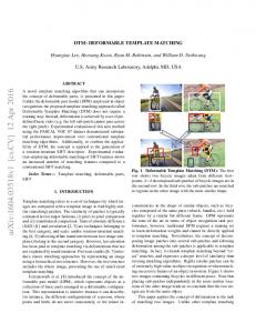
Fig. 1. An ensemble of manually segmented lung nodules taking various sizes and shapes.
for different orientations.
OA24
R 1o0
50
0
15D
200
250
Fig. 4. The gray level distrib ion for a circular template where a typical nodule histogram with exponential-like form is shown (e.g., [3][11][12]). 0
Fig. 2.
15 io¶ 20 25 DJi lawnc, Ifbro t~ hrnoduid denter mea
30
35
tue
com
40 in
The radial gray level distribution of the ensemble
of nodules in Fig 1. The bars show the variations from the
mean value.
II. PARAMETRIC TEMPLATE DESIGN
s - u
t
e
nodule models used in our study. In [13] we used the Gaussian symmetric model (see also Lee, 2001 [3]) to automatically generate the gray level
distribution of the nodules given the radius R and the histogram of typical nodule prototypes which were obtained by expert radiologists. The equations for the template gray level distribution are given by:
R(ln(qma,,,) -ln(qmjmi)