Neuro-Oncology
Expression of the neurogenic basic helix-loop-helix transcription factor NEUROG1 identifies a subgroup of medulloblastomas not expressing ATOH1 Ettore Salsano, Laura Croci, Emanuela Maderna, Linda Lupo, Bianca Pollo, Maria Teresa Giordana, G. Giacomo Consalez, and Gaetano Finocchiaro Unit of Experimental Neuro-Oncology (E.S., L.L., G.F.) and Division of Neuropathology (E.M., B.P.), “Carlo Besta” Neurological Institute Foundation, 20133 Milan; San Raffaele Scientific Institute, 20132 Milan (L.C., G.G.C.); and Department of Neurosciences, University of Turin, 10126 Turin (M.T.G.); Italy
To gain insight into the lineage of origin of medulloblastomas, the mRNA expression of NEUROG1, a gene encoding a proneural transcription factor transiently detected during nervous system development, was investigated in 27 human medulloblastomas characterized for mRNA expression of ATOH1, a marker of cerebellar granule precursors and corresponding medulloblastomas. Expression of Ngn1, the mouse homolog of NEUROG1, was also analyzed in the mouse cerebellar primordium. In addition, we studied mRNA expression of GLI1 as a marker of the SHH pathway activation, and nuclear b-catenin staining, b-catenin mutations, and mRNA expression of MYC as indicators of the WNT pathway status. In 15 cases, we also examined expression of OTX2, a transcription factor recently indicated as a positive marker of medulloblastomas originating from cerebellar granule precursors. The mRNA expression of NEUROG1 and Ngn1 was selectively found in medulloblastomas not expressing ATOH1 and in progenitors of the cerebellar ventricular zone, respectively. GLI1 transcript was expressed in medulloblastomas with ATOH1 transcript, whereas high levels of MYC transcript were Received September 13, 2006; accepted November 15, 2006. Address correspondence to Ettore Salsano, Unit of Experimental Neuro-Oncology, “Carlo Besta” Neurological Institute Foundation, via Celoria 11, 20133 Milan, Italy (
[email protected]).
Copyright Neuro-Oncology 2007 by the Society■for Neuro-Oncology 298 JU LY 2 0 0 6
unrelated to NEUROG1 or ATOH1 expression. No clear association between MYC overexpression and nuclear b-catenin staining was found. Finally, OTX2 mRNA was expressed in all medulloblastomas with NEUROG1 transcript, but also in a subset of these malignancies with ATOH1 transcript. These observations may help to define the lineage of origin of medulloblastomas, and support a role for ATOH1 and NEUROG1 in the classification of these malignancies. Neuro-Oncology 9, 298–307, 2007 (Posted to Neuro-Oncology [serial online], Doc. D06-00162, May 23, 2007. URL http://neuro-oncology .dukejournals.org; DOI: 10.1215/15228517-2007-014) Keywords: ATOH1 (MATH-1), cerebellum, GLI1, medulloblastoma, MYC, NEUROG1 (neurogenin-1), OTX2
M
edulloblastomas (MBs) are malignant tumors of the cerebellum. They commonly affect children, no more than 30% occurring in individuals older than 16 years, and are considered embryonic tumors since they originate from precursors present during cerebellar development. In particular, MBs may derive from cerebellar granule cell precursors (GCPs), which form a layer of proliferating committed cells in the subpial surface of the cerebellum during the fetal and the postnatal period. Neuroepithelial cells of the cerebellar ventricular zone (CVZ), displaced around the midline of
Salsano et al.: NEUROG1 is expressed in a medulloblastoma subgroup
the neuroectodermal tube, may constitute the other cells of origin of MBs.1 The idea that MBs have a double origin is molecularly supported by the expression of the neurotrophin receptor p75NTR, 2 the calcium-binding protein calbindin-D28k, 3 and the human atonal homolog 1 (ATOH1) gene in distinct subsets of these tumors. ATOH1 is a basic helix-loop-helix (bHLH) transcription factor (TF), and its mouse homolog, Math1, is exclusively expressed in proliferating GCPs during cerebellar development. Math1 expression seems necessary to uncommitted precursors of the CVZ to differentiate first as GCPs and finally as postmitotic, cerebellar granule cells.4 ATOH1 is expressed only in a subgroup of MBs, suggesting that not all these tumors originate from the GCPs. 5–7 Interestingly, MBs with ATOH1 transcript are commonly located in the hemispheres rather than in the vermis, as should be expected if they effectively originate from the GCPs. 8 Since MBs expressing ATOH1 afflict almost exclusively adult patients, GCPs may be the cells of origin of adult MBs. On the other hand, there is no molecular marker, functionally analogous to ATOH1, identifying MBs deriving from precursor cells of the CVZ. Indeed, calbindinD28k, which is considered a marker of MBs originating from the CVZ, is a calcium-binding protein, and it is also expressed in mature neurons. In contrast, neurogenin-1 (NEUROG1) is a proneural bHLH TF that functions as a determinant of neuronal identity and is expressed in a subset of interneuronal precursors of the murine spinal cord in a pattern complementary to Math1.9 Its role in cerebellar development, if any, was not yet investigated, but the expression of the human homolog, NEUROG1, was detected in a subset of human MBs and associated with a poor prognosis.10 Moreover, during development of the murine brain, Ngn1 is transiently expressed, and no transcript is detectable in adult nervous tissue, reinforcing its similarity to Math1.11 A distinct histogenesis may be associated with activation of different proliferative signaling pathways, as the SHH and WNT pathways. SHH is a glycoprotein secreted from Purkinje cells during development of the cerebellar cortex that interacts with the transmembrane PTCH receptor expressed by GCPs, stimulating their proliferation. PTCH mutations were found in sporadic MBs and in Gorlin syndrome, an autosomaldominant cancer syndrome characterized by the propensity to develop multiple neoplasms, including basal cell carcinomas and MBs. The final common effector of the SHH-dependent signaling pathway is TF GLI1.12 In murine models, Gli1 plays an important role in the formation of MBs,13 even though its alteration does not appear to be a general requirement.14 On the other hand, the role of the SHH pathway in the development of neuroepithelial cells of the CVZ is unclear.15,16 Since MB cells conserve many characters of their cells of origin, it is likely that the SHH pathway does not play a relevant role in MBs originating from progenitors of the CVZ. The WNT pathway, on the contrary, may play a pivotal role in the pathogenesis of this latter subgroup. Dysregulation of the WNT-dependent
signaling pathway has been involved in sporadic MBs and in Turcot syndrome, another autosomal-dominant cancer syndrome characterized by malignant tumors of the CNS associated with familial polyposis of the colon.17 Many WNT proteins and corresponding human frizzled-homolog receptors (FZDs) have been described, but much less is known about their role in the development of the human cerebellum. The WNT pathway is activated when WNT ligands bind to FZDs, causing stabilization of b-catenin and its translocation into the nucleus. In the nucleus, b-catenin activates transcription of a number of oncogenic targets, including MYC, although transcriptional activation by a b-catenin– independent pathway may be a mechanism for MYC overexpression in a subset of MBs.18 Here, we examined the mR NA expression of NEUROG1 in human MBs that were characterized for the expression of ATOH1, and the mRNA expression of Ngn1 in mouse embryonal cerebellum. In addition, we investigated the mRNA expression of GLI1 as marker of the SHH pathway activation, and nuclear b-catenin staining, b-catenin mutations, and/or mRNA expression of MYC as indicators of the WNT pathway activation. A link between the activation of these pathways and the mRNA expression of ATOH1 and NEUROG1 was assessed. Finally, in a smaller series of human MBs, we analyzed mRNA expression of OTX2, a TF that has been shown to be a further positive marker for MBs that originated from GCPs.19
Materials and Methods Patients Tumor specimens were from 27 patients (16 children and 11 adults) diagnosed with MB at the Division of Neuropathology, “C. Besta” Neurological Institute (Milan, Italy) or at the Department of Neurosciences, University of Turin (Turin, Italy). Sixteen tumors were Carnoy-fixed paraffin-embedded specimens archived in the Division of Neuropathology at the “C. Besta” Neurological Institute (n 5 9) or the University of Turin Department of Neurosciences (n 5 7). The remaining 11 tumors, all from the “C. Besta” Neurological Institute, were fresh-frozen specimens stored at –80°C. In 4 of the 11 fresh-frozen tumors, RNA was also obtained from corresponding Carnoy-fixed, paraffin-embedded tissues; 4 of the 16 Carnoy-fixed paraffin-embedded specimens were obtained in duplicate. Sixteen of these 27 MB samples (from eight children and eight adults) were used for b-catenin immunohistochemistry, and genomic DNA for b-catenin mutation analysis was also isolated from 15 of these 16 samples. RNA Extraction and cDNA Synthesis Total RNA from tumor specimens was isolated using TRIZOL as indicated by the producer (Invitrogen, San Giuliano Milanese, Italy). Briefly, 1 ml of TRIZOL was added to ~30 mg of fresh-frozen or deparaffinized Car-
Neuro-Oncology • july 2 0 0 7 299
Salsano et al.: NEUROG1 is expressed in a medulloblastoma subgroup
noy-fixed paraffin-embedded tumor sample in 1.5 ml tubes (Safelock-Plus, item 87377, International Pbi S.p.a., Milan, Italy), and homogenization was accomplished by manual grinding using a micropestle (item F199220001, Bel-Art Products Inc., Pequannock, NJ, USA). Tumor fragments from paraffin block were selected by light microscopy and drawn with a surgical blade. For deparaffination, they were incubated, and periodically vortexed, in 1 ml octane at room temperature for 20 min. After addition of 75 ml methanol to each tube and vortexing, samples were centrifuged at 7,000g for 2 min; subsequently, octane (upper layer) and methanol (lower layer) were removed with a fine-tip pipette, the pellet was air dried for a few minutes, and TRIZOL was added. Total RNA from human cerebellum was from a commercial pool of 24 adult individuals (16–70 years of age; Clontech, Saint-Germain-en-Laye, France). To degrade genomic DNA, RNA samples were treated with 2.5 ml of 10 U/ml DNase I (Roche Applied Science) in a 50 ml reaction volume, containing 10 ml of 53 Expand reverse transcriptase (RT) buffer (Roche Applied Science, Monza, Italy) and 2.5 ml of 20 U/ml SUPERase-In RNAse inhibitor (Ambion, Austin, TX, USA). They were successively cleaned up with the Qiagen RNeasy Mini Kit (Invitrogen), and RNA yield was measured by spectrophotometry. Two estimated micrograms of total RNA, when available, were reverse transcribed in a final volume of 40 ml containing 2 ml of 20 U/ml SUPERase-In RNAse inhibitor, 8 ml of 53 Expand RT buffer, 4 ml of 25 mM random hexanucleotides (Roche Applied Science), 4 ml of a dNTP mix (10 mM each), 4 ml of 100 mM dithiothreitol (Roche Applied Science), and 2 ml of 50 U/ml Moloney murine leukemia virus (M-MuLV) RT (Applied Biosystems, Monza, Italy). Reverse transcription was performed in an Applied Biosystems 9600 PCR System (65°C for 10 min, 10°C for 10 min, 42°C for 30 min, 95°C for 5 min, followed by 4°C soaking). M-MuLV was added after the first stage at 65°C. Total RNA isolated from each Carnoy-fixed paraffin-embedded specimen was usually reverse transcribed all at once. To check for the presence of residual genomic DNA after DNase treatment, 40 cycles of PCR with a 55°C annealing temperature were done on the cDNA of each sample, using a primer pair (5' primer sequence AGACCATGCAGGCTACCATC, 3' primer sequence GATGTTTTTGGCTCCTTTGC), which amplifies a 261-bp intron-containing sequence within the housekeeping hydroxymethylbilane synthase (HMBS) gene, and a 173-bp region within the corresponding HMBS transcript. HMBS was selected because no analogue pseudogene exists, unlike other housekeeping genes such as ACTB or GAPDH. Ten microliters of each sample was then analyzed with 2% agarose gel electrophoresis and ethidium bromide staining. Real-Time PCR for NEUROG1, GLI1, and MYC Real-time PCRs for the target genes—NEUROG1, GLI1, and MYC—were performed in duplicate on 22 MB cDNA samples (from 11 children and 11 adults) pre-
300 Neuro-Oncology • july 2 0 0 7
viously analyzed for the expression of ATOH1. In one child’s sample, RNA was sufficient only for NEUROG1 and MYC analysis. Results of real-time PCR were expressed as Ct values, where Ct is defined as the threshold PCR cycle number at which an amplified signal above the baseline is detected. To normalize the amount of total cDNA present in each reaction, we amplified the housekeeping 18S cDNA, which is assumed to be constant in both normal samples and MBs. We used the ABI GeneAmp 5700 cycler, the TaqMan Universal PCR Master Mix, and the NEUROG1, GLI1, MYC, and 18S Assays on Demand (assays Hs00246211_s1, Hs00171790_m1, Hs00153408_m1, and Hs99999901_s1, respectively), all from Applied Biosystems. Gene expression changes were assessed with the 2 –Ct method, according to the protocol of the manufacturer (http://docs.applied biosystems.com/pebiodocs/04333458.pdf). Briefly, for any sample, we first calculated the relative expression levels of NEUROG1, GLI1, and MYC compared to that of the housekeeping 18S using the formula 2 –Ct , where C t corresponds to the mean of the duplicate C t values for the target gene minus the mean of the duplicate C t values for 18S. Then, these relative expression values calculated for each target gene in any sample were divided by the relative expression values calculated for the corresponding target gene in a sample called “calibrator,” which is chosen to represent the 13 expression of this gene. Hence, all quantities were expressed as n-fold relative to the calibrator. Standard PCR for NEUROG1, ATOH1, and OTX2 Standard PCRs for NEUROG1, ATOH1, and OTX2 were performed on 15 MBs (from seven children and eight adults), five of which were tumor samples not previously investigated for ATOH1 expression. mRNA expression of OTX2 was directly investigated using standard RT-PCR because it was proved to be expressed in an allor-nothing manner in a large panel of MBs.19 We used the primer pairs 5'-AGCTCACCAAAATCGAGACG-3' (forward) and 5'-GGGCTACTGGGGTCAGAGAG-3' (reverse) for NEUROG1, 5'-CCGCCCAGTATTTG C TAC AT-3' (for wa rd) and 5'- C AT TC ACC TGTTTGCTGGAA-3' (reverse) for ATOH1, and 5'AGAGGAGGTGGCACTGAAAA-3' (forward) and 5'-ATTGGCCACTTGTTCCACTC-3' (reverse) for OTX2. These primers gave products of 226 bp, 234 bp, and 188 bp, respectively. Primers for OTX2 cross the intron sequence between exons 4 and 5, thus amplifying both variants of the OTX2 transcript. NEUROG1 and ATOH1 are monoexonic genes; thus, genomic DNA, if present, is amplified with all primer pairs we synthesize. Hence, to avoid “noise” in gene expression studies, genomic DNA was completely removed with DNase treatment, as previously specified. Primer pairs were synthesized with Primer3 software (http://frodo.wi.mit .edu/cgi-bin/primer3/primer3_www.cgi). All PCRs were carried out in a final volume of 40 ml, containing 4 ml GeneAmp 103 PCR buffer (Applied Biosystems), 5 ml of a dNTP mix (2 mM each), 2 ml of 10 mM concentration of each primer, and 0.4 ml of 5 U/ml AmpliTaq
Salsano et al.: NEUROG1 is expressed in a medulloblastoma subgroup
Gold (Applied Biosystems). The PCR for the detection of NEUROG1 was optimized with 2 ml of dimethyl sulfoxide using genomic DNA as positive control. The PCR program consisted of initial denaturation at 95°C for 10 min, followed by 40 cycles of 95°C for 30 s, 55°C for 30 s, 72°C for 30 s, and a final extension step at 72°C for 10 min. Ten microliters of each PCR product was then analyzed with agarose gel electrophoresis and ethidium bromide staining. Expression of NEUROG1, ATOH1, and OTX2 was scored as positive or negative.
b-Catenin Immunohistochemistry and Sequencing For b-catenin immunohistochemistry, Carnoy-fixed paraffin-embedded samples were sectioned at a thickness of 4 mm, mounted on slides, and deparaffinized in xylene. Endogenous peroxidase activity was blocked in 10% hydrogen peroxide. Then, the slides were immersed in a 3 M guanidine thiocyanate solution for 20 min, rinsed in Tris buffer, and incubated for 20 min with normal goat serum, before being exposed to the mouse anti–b-catenin IgG1 (1:50, BD Transduction Laboratories, Milan, Italy) in a moist chamber overnight. They were subsequently washed three times with Tris buffer and incubated with the secondary antibody conjugated to a horseradish-peroxidase–labeled polymer (EnVision Detection System, DAKO, Milan, Italy), at room temperature for 1 h. Visualization as a brown-colored precipitate at the antigen site was performed using 3,3'-diaminobenzidine (DAKO) as a chromogen at room temperature for 5–10 min. Finally, sections were counterstained with hematoxylin and observed under light microscopy. For CTNNB1 (encoding b-catenin) sequencing, DNA was simultaneously extracted by TRIZOL reagent from samples used for RNA isolation, as indicated by the producer. A 225-bp fragment of exon 3 was amplified by 35 cycles of PCR with a 55°C annealing temperature (5' primer sequence TGATTTGATGGAGTTGGACA, 3' primer sequence GCTACTTGTTCTTGAGTGAAG), 20 and PCR products were sequenced on both strands using a capillary sequencer (ABI PRISM 3100 Genetic Analyzer, Applied Biosystems). In Situ Hybridization for Ngn1 Transcript and Immunohistochemistry for Ki-67 Wild-type mouse embryos at embryonic stage 12.5 (E12.5) were fixed overnight by immersion in 4% paraformaldehyde, rinsed three times in 13 phosphatebuffered saline, cryoprotected in 30% sucrose overnight, embedded in optical cutting temperature compound (OCT; BioOptica, Milan, Italy), and stored at –80°C before sectioning on a cryotome (20 mm). For in situ hybridization, a digoxygenin-labeled riboprobe was transcribed from a plasmid containing Ngn1 cDNA. The experiment was performed as described by Pringle. 21 For immunohistochemistry, cryosections were processed for high-temperature antigen retrieval and immunostained with the rabbit polyclonal anti–Ki-67 (1:800; Novocastra Laboratories, Newcastle-upon-Tyne, UK), as recom-
mended (ABC Elite kit, Vector Laboratories, Burlingame, CA, USA).
Results mRNA Expression of NEUROG1 By real-time PCR, NEUROG1 expression was high (mean C t value 5 26 6 2 SD), in 10 of 10 ATOH1 mRNA–negative MBs but very low or hardly measurable (mean C t value 5 36 6 2 SD) in 12 of 12 ATOH1 mRNA–positive MBs (p , 0.0001, Fisher’s exact test; Fig. 1). No NEUROG1 expression was found in normal cerebellum (mean C t value . 40), and all NEUROG1 expression values (18S normalized) were expressed as n-fold relative to the tumor sample with the lowest expression, chosen as a calibrator. All ATOH1 mRNA– negative MBs were from children, whereas all except one ATOH1 mRNA–positive MB were from adults. In this panel, all pediatric MBs were located in the midline, while all adult MBs but one were located in a cerebellar hemisphere. By standard RT-PCR, we confirmed realtime PCR results for NEUROG1 and ATOH1 expression in 10 of 27 MBs, and we found analogous results in five new MBs (Fig. 2). In this novel panel of samples, we also found one adult NEUROG1 mRNA–positive MB that was located in the midline. mRNA Expression of GLI1 GLI1 was found to be expressed (mean Ct value 5 32 6 2 SD) in all (12 of 12) MBs expressing ATOH1, while no or hardly measurable GLI1 transcript was detected in nine of nine ATOH1 mRNA–negative MBs (mean Ct values . 40 in all MBs except two, where they were around 36–37 cycles; p 5 0.0000034, Fisher’s exact test). In GLI1 mRNA–positive MBs, GLI1 expression quantities (18S normalized) were expressed as n-fold relative to the normal cerebellum, which expressed GLI1 transcript at very low levels. mRNA Expression of MYC Reliable data on MYC expression were available for 20 of 22 MBs (from 10 children and 10 adults), since in two tumors the C t value of the housekeeping 18S was too high to be considered acceptable (mean Ct value . 20), and no RNA was available for further reverse transcriptions. MYC expression, normalized to 18S, varied from 0.31- to 44.63-fold relative to normal cerebellum, which expressed traces of MYC transcript and was chosen as calibrator. The values of MYC expression were categorized as high if they were above 6.34, the upper quartile limit of expression distribution. Analogous results were obtained using as a threshold the mean value of 9.19. Five of 20 tumors (25%) showed high MYC expression (mean C t value 5 29 6 2 SD), whereas the remaining 15 tumors (75%) had lower MYC expression (mean C t value 5 34 6 2 SD). In 4 of these 15 tumors, mRNA expression of MYC was lower than in normal cerebel-
Neuro-Oncology • july 2 0 0 7 301
Salsano et al.: NEUROG1 is expressed in a medulloblastoma subgroup
Fig. 1. Real-time reverse transcriptase PCR analysis of NEUROG1, GLI1, and MYC mRNA expression in 22 medulloblastomas. Data for ATOH1, reported previously,5 are shown to facilitate comparisons. NEUROG1 mRNA was detected in tumor samples without ATOH1 transcripts (p , 0.0001), while GLI1 mRNA expression overlapped that of ATOH1 (p , 0.0001). High levels of MYC mRNA appeared more common in the group of pediatric NEUROG1 mRNA–positive medulloblastomas, though the difference was not statistically significant (p 5 0.3). 18S was used as housekeeping gene. Boxes represent age (gray box, ,18 years; white box, >18 years), tumor location (gray box, vermian; white box, hemispheric), and histology (gray box, classic; white box, desmoplastic) for each patient; n/a, not available. Relative expression values of the NEUROG1 and ATOH1 transcripts are 31,000 and 3100, respectively.
lum, while in the remaining 11 it was, on average, threefold higher than in normal cerebellum. High levels of MYC transcripts were more common in pediatric, NEUROG1 mRNA–positive MBs (4 of 10 vs. 1 of 10), but the difference was not statistically significant (p 5 0.3, two-tailed Fisher’s exact test). No relevant difference was found in the analysis of mRNA expression when we compared data from four fresh-frozen samples and their corresponding Carnoy-fixed paraffin-embedded counterparts (data not shown). mRNA Expression of OTX2 Using RT-PCR, the OTX2 transcript was detected in 10 of 15 MBs: six were vermian, NEUROG1 mRNA– positive tumors, whereas the remaining four were hemispheric, ATOH1 mRNA–positive MBs. All OTX2 mRNA–negative MBs (n 5 5) expressed ATOH1. The OTX2 transcript was also detected in normal cerebellum.
302 Neuro-Oncology • july 2 0 0 7
Nuclear b-Catenin Staining and CTNNB1 Mutations Widespread cytoplasmic/nuclear b-catenin staining was found in one (12.5%) of eight childhood MBs and in none of eight adult MBs. Scattered single cells with a cytoplasmic/nuclear b-catenin staining were found in two (25%) of eight childhood MBs and in three (37.5%) of eight adult MBs. In the remaining 10 MBs (five children and five adults), b-catenin immunoreactivity was limited to the cytoplasm in eight and completely absent in one. A b-catenin missense point mutation at codon 33 (TCT ➝ GCT; SER ➝ ALA) was identified in the tumor with widespread cytoplasmic/nuclear b-catenin immunopositivity (Fig. 3). No significant association was found between MYC overexpression and nuclear b-catenin immunoreactivity. However, the sole MB with mutant b-catenin gene and strong nuclear b-catenin immunoreactivity expressed MYC at high levels.
Salsano et al.: NEUROG1 is expressed in a medulloblastoma subgroup
Fig. 2. Standard reverse transcriptase PCR analysis of NEUROG1, ATOH1, and OTX2 mRNA expression in medulloblastomas. A paradigmatic example of this analysis is shown here for 4 of 15 tumors examined. OTX2 mRNA was detected in all NEUROG1 mRNA– positive medulloblastomas and in only a part of those expressing ATOH1. Gray or white boxes are as in Fig. 1. Abbreviations: CA, FL, GD, and MM, patients’ initials (family name, given name); VI, DNA Molecular Weight Marker VI (Roche Applied Science).
mRNA Expression of Ngn1 in the Mouse Cerebellar Primordium By in situ hybridization, we have analyzed the distribution of the Ngn1 transcript in the mouse cerebellar primordium at 12.5 days of embryonic development (E12.5). At this stage, the CVZ hosts mitotic progenitors fated to give rise to radial glia and GABAergic but not glutamatergic neurons. In fact, cerebellar granules and other glutamatergic neurons originate in the upper rhombic lip (see Discussion). As shown in Fig. 4, the Ngn1 transcript occupies the CVZ, in a territory that
Fig. 3. b-catenin immunohistochemistry and b-catenin sequencing: strong combined cytoplasmic and nuclear staining (A), scattered single cells with cytoplasmic/nuclear b-catenin reactivity (B), and b-catenin immunoreactivity limited to cytoplasm (C) in three human medulloblastomas. Arrows indicate b-catenin–immuno positive nuclei. Direct DNA sequencing (D) unveils a b-catenin mutation at codon 33 (TCT ➝ GCT; SER ➝ ALA) detected in the classic medulloblastoma shown in A. Magnification: 40.
Neuro-Oncology • july 2 0 0 7 303
Salsano et al.: NEUROG1 is expressed in a medulloblastoma subgroup
Fig. 4. Neurogenin-1 expression in the embryonic cerebellar primordium: sagittal and frontal sections of E12.5 mouse cerebella. (A, D, G) Sagittal sections: anterior shown on the left; from medial (A) to lateral (G). (B, C, E, F, H, I) Frontal sections: hemicerebella, midline on the left, from anterior (B, C) to posterior (H, I). (A, B, D, E, G, H) Nonradioactive in situ hybridization using an Ngn1 riboprobe. (C, F, I) Immunostaining for Ki-67. Ngn1 is expressed discontinuously in the ventricular zone (arrows in A, B, D, E). In comparison, Ki-67, a global cell proliferation marker, is expressed throughout the ventricular zone and into the rhombic lip. Ngn1 expression excludes the rhombic lip (arrowheads in B, E, H), which is the source of cerebellar glutamatergic neurons. Abbreviations: chp, choroid plexus; hb, hindbrain; mes, mesencephalon; rl, rhombic lip; vz, ventricular zone. Scale bar 5 100 mm.
overlaps partially with the distribution of the cell proliferation marker Ki-67. Ngn1 expression is split anteriorly in two discontinuous domains, a medial one and a more lateral one, whereas a single domain can be detected in the posterior half of the cerebellar primordium. Ngn1, however, is excluded from the rhombic lip.
Discussion The precise cell of origin of MBs is still a matter of debate.1,2 Two types of mitotically active, neuronal restricted precursors have been proposed: (1) progenitor cells of the external germinal layer (EGL), that is, GCPs, giving rise to glutamatergic cerebellar granule cells; and (2) progenitor cells of the CVZ that generate GABAergic neurons, including Purkinje cells. In previous work, expression of ATOH1, the earliest known gene specifically expressed in the GCPs, was detected in a subgroup of MBs. 5 Tumors are characterized by expression of stem/progenitor cell-like markers because they probably originate from a stem/progenitor cell that becomes aber-
304 Neuro-Oncology • july 2 0 0 7
rantly activated. Hence, considering ATOH1 expression in the normal cell counterpart, we proposed that MBs with ATOH1 transcript arise from GCPs. In the present work, we found that NEUROG1, another bHLH TF implicated in neuronal determination and differentiation, is expressed in the subgroup of MBs without ATOH1. To date, no information has been provided on the role of NEUROG1 in the context of cerebellar neurogenesis. During cerebellar development, glutamatergic and GABA- ergic neurons are produced from separate progenitor compartments. Our results in mouse cerebellar primordium suggest that Ngn1 mRNA labels a separate lineage, that colocalizes with the CVZ from which GABAergic neuron progenitors, including Purkinje cells, originate. Notably, in the lower rhombic lip, a germinal zone productive of mossy and climbing fiber neurons, Math1 identifies the primordium for mossy fiber neurons, which project to cerebellar granule cells, while Ngn1 identifies the primordium for climbing fiber neurons, which project to Purkinje cells. 22 This observation may further
Salsano et al.: NEUROG1 is expressed in a medulloblastoma subgroup
suggest that NEUROG1 is expressed by progenitors of the CVZ, from which Purkinje cells derive, because the same TF may define specific subsets of afferent and target neurons, such as climbing fibers and Purkinje cells. 23 Based on these data, we propose that mRNA expression of NEUROG1 is a positive marker of MBs deriving from progenitor cells of the CVZ. These MBs have origin in the midline, arise during childhood, and are usually the classic type. Obviously, the presence of NEUROG1 in MBs not expressing ATOH1 does not exclude an origin from GCPs with absolute certainty. We cannot rule out that the expression of NEUROG1, just as of other TFs, may be an epiphenomenon of malignant transformation, rather than a marker of a particular lineage of origin. Recent data from de Haas et al.19 suggest that hemispheric and some of vermian MBs originate from GCPs. Vermian MBs presented a strong positivity for OTX2, which was expressed in EGL cells during development, but not in cells of the ventricular zone. Here, we found that vermian MBs always express OTX2, while a subset of hemispheric MBs do not. This may be due to origin from GCPs of different regions, since OTX2 expression during human cerebellar development may present an anteroposterior pattern, with the highest levels of OTX2 expression in the most posterior regions of the cerebellum. However, it remains unclear why ATOH1, a marker of GCPs of any region, is not always coexpressed with OTX2. One hypothesis is that OTX2 is not a selective marker of GCPs, as also suggested by de Haas et al.19 Indeed, they detected OTX2 expression both in Purkinje cells, the progeny of the cerebellar ventricular neuroepithelium, and in the human MB D283 Med cell line, which highly expresses calbindin-D28k, a positive marker of Purkinje cells and their precursors. Strikingly, D283 Med cells also express NEUROG1.10 Thus, MBs with NEUROG1 transcripts might present a differentiation status of Purkinje-like cells and express OTX2, while MBs with ATOH1 transcripts might present a differentiation status of granule-like cells and express OTX2 only if they originate in the most posterior regions of the cerebellum. Precursor markers such as calbindin-D28k are much less attractive than NEUROG1 or ATOH1 in RT-PCR studies, since they are not transiently expressed in precursor cells but are also found in their mature progeny. Thus, their detection in a tumor sample using RT-PCR may be due to normal cells intermingled with the malignant population. Our results suggest that expression of OTX2 is also retained in adult human cerebellum, as observed by others in the rat. 24 In contrast, de Haas et al.19 found that OTX2 expression was undetectable in adult human cerebella; thus, further studies are necessary to exclude that OTX2 expression is not due to sequestered normal cells. The expression pattern of GLI1, a marker of SHH signaling activity, overlaps with that of ATOH1 transcript, as previously noted by Lee et al. 25 This observation further supports the hypothesis that MBs expressing ATOH1 transcripts derive from GCPs. 26 In fact, during the development of the mouse cerebellum, Gli1 expression is restricted to proliferating GCPs. Since GLI1 is
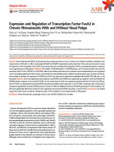
Fig. 5. Schematic representation of cerebellar development showing the sites of origin of medulloblastoma (sagittal midline view). Progenitor cells located in the ventricular zone (VZ) migrate radially to give rise to GABAergic neurons, including Purkinje cells, while progenitor cells located in the rhombic lip (RL) migrate tangentially to the subpial surface of the cerebellum, populate the external granular layer (EGL), and give rise to cerebellar granule neurons. ATOH1 and GLI1 are markers of the cerebellar granule neuron precursors (white circles) and corresponding medulloblastomas, while NEUROG1 should be a marker of progenitors of the cerebellar ventricular zone (black circles) and corresponding medulloblastomas (adapted from Hamilton WJ, Mossman HW. Embriologia Umana. Padova: Piccin Editore; 1977:463).
a target of the SHH-dependent pathway, it is plausible that this pathway is involved only in the formation of MBs originating from the GCPs. In contrast, MYCN, another TF regarded as a target of hedgehog signaling in GCPs and corresponding MBs, may be up-regulated even in MBs not expressing ATOH1 and GLI127 (E. Salsano et al., unpublished data), in agreement with its essential role also for the proliferation of progenitor cells of the CVZ. 28 It should be stressed that MBs expressing ATOH1 and GLI1 typically arise in adulthood from GCPs of the EGL, are of the desmoplastic type, and may represent a distinct minority of all MBs; 29 in contrast, most MBs arising in children, probably from CVZ precursors, are of the classic type, occur in the midline, and may be biologically characterized by the expression of NEUROG1 and by the absence of ATOH1 and GLI1 transcripts. A diagram of the sites of origin of MBs (CVZ and EGL) and the corresponding TFs is shown in Fig. 5. MYC is a TF allowing cells to enter the proliferative state, although in rapidly dividing cells it may be expressed at high or low levels. 30 Our data suggest that overexpression of MYC transcript occurs more commonly in pediatric MBs, which normally express NEUROG1 transcripts. A similar observation is present in raw data published by Eberhart et al., 31 who found that five of five MBs with stronger MYC expression were from patients younger than 18 years. However, this association is not statistically significant. Overexpression of MYC likely reflects alterations in its transcriptional regulation because the low frequency of MYC amplification in MBs clearly suggests that mechanisms other than
Neuro-Oncology • july 2 0 0 7 305
Salsano et al.: NEUROG1 is expressed in a medulloblastoma subgroup
gene amplification lead to MYC overexpression. 32 Transcription of MYC may be under control of the canonical WNT pathway through the b-catenin nuclear translocation, a marker of WNT signaling activity. However, our data do not support this hypothesis. In fact, although the only MBs with a strong nuclear b-catenin staining and a stabilizing mutation of CTNNB1 (encoding b-catenin) express MYC at high levels, no clear correlation was found between elevated levels of MYC transcript and nuclear b-catenin staining in the remaining MBs. Accordingly, Siu et al.18 found that in MB cell lines, specific regions of the MYC promoter, independent of b-catenin binding sites, were responsible for its activation. Hence, at least in a subset of MBs with high MYC transcript levels, a b-catenin–independent pathway is the candidate mechanism for transcriptional activation of MYC. This is also in agreement with suggestions that in MBs, nuclear accumulation of b-catenin is a marker of favorable outcome, 33 while elevated MYC expression is associated with aggressive disease. 32 Together, these data suggest that high levels of MYC are a poor marker of WNT pathway status and do not clearly correlate with NEUROG1 or ATOH1 expression. We believe that our observations help to define the lineage of origin of MBs and support a classification for these malignancies based on molecular markers involved in cerebellar development. However, limitations must be considered. First, while we have studied pediatric MBs arising in the vermis, we do not know if expression patterns of ATOH1 and NEUROG1 are similar in pediatric MBs arising from cerebellar hemispheres (approx. 25% of all pediatric MBs). Second, we analyzed only one MB in a child younger than three years; based on our data, we cannot confirm that very precocious MBs can be dichotomized by the expression of NEUROG1 and ATOH1, also because GCPs persist in the human cerebellum for 12–15 months after birth, and ATOH1 expression may be due to the presence of GCPs trapped among the tumor cells. Our data may also have implications for normal development. The molecules that regulate neuroepithelial cell differentiation and determine the cell fate in
the mammalian cerebellum are poorly defined, except for the GCPs, whose genesis in mice is dependent on Math1.4 Math1 and Neurog1 transcripts have a distinct function in the formation of neuronal subpopulations in the developing spinal tube of mice;9 analogously, we have found that their human homologs, ATOH1 and NEUROG1, are expressed in distinct subgroups of MBs that may arise from GCPs and progenitors of the CVZ, respectively. Since MB cells conserve molecular features of their cells of origin, we hypothesize that during cerebellar development NEUROG1 functions in specifying neuronal subtypes different from GCPs, as suggested by the Ngn1 expression pattern in the mouse cerebellar primordium. In particular, neural precursors of the dorsal neuroepithelium may be committed to become neuronal precursors by the expression of NEUROG1. 34 Successively, cells immediately adjacent to the roof plate of the fourth ventricle may be induced to express ATOH1 by bone morphogenetic protein signals, thus acquiring the rhombic lip identity. 35 ATOH1 may inhibit NEUROG1 expression and direct these cells to become GCPs. In contrast, the cells away from the roof plate may continue to express NEUROG1. This proneural gene, alone or in combination with other TFs, may orchestrate the genesis of other interneurons of the cerebellum. Specifically, the property of NEUROG1 to differentiate neural stem cells as GABAergic neurons when transplanted into the cerebral cortex36 is consistent with the idea that NEUROG1 is necessary for generating GABAergic interneurons (i.e., stellate, basket, Golgi, or Purkinje neurons) also in the cerebellar anlage.
Acknowledgments We thank the reviewers whose suggestions significantly improved the manuscript; Marica Eoli and Rossella Galli for help and suggestions during the study; and the personnel of the Tumor Registry of Istituto Neurologico Besta for sharing information on patients examined in this study. This work has been supported by funds from Fondazione Pierfranco e Luisa Mariani ONLUS Neurologia Infantile.
References 1. Katsetos CD, Del Valle L, Legido A, de Chadarevian JP, Perentes E,
4. Ben-Arie N, Bellen HJ, Armstrong DL, et al. Math1 is essential for genesis of cerebellar granule neurons. Nature. 1997;390:169–172.
Mork SJ. On the neuronal/neuroblastic nature of medulloblastomas: a tribute to Pio del Rio Hortega and Moises Polak. Acta Neuropathol
5. Salsano E, Pollo B, Eoli M, Giordana MT, Finocchiaro G. Expression
(Berl). 2003;105:1–13.
of MATH1, a marker of cerebellar granule cell progenitors, identifies different medulloblastoma sub-types. Neurosci Lett. 2004;370:180–
2. Buhren J, Christoph AH, Buslei R, Albrecht S, Wiestler OD, Pietsch T. Expression of the neurotrophin receptor p75NTR in medulloblastomas is correlated with distinct histological and clinical features: evidence
185. 6. Lee A, Kessler JD, Read TA, et al. Isolation of neural stem cells from
for a medulloblastoma subtype derived from the external granule cell layer. J Neuropathol Exp Neurol. 2000;59:229–240. 3. Katsetos CD, Herman MM, Krishna L, et al. Calbindin-D28k in subsets of medulloblastomas and in the human medulloblastoma cell line D283 Med. Arch Pathol Lab Med. 1995;119:734–743.
306 Neuro-Oncology • july 2 0 0 7
the postnatal cerebellum. Nat Neurosci. 2005;8:723–729. 7.
Schuller U, Koch A, Hartmann W, et al. Subtype-specific expression and genetic alterations of the chemokine receptor gene CXCR4 in medulloblastomas. Int J Cancer. 2005;117:82–89.
Salsano et al.: NEUROG1 is expressed in a medulloblastoma subgroup
8. Yachnis AT, Rorke LB, Trojanowski JQ. Cerebellar dysplasias in
23. Logan C, Wingate RJ, McKay IJ, Lumsden A. Tlx-1 and Tlx-3 homeo-
humans: development and possible relationship to glial and primitive
box gene expression in cranial sensory ganglia and hindbrain of
neuroectodermal tumors of the cerebellar vermis. J Neuropathol Exp
the chick embryo: markers of patterned connectivity. J Neurosci.
Neurol. 1994;53:61–71. 9. Gowan K, Helms AW, Hunsaker TL, et al. Crossinhibitory activities of
1998;18:5389–5402. 24. Frantz GD, Weimann JM, Levin ME, McConnell SK. Otx1 and Otx2
Ngn1 and Math1 allow specification of distinct dorsal interneurons.
define layers and regions in developing cerebral cortex and cerebel-
Neuron. 2001;31:219–232.
lum. J Neurosci. 1994;14:5725–5740.
10. Rostomily RC, Bermingham-McDonogh O, Berger MS, et al. Expression of neurogenic basic helix-loop-helix genes in primitive neuro ectodermal tumors. Cancer Res. 1997;57:3526–3531. 11. Ma Q, Kintner C, Anderson DJ. Identification of neurogenin, a vertebrate neuronal determination gene. Cell. 1996;87:43–52. 12. Stecca B, Ruiz i Altaba A. Brain as a paradigm of organ growth: Hedge-
25. Lee Y, Miller HL, Jensen P, et al. A molecular fingerprint for medulloblastoma. Cancer Res. 2003;63:5428–5437. 26. Corrales JD, Rocco GL, Blaess S, Guo Q, Joyner AL. Spatial pattern of sonic hedgehog signaling through Gli genes during cerebellum development. Development. 2004;131:5581–5590. 27. Pomeroy SL, Tamayo P, Gaasenbeek M, et al. Prediction of central
hog-Gli signaling in neural stem cells and brain tumors. J Neurobiol.
nervous system embryonal tumour outcome based on gene expres-
2005;64:476–490.
sion. Nature. 2002;415:436–442.
13. Kimura H, Stephen D, Joyner A, Curran T. Gli1 is important for medul-
28. Knoepfler PS, Cheng PF, Eisenman RN. N-myc is essential during neuro
loblastoma formation in Ptc1+/- mice. Oncogene. 2005;24:4026–
genesis for the rapid expansion of progenitor cell populations and the
4036. 14. Weiner HL, Bakst R, Hurlbert MS, et al. Induction of medulloblastomas in mice by sonic hedgehog, independent of Gli1. Cancer Res. 2002;62:6385–6389. 15. Dahmane N, Ruiz i Altaba A. Sonic hedgehog regulates the growth and patterning of the cerebellum. Development. 1999;126:3089– 3100. 16. Cayuso J, Ulloa F, Cox B, Briscole J, Marti E. The Sonic hedgehog pathway independently controls the patterning, proliferation and sur-
inhibition of neuronal differentiation. Genes Dev. 2002;16:2699– 2712. 29. Giordana MT, D’Agostino C, Pollo B, et al. Anaplasia is rare and does not influence prognosis in adult medulloblastoma. J Neuropathol Exp Neurol. 2005;64:869–874. 30. Ruppert C, Goldowitz D, Wille W. Proto-oncogene c-myc is expressed in cerebellar neurons at different developmental stages. EMBO J. 1986;5:1897–1901. 31. Eberhart CG, Kratz J, Wang Y, et al. Histopathological and molecu-
vival of neuroepithelial cells by regulating Gli activity. Development.
lar prognostic markers in medulloblastoma: c-myc, N-myc, TrkC, and
2006;133:517–28.
anaplasia. J Neuropathol Exp Neurol. 2004;63:441–449.
17. Reya T, Clevers H. Wnt signalling in stem cells and cancer. Nature. 2005;434:843–850. 18. Siu IM, Lal A, Blankenship JR, Aldosari N, Riggins GJ. c-Myc promoter activation in medulloblastoma. Cancer Res. 2003;63:4773–4776. 19. de Haas T, Oussoren E, Grajkowska W, et al. OTX1 and OTX2 expres-
32. Herms J, Neidt I, Luscher B, et al. C-MYC expression in medulloblastoma and its prognostic value. Int J Cancer. 2000;89:395–402. 33. Ellison DW, Onilude OE, Lindsey JC, et al.; United Kingdom Children’s Cancer Study Group Brain Tumour Committee. Beta-catenin status predicts a favorable outcome in childhood medulloblastoma:
sion correlates with the clinicopathologic classification of medullo-
the United Kingdom Children’s Cancer Study Group Brain Tumour
blastomas. J Neuropathol Exp Neurol. 2006;65:176–186.
Committee. J Clin Oncol. 2005;23:7951–7957.
20. Eberhart CG, Tihan T, Burger PC. Nuclear localization and mutation
34. Sun Y, Nadal-Vicens M, Misono S, et al. Neurogenin promotes neuro-
of beta-catenin in medulloblastomas. J Neuropathol Exp Neurol.
genesis and inhibits glial differentiation by independent mechanisms.
2000;59:333–337.
Cell. 2001;104:365–376.
21. Pringle N. Double in situ hybridization. London: Wolfson Institute for
35. Alder J, Lee KJ, Jessell TM, Hatten ME. Generation of cerebellar gran-
Biomedical Research, University College London. Available at: http://
ule neurons in vivo by transplantation of BMP-treated neural progeni-
www.ucl.ac.uk/~ucbzwdr/double%20in%20situ%20protocol.htm. Accessed September 2, 2006. 22. Landsberg RL, Awatramani RB, Hunter NL, et al. Hindbrain rhombic lip is comprised of discrete progenitor cell populations allocated by
tor cells. Nat Neurosci. 1999;2:535–540. 36. Muramatsu D, Sato Y, Hishiyama S, Miyamoto Y, Hisatsune T. Transplantation of GABAergic neurons into adult mouse neocortex. Exp Neurol. 2005;194:1–11.
Pax6. Neuron. 2005;48:933–947.
Neuro-Oncology • july 2 0 0 7 307