JOURNAL OF VIROLOGY, Dec. 2001, p. 11948–11960 0022-538X/01/$04.00⫹0 DOI: 10.1128/JVI.75.24.11948–11960.2001 Copyright © 2001, American Society for Microbiology. All Rights Reserved.
Vol. 75, No. 24
Functional Mapping of the DNA Binding Domain of Bovine Papillomavirus E1 Protein MICHAEL WEST, DAVID FLANERY, KELLY WOYTEK, DHANDAPANI RANGASAMY,† AND VAN G. WILSON* Department of Medical Microbiology and Immunology, Texas A&M University System Health Science Center, College Station, Texas 77843-1114 Received 25 May 2001/Accepted 5 September 2001
Bovine papillomavirus type 1 (BPV-1) requires viral proteins E1 and E2 for efficient DNA replication in host cells. E1 functions at the BPV origin as an ATP-dependent helicase during replication initiation. Previously, we used alanine mutagenesis to identify two hydrophilic regions of the E1 DNA binding domain (E1DBD), HR1 (E1179–191) and HR3 (E1241–252), which are critical for sequence-specific recognition of the papillomavirus origin. Based on sequence and structure, these regions are similar in spacing and location to DNA binding regions A and B2 of T antigen, the DNA replication initiator of simian virus 40 (SV40). HR1 and A are both part of extended loops which are supported by residues from the HR3 and B2 ␣-helices. Both elements contain basic residues which may contact DNA, although lack of cocrystal structures for both E1 and T antigen make this uncertain. To better understand how E1 interacts with origin DNA, we used random mutagenesis and a yeast one-hybrid screen to select mutations of the E1DBD which disrupt sequence-specific DNA interactions. From the screen we selected seven single point mutants and one double point mutant (F175S, N184Y/K288R, D185G, V193M, F237L, K241E, R243K, and V246D) for in vitro analysis. All mutants tested in electrophoretic mobility shift assays displayed reduced sequence-specific DNA binding compared to the wild-type E1DBD. Mutants D185G, F237L, and R243K were rescued in vitro for DNA binding by the replication enhancer protein E2. We also tested the eight mutations in full-length E1 for the ability to support DNA replication in Chinese hamster ovary cells. Only mutants D185G, F237L, and R243K supported significant DNA replication in vivo which highlights the importance of E1DBD-E2 interactions for papillomavirus DNA replication. Based on the specific point mutations examined, we also assigned putative roles to individual residues in DNA binding. Finally, we discuss sequence and spacing similarities between E1 HR1 and HR3 and short regions of two other DNA tumor virus origin-binding proteins, SV40 T antigen and Epstein-Barr virus nuclear antigen 1 (EBNA1). We propose that all three proteins use a similar DNA recognition mechanism consisting of a loop structure which makes base-specific contacts (HR1) and a helix which primarily contacts the DNA backbone (HR3). Papillomaviruses are members of the papovavirus family and comprise both human and animal strains. In addition to causing common cutaneous warts, papillomaviruses can induce various skin and mucosal lesions (35, 36, 39). Some of these lesions may progress to malignant carcinomas, depending on the particular viral strain involved as well as environmental factors. Malignancies which are highly associated with papillomavirus infection include laryngeal, cervical, and other anogenital cancers, and it is noteworthy that human papillomavirus (HPV) types 16, 18, 31, 33, and 45 pose an especially high risk for females, as they are estimated to be present in at least 90% of cervical cancers (17, 27). Bovine papillomavirus (BPV) has been used as a model for studying the biology of papillomavirus replication, particularly the DNA replication components. Upon entry of a host cell by wounding or abrasion, BPV replicates its small 8-kb genome to a multicopy episome which is maintained at steady-state levels of up to several hundred copies per cell (14). After a steady* Corresponding author. Mailing address: Department of Medical Microbiology and Immunology, Texas A&M University System Health Science Center, College Station, TX 77843-1114. Phone: (979) 8455207. Fax: 979-845-3479. E-mail:
[email protected]. † Present address: Division of Molecular Biology, John Curtin School of Medical Research, ANU Campus, Canberra City, ACT 2613, Australia.
state level of replication has been achieved, the viral DNA then replicates approximately once during each host cell cycle and is likely regulated by cell cycle controls (8, 18, 21). In order to initiate viral DNA replication, the E1 protein forms a multimeric complex with the E2 protein, the viral origin of replication, and several host cell factors (2, 6, 8, 13, 15, 26, 28, 33). The minimal BPV origin of replication is approximately 60 bp in length and contains, in order from 5⬘ to 3⬘, a 23-bp AT-rich element, an 18-bp imperfect palindrome which serves as an E1 binding site, and a 12-bp palindromic E2 binding site (20). E1 functions as the initiator of papillomavirus DNA replication in vivo and plays several roles in this capacity. It is a multifunctional 68-kDa phosphoprotein which cooperates with the E2 protein to bind sequence-specifically to its cognate E1 binding element in the viral origin, facilitates origin DNA unwinding by acting as an ATP-dependent helicase, recruits the host cell DNA polymerase ␣-primase complex, which then begins de novo DNA synthesis, and interacts with regulatory host cell factors such as cyclin E (8, 18, 23, 28, 32, 38). In addition, the phosphorylation state of E1 has been suggested as a regulatory device and link to the cell cycle control apparatus, while sumoylation is required for nuclear localization (8, 18, 19, 21, 24, 25, 39). How initiation proteins such as E1 recognize and interact with specific origin sequences is a fundamental question in
11948
VOL. 75, 2001
DNA BINDING BY E1 MUTANTS
DNA replication. We previously demonstrated that an isolated E1 polypeptide, E1121–311, retained specific origin recognition capacity similar to full-length E1 (16). The origin recognition activity of E1121–311 indicated that the double-stranded DNA binding function of E1 resides in an independent domain that could be investigated in the absence of other E1 sequences. The precise demarcation of this functional domain has not yet been established, but the N-terminal boundary must lie near residue 160, as both E1159–303 and E1162–422 possess originbinding activity in vitro while E1162–308 does not (6, 10, 26). Using alanine mutagenesis of E1121–311 we identified two short hydrophilic regions, HR1 and HR3, that are critical for origin binding (12). These results were consistent with a previous mutational study by Thorner et al. that identified DNAbinding-negative mutants of E1 mapping to these same regions (34). By comparison with simian virus 40 (SV40) T antigen, we suggested that HR1 and HR3 were likely juxtaposed to form the DNA interaction surface of the E1 DNA binding domain (E1DBD). The subsequently determined crystal structure for this region confirmed this prediction and revealed that HR1 was located in a structured loop region positioned adjacent to ␣-helix 4, which corresponds to HR3 (Fig. 1) (10). In addition, the crystal structure revealed an overall conformational similarity between the E1DBD and the T antigen DBD. To further define critical residues for E1DBD function, we used a random mutagenesis procedure and an in vivo yeast one-hybrid screen to identify nonbinding mutants. The roles of individual residues in E1-DNA interaction are discussed, and sequence similarities between E1, T antigen, and EBNA1 are presented. MATERIALS AND METHODS E1DBD mutant library construction. The yeast expression construct pGAD424-E1DBD containing E1121–311 has been described previously (12). Primers 5⬘GAD424 (5⬘-CCACTACAATGGATGATGTA-3⬘) and 3⬘GAD424 (5⬘-TGCACAGTTGAAGTGAACTTG-3⬘) were used to PCR amplify the E1DBD region in the presence of the nucleotide analog dITP. A 50-l reaction contained 10 mM Tris-HCl (pH 9.0) at 25°C, 50 mM KCl, 0.1% Triton X-100, 5 mM MgCl2, 200 M dCTP, 200 M dGTP, 200 M dTTP, 20 M dATP, 200 M dITP (Sigma), 50 ng of template pGAD424-E1DBD, 25 pmol of primer 5⬘GAD424, 25 pmol of primer 3⬘GAD424, and 1.25 U of Taq polymerase (Promega). The reaction was run in an MJR PTC-200 thermocycler and used an initial denaturation at 94°C for 1 min followed by 30 cycles of 94°C for 15 s, 55°C for 30 s, and 72°C for 1 min. The single PCR product was purified using a Qiaquick gel extraction kit (Qiagen) and quantified by UV spectroscopy. A second PCR was run under standard conditions to generate products lacking dITP. The 50-l reaction contained 10 mM Tris-HCl (pH 9.0) at 25°C, 50 mM KCl, 0.1% Triton X-100, 3 mM MgCl2, 200 M dATP, 200 M dCTP, 200 M dGTP, 200 M dTTP (Sigma), 5 ng of purified PCR product from the previous mutagenic reaction, 25 pmol of primer 5⬘GAD424, 25 pmol of primer 3⬘GAD424, and 1.25 U of Taq polymerase (Promega). The reaction was run under conditions identical to the previous reaction. The single PCR product was gel extracted, quantified by UV spectroscopy, and digested with EcoRI and SalI (Roche Molecular Biochemicals). The product was ligated into pGAD424 which had been cut with EcoRI and SalI, dephosphorylated with shrimp alkaline phosphatase (Roche Molecular Biochemicals), and gel purified. The ligation product was electroporated into Escherichia coli (Epicurean Coli XL1-Blue; Stratagene). Resulting colonies were scraped into Luria-Bertani (LB) broth, and plasmid DNA was extracted with a Qiaprep spin miniprep kit (Qiagen). Evaluation of clones. (i) -Galactosidase colony-lift filter assay. The mutant library plasmid DNA was transformed into yeast strain YM4271[E1BST-LacZi] (hereafter referred to as YM4271[E1BST]) using a Frozen-EZ Yeast Transformation II kit (Zymo Research). Colonies were grown at 30°C on synthetic dropout medium plates lacking uracil and leucine and then lift-transferred onto nitrocellulose filters (Schleicher & Schuell). Filters were frozen in liquid nitrogen, thawed, and incubated on top of filter paper soaked in buffer containing 60
11949
mM Na2HPO4 䡠 7H2O, 40 mM NaH2PO4 䡠 H2O, 10 mM KCl, 1 mM MgSO4 䡠 7H2O, 0.2% -mercaptoethanol, and 265 g of 5-bromo-4-chloro-3-indolyl--Dgalactopyranoside (X-gal) at 30°C for 3 h. The E1DBD open reading frame (ORF) was sequenced in reporter-negative colonies as described below. Selected reporter-negative clones were transformed into the control yeast reporter strain YM4271[p53BLUE] and assayed on nitrocellulose filters for reporter activity. (ii) SSCP assay. Subsequent to filter assays, 60 reporter-positive yeast colonies were cultured and plasmid DNA was extracted with a Zymoprep yeast plasmid miniprep kit (Zymo Research). Two sets of primers were used in separate 50-l PCRs to amplify and label the 5⬘ and 3⬘ halves of the E1DBD ORF from each clone. Oligonucleotide primers SSCP1 (5⬘-CCAAAAAAAGAGATC-3⬘), SSCP2 (5⬘-ATGAGATCTTTTTTGC-3⬘), SSCP3 (5⬘-GTTTCGAACTCCTAA-3⬘), and SSCP4 (5⬘-TTCATAGATCTCTGC-3⬘) were purchased from the Texas A&M University Veterinary Pathobiology Core Facility. Reactions contained 10 mM Tris-HCl (pH 9.0) at 25°C, 50 mM KCl, 0.1% Triton X-100, 3 mM MgCl2, 200 M dATP, 200 M dCTP, 200 M dGTP, 200 M dTTP (Sigma), 1.25 l of template plasmid DNA extracted from yeast, 1.5 pmol of each primer from the SSCP1/SSCP2 primer pair or SSCP3/SSCP4 primer pair (Sigma-Genosys), and 0.25 U of Taq polymerase (Promega) in a 12.25-l reaction volume. Ten microliters of each reaction product was added to 2 l of single-strand conformation polymorphism (SSCP) sample buffer, which was made by combining 9.5 ml of deionized formamide, 0.4 ml of 0.5 M EDTA (pH 8.0), 0.05 ml of 10% bromophenol blue, and 0.05 ml of 10% xylene cyanol. Samples were boiled in a water bath for 5 min and immediately transferred to ice for 2 min. Samples were electrophoresed for 8 h at 350 V on an 8% polyacrylamide gel containing 5% glycerol and maintained at 19°C. The gel was dried and imaged using a Molecular Dynamics Phosphorimager. Samples which exhibited altered gel mobilities compared to a control were sequenced as described below. (iii) Temperature-sensitive screens. The mutant libraries of pGAD-E1DBD were transformed into yeast strain YM4271[E1BST] and plated on synthetic dropout medium lacking uracil and leucine. Colonies were grown at 39°C for 1 week and then assayed for reporter activity by the filter assay described above. The same colonies were then grown for 3 additional days at 30°C and examined again for reporter activity by filter assay. Results at 30 and 39°C were compared, and colonies which were reporter positive at 30°C and reporter negative at 39°C were selected. Plasmid DNA was then extracted and sequenced as described below. (iv) DNA sequencing. Yeast colonies were cultured and plasmid DNA was extracted using a Zymoprep yeast plasmid miniprep kit (Zymo Research). For reporter-negative clones, PCR was used prior to DNA extraction to confirm the presence of E1DBD ORF in the plasmid. After extraction, plasmid DNA was electroporated into E. coli (Epicurean Coli XL1-Blue; Stratagene). The E1DBD ORF was sequenced using an ABI Big Dye Terminator DNA sequencing kit (PE Biosystems) with primers against the pGAD424 plasmid, 5⬘GAD424, and 3⬘GAD424 (sequences listed above). Purification of GST-E1DBD and E2 protein. The E1DBD coding region from pGAD424-E1DBD mutants F175S, N184Y/K288R, D185G, V193M, F237L, K241E, R243K, and V246D was subcloned into pGEX-5X-1 (Pharmacia) using the same EcoRI and SalI sites used to create the pGAD424-E1DBD mutant library. The subclones were electroporated into E. coli strain BL21 and verified by PCR and restriction digestion. Fusion proteins were purified as previously described (12). For E2 purification, BL21 E. coli containing pGEX-4T-E2 (expresses glutathione S-transferase [GST]-E2 fusion protein with thrombin cleavage site) was grown in 200 ml of 2XYT broth containing 100 g of ampicillin, which was inoculated with a 20-ml overnight culture and grown for 3 h at 37°C with shaking at 225 rpm. The culture was induced with 100 mM IPTG (isopropylthiogalactopyranoside) to a final concentration of 1 mM and grown for another 2 h at 37°C with shaking at 225 rpm. Cells were centrifuged at 5,000 ⫻ g for 10 min, and the pellet was frozen at ⫺20°C overnight. The pellet was thawed for 30 min on ice and resuspended in B-PER reagent (Pierce) with 5 mM dithiothreitol (DTT) and 1 mM phenylmethylsulfonyl fluoride (PMSF). Lysozyme was added to a concentration of 100 g/ml, and the suspension was rotated end over end at 4°C for 1 h. The sample was sonicated twice for 15 s using an Ultrasonics sonicator with the microtip at maximum power and then centrifuged at 20,000 ⫻ g for 30 min at 4°C. Two hundred microliters of glutathione-Sepharose 4B beads (Pharmacia) was added to the supernatant, which was then rotated overnight at 4°C. Beads were washed twice with GST-C buffer (50 mM Tris-HCl [pH 7.9], 250 mM NaCl, 5 mM EDTA, 10% glycerol) plus 5 mM DTT and 10 mM PMSF, twice with GST-E buffer (50 mM Tris-HCl [pH 8.0], 1 M NaCl, 5 mM EDTA, 10% glycerol) plus 5 mM DTT and 10 mM PMSF, and a final wash twice in GST-C buffer. GST-E2 bound to glutathione-Sepharose beads was cleaved using 5 NIH units of thrombin in 425 l of phosphate-buffered saline overnight at room temperature. Beads
11950
WEST ET AL.
J. VIROL.
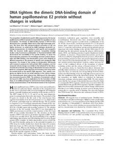
FIG. 1. Summary of E1DBD mutations. (A) E1 amino acid sequence from residues 150 to 304. The locations of ␣-helices and -sheets derived from the crystal structure (10) are diagramed above the sequence, along with the positions of the previously described hydrophilic regions HR1 and HR3 (12). Below the sequence are the origin-nonbinding (⫺ row, squares) and origin-binding (⫹ row, triangles) mutants from this study (solid symbols) and our previous study (open symbols). (B) Three-dimensional ribbon diagram of E1 protein residues 159 to 303 from Enemark et al. (10). Residues whose mutation yields a nonbinding phenotype in the yeast one-hybrid assay are indicated in red on the left monomer, and binding-positive mutations are in blue on the right monomer. The boundaries of the HR1 loop region are indicated with red arrows, and the HR3 region boundaries in ␣-helix 4 are indicated with blue arrows. Specific amino acid positions characterized in detail in the text are indicated (note that V246 is on the posterior side of ␣-helix 4 and is not visible in this figure).
were centrifuged at low speed, and supernatant containing cleaved E2 was removed and stored at ⫺20°C. Cleavage and integrity of the E2 protein were verified by sodium dodecyl sulfate-polyacrylamide gel electrophoresis (SDSPAGE) analysis. In vitro DNA binding assays. The concentration of wild-type GST-E1DBD protein was determined by the Bradford assay (5). The concentrations of mutant GST-E1DBD proteins were estimated by Coomassie blue staining of acrylamide gels followed by densitometry and comparison to a known amount of wild-type protein. In vitro binding was assessed by a gel shift assay using a blunt-ended,
double-stranded oligonucleotide comprising BPV-1 nucleotides 7926 to 29 as previously described (12, 37). Except where indicated otherwise, 10 ng of each wild-type or mutant GST-E1DBD protein was used in the binding assays; where E2 was also included, typically 20 ng of purified protein was used. In vivo DNA replication assays. Mutations were constructed in heterologous expression vector pCGE1 for expression of full-length E1 mutants F175S, N184Y, D185G, V193M, F237L, K241E, R243K, and V246D. Heterologous expression vectors pCGE1 and pCGE2 at 1.0 and 0.1 g, respectively, were mixed with 1.0 g of the BPV origin-containing vector pBOR or 1.0 g of pUC18
VOL. 75, 2001
DNA BINDING BY E1 MUTANTS
for positive and negative controls, respectively. Similar mixtures were made with each pCGE1 mutant, pCGE2, and pBOR. The combined DNAs were transected into Chinese hamster ovary (CHO) cells using the Lipofectamine transfection reagent (Gibco-BRL). At 48, 72, and 96 h posttransfection, plasmid DNA was harvested by Hirt extraction and digested with DpnI and HindIII (New England Biolabs). DNA samples were electrophoresed on an 8% agarose gel, transferred to a Nytran Supercharge nylon membrane (Schleicher & Schuell) by Southern transfer, and cross-linked to the membrane using a Stratalinker (Stratagene). A pBOR probe was radiolabeled using the Prime-a-Gene labeling system (Promega) and hybridized to the membrane using Rapid Hyb buffer (Amersham). The membrane was analyzed by Phosphorimager analysis.
RESULTS Mutations in and near HR1 and HR3 decrease E1DBDdependent reporter activity in yeast cells. We previously described the development of a yeast one-hybrid system for monitoring sequence-specific interactions of both full-length E1 and the E1DBD with chromosomal DNA containing three tandem E1 binding sites (E1BST) upstream of the lacZ gene (12) (Fig. 2A). Reporter gene expression in this system requires specific E1-E1BS interaction, as neither the pGAD424 parental in the YM4271[E1BST] strain nor the pGAD424E1DBD construct in YM4271[p53BLUE] (which contains a p53 binding site instead of the E1 binding sites) is sufficient for -galactosidase production. In order to generate unbiased genetic data of the amino acid requirements for E1 DNA binding, we generated two libraries of mutations in the E1DBD ORF by a PCR random mutagenesis procedure (31). After cloning the PCR products into pGAD424, the libraries were screened using the yeast onehybrid system, and mutant clones which failed to activate the reporter gene, suggesting possible defects in DNA binding, were isolated (Fig. 2B). Our first library was made using limiting amounts of dATP to drive dITP incorporation and resulted in clones with an average substitution frequency that was too high for easy identification of the critical residues (Table 1, A clones). Our second library was made using limiting dGTP and resulted in a lower substitution frequency of about one to two mutations per clone (Table 1, G clones, and Table 2, BG clones). This second library was used for the remaining studies reported here. Approximately half of 84 reporter-negative colonies that we selected from filter screens of the second library tested positive for the E1DBD insert by PCR, and approximately 65% of the insert-containing clones had appropriate restriction digestion profiles. These clones were sequenced to identify the mutations present (Table 1), and the mutant residues are depicted on the E1DBD structure in Fig. 1. Additionally, mutant clone G75 (V193M) was isolated in a yeast two-hybrid screen of the E1DBD library for mutants which fail to interact with an E1 fragment, E11–311, which normally interacts with the wild-type E1DBD (K. Woytek, unpublished data). This mutant was subsequently assayed in our yeast one-hybrid system and failed to activate the E1DBD-dependent reporter. All mutants were also transferred into strain YM4271[p53BLUE] to confirm that they are unable to activate the p53-dependent reporter gene by any spurious activity. Considering only single-, double-, and triple-point mutants, we identified 22 new mutants with 34 amino acid positions altered and 36 total substitutions. Eleven of 34 positions (34%) mapped to the HR1 and HR3 regions. However, this number
11951
may be an underestimate, since some mutations present in double and triple mutants are likely nonessential for DNA binding. Consequently, if only single and double mutants are considered, we have 19 mutations and can discount substitutions at positions 137, 139, and 288 because 137 and 139 are outside the sequences required for DNA binding and 288 is functional when tested separately (not shown). Thus, we are left with 16 mutations causing DNA binding defects, of which 8 (50%) are located in HR1 and HR3. Nonessential residues are rare in and near HR1 and HR3. We also screened the libraries for binding-functional E1DBD mutants using two different methods. First, we examined reporter-positive yeast clones for SSCP. Following plasmid DNA extraction from the yeast clones, we generated PCR products from the E1DBD ORF, which were then processed as described in Materials and Methods and examined on native polyacrylamide gels (Fig. 2C). Of 60 clones screened, 21 (35%) were found to display SSCP. Second, we used a temperature sensitivity screen by performing reporter assays at both 30 and 39°C. Using this method, we screened approximately 300 clones and found four mutant candidates (1.3%). Upon sequence analysis of clones which displayed conformation polymorphisms or temperature sensitivity, we identified a variety of mutations which were scattered throughout the E1DBD (Table 2 and Fig. 1). These binding-functional mutations were largely absent from the HR1 and HR3 regions, with a few exceptions which may represent functional papillomavirus polymorphisms or simply nonconserved residues (see Discussion). Furthermore, the paucity of nonsense and frameshift mutations in reporter-positive clones compared to reporternegative clones is consistent with this tested E1 polypeptide being a minimized DNA binding domain. From our screens of reporter-positive mutants, we isolated 13 new mutants with 20 amino acid positions altered and 20 total substitutions. Between residues 140 and 311, we found 19 mutations, for an average of one substitution every nine residues, only one (5%) of which mapped to HR1 or HR3, D185N in clone BG14. This is a conservative alteration at a residue nonessential for in vitro DNA binding, as shown by alanine substitution (12). Using a chi-square test with 1 degree of freedom, we compared the frequency of HR1 and HR3 mutations in reporter-negative and reporter-positive clones based on white clones with 8 of 16 mutations in HR1 or HR3 and blue clones with 1 of 19 mutations in HR1 or HR3. The higher number of HR1 and HR3 mutations in the nonbonding clones compared to the functional clones was statistically significant, with a P value of ⬍0.005. This strongly supports the previously demonstrated importance of these two regions for origin recognition by the E1 protein. HR1- and HR3-associated mutations affect sequence-specific DNA binding in vitro. Since we isolated the mutants by in vivo screening, it was important to test them in vitro for confirmation of their binding negativity as well as to control protein amounts, which may vary with expression in vivo. In a previous study we examined five substitution mutants of HR1 (K183A, D185A, K186A, T187A, and T188A) but only one mutant of HR3 (K241A). Our current yeast screens yielded two new mutations in HR1 (N184Y and D185G) and three new mutations in HR3 (K241E, R243K, and V246D), which gave us the opportunity to examine HR3 in more depth. In
11952
WEST ET AL.
J. VIROL.
FIG. 2. Yeast one-hybrid screening system and representative data. (A) Expression of yeast GAL4 transactivation domain (GAL4AD) fused to the E1 DNA binding domain (E1DBD) activates a lacZ reporter gene upon interaction of the E1DBD with three tandem copies of the E1 binding site (E1BST). (B) Nitrocellulose filter assay for reporter activity in yeast colonies harboring pGAD424-E1DBD constructs. YM4271[p53BLUE] is a control strain containing a p53 binding site in lieu of E1 binding sites, which confirms that interaction between the E1DBD and E1BST is sequence specific. Shown are both reporter-positive and reporter-negative mutants from our screens along with their genotypes. (C) SSCP assay was used to detect mutations in the E1DBD coding regions of reporter-positive yeast colonies. PCR was used to amplify and radiolabel the 5⬘ and 3⬘ halves of the 573-bp E1DBD open reading frame of wild-type pGAD424-E1DBD and candidate mutants. PCR products were then processed as described in Materials and Methods and electrophoresed on native polyacrylamide gels. Samples from the 5⬘ and 3⬘ amplimers are shown in the right and left panels, respectively. Lane designations are the clone names as listed in Table 2. Lanes marked C were amplified from the parental wild-type E1DBD clone.
addition, the screen also identified several nonfunctional mutations which resided outside the HR1 and HR3 sequences. Mutants F175S, V193M, and F237L neighbor HR1 or HR3 and were chosen for additional study. Mutations at positions 210, 265, and 293 involved adding or removing a proline, which is likely to disrupt structure, and were not investigated further. Mutation S207C lies in the ␣3 helix and may be critical for
dimerization of the E1DBD (10), and will be characterized as part of a later study. The reporter-negative mutants F175S, N184Y/K288R, D185G, V193M, F237L, K241E, R243K, and V246D from our yeast screen were subcloned into pGEX (Pharmacia) for expression of GST fusion proteins in E. coli. The only double mutant included for in vitro study was selected because of the
VOL. 75, 2001
DNA BINDING BY E1 MUTANTS
TABLE 1. Mutations in the E1DBD ORF of reporter-negative yeast clonesa Mutation class
Clone
Mutation(s)
One substitution
G2 G6 G13 G21 G22 G31 G34 G51 G56 G67 G75 A33 G1 G26 G38 G44 A11 A34 G3 G17 G25 G30 A37
P293H L210P F237L S207C F175S P265S K241E D185G R243K V246D V193M E139D, T187S R137W, K186E N184Y, K288R R247Q, W277G F203V, P265L H150P, K155I, L172P Q144R, T188P, N189D A159T, N184K, A206V F203V, Q219L, Q299R C173S, N252S, L261S F203V, W277R, L306S R123H, T187A, F204S, Q307R
A3
D185G, T187A, S224F, V256I, S281G, K288E 221STOP
Two substitutions
Three substitutions
More than three substitutions Premature termination codon
Deletion/frameshift Insertion/frameshift a
A32 G10 G24 G46 G48 G62 A1 G11 G69 None
212STOP 144STOP 152STOP 264STOP 219STOP Codon 241 Codon 158 Codon 265
Underlined mutants were selected for in vitro analyses.
presence of substitution K288E in a binding-functional mutant (clone BG28, Table 2), which makes it likely that N184Y is the mutation critical for DNA binding. Purified wild-type and mutant E1DBD proteins were compared for DNA binding activity using a radiolabeled oligonucleotide substrate (BPV nucleotides 7926 to 29) containing the E1 binding site and E2 binding site 12. As previously described, wild-type E1DBD bound this substrate and produced multiple E1DBD-DNA complexes (12). All E1DBD mutants were observed to have greatly reduced binding affinity for the probe compared to the wild type, confirming the in vivo results that the mutated residues were critical for sequence-specific DNA interaction (Fig. 3). However, mutants D185G, F237L, and R243K formed a detectable though still not wild-type level of complex with the probe at higher protein concentrations (data not shown and Fig. 4). The weak binding activity of these three mutants suggested that they may still be capable of cooperative interaction with E2, so the in vitro DNA binding assays were repeated in the presence of bacterially purified E2 (Fig. 4). E1DBD-E2-DNA complexes were detected for all three of the mutants, though not for N184Y/K288R, which does not exhibit any origin binding in the presence or absence of E2. These results indicate, at least in some cases, that protein-protein interactions between
11953
TABLE 2. Mutations in the E1DBD ORF of reporter-positive yeast clonesa Mutation class
One substitution
Two substitutions Three substitutions Premature termination codon Deletion/frameshift Insertion/frameshift a b
Clone
Mutation(s)
BG3 BG13 BG16 BG20 BG30 BG33 BG37 BG47 BG4 BG28 GTS4 BG14 GTS3 GTS1
E205V F175Y S174R A291T I268S C236S Q214L S283L G136E, A159V F197L, K288E N238I, L275P D185N, E258G, F278S G227E, E259V, H289R Codon 190b
None None
Underlined mutants represent potential papillomavirus polymorphisms. This mutant is suspected to display terminator codon readthrough.
the E1DBD and E2 are sufficient to stabilize very poor interactions between the E1DBD and its binding sequence. E1DBD mutants positive for E2 interaction support partial in vivo DNA replication. The ability of some binding-defective mutants to be rescued by E2 suggested that they may retain sufficient function to support DNA replication. Single point mutations F175S, N184Y, D185G, V193M, F237L, K241E, R243K, and V246D were constructed in the vector pCGE1, which expresses full-length E1 in mammalian cells for in vivo DNA replication assays. The N184Y mutation was separated from K288R in this experiment so that substitution at residue 184 was the only change present in E1. Heterologous expression vectors pCGE1 (wild-type or mutant) and pCGE2 were transfected along with the BPV-1 origin-containing plasmid pBOR into Chinese hamster ovary cells. Plasmid DNA was Hirt extracted at 48, 72, and 96 h posttransfection and examined by Southern blotting as described in Materials and Methods (Fig. 5A). DpnI-resistant replication products were quantified by Phosphorimager analysis, and a minimum of two independent transfections were performed for each mutant construct. From each experiment, replication efficiencies were calculated as the percentage of replication product made in the presence of wild-type E1, and the average replication efficiencies are plotted in Fig. 5B. Mutants E1D185G, E1F237L, and E1R243K supported 19, 51, and 72% of wild-type replication, respectively. In contrast to these three mutants, which supported significant levels of DNA replication, mutants E1F175S, E1N184Y, E1V193M, E1K241E, and E1V246D, which were unable to form E1-E2-DNA complexes in vitro, supported little if any replication. Thus, only those E1DBD mutants which retained functional interaction with E2 in vitro could support replication activity as full-length E1 mutants. This result is consistent with the recent report that E2 mutations which specifically prevent interaction with the E1DBD also reduce in vivo DNA replication (7, 11). HR3 is unlikely to make the majority of base contacts with DNA. The distribution of functional versus nonfunctional mutations described above strongly implicates HR1 and HR3 as
11954
WEST ET AL.
J. VIROL.
FIG. 3. In vitro analysis of GST-E1DBD interactions with a 50-bp double-stranded, sequence-specific DNA probe. (A) GST-E1DBD fusion proteins were purified by glutathione-Sepharose affinity chromatography. Shown are 10% polyacrylamide gels containing wild-type and mutant proteins and stained with Coomassie blue. Lanes M, size markers. (B) Electrophoretic mobility shift assay of wild-type (WT) and mutant GST-E1DBD fusion proteins. For each reaction, 10 ng of protein was incubated with the radiolabeled DNA except for mutant V246D, for which 3 ng was used in this experiment.
critical elements for sequence-specific E1-DNA interaction, but does not discriminate the individual roles of each element. We noted previously that the organization of these two E1 elements resembled that of the A and B2 regions of SV40 T antigen, which are similarly critical for sequence-specific DNA binding (12). The recent crystal data for the E1DBD confirmed an overall structural similarity between the T antigen and E1 DNA binding domains (10), and a manual alignment of the primary sequences shows a remarkable relatedness, particularly in the DNA interaction elements (Fig. 6A). Interestingly, the EBNA1 protein also uses two motifs separated by approximately 50 residues in the linear sequence, called the flanking domain and recognition helix, for specific
binding to an 18-bp sequence in the Epstein-Barr virus origin (1, 4, 9). The EBNA1 flanking domain is an extended loop which makes base contacts, while the recognition helix makes nonspecific phosphate contacts (4). Thus, EBNA1 achieves sequence-specific DNA binding using a site-specifying base interactor and a nonspecific phosphate interactor. Though less distinctive than between E1 and T antigen, there is intriguing sequence similarity between the EBNA1 flanking domain and HR1/A, and between the EBNA1 recognition helix and HR3/B2 (Fig. 6B). In addition to the sequence similarity, the corresponding elements have common secondary structures, with the flanking domain/HR1/A elements all located in loops while the distal elements for all three proteins form helices.
VOL. 75, 2001
DNA BINDING BY E1 MUTANTS
11955
FIG. 4. In vitro E2-mediated rescue of DNA binding by E1DBD mutants. Electrophoretic mobility shift assays were performed as in Fig. 3 using the amounts of proteins (in nanograms) indicated. The other single-substitution mutations shown in Fig. 3 were all negative for E2 rescue in this assay (not shown).
These physical parallels suggest common functions and implicate HR3 as the DNA backbone binding unit rather than the base-specific contact element. To assess the functional contribution of HR3 to sequencespecific DNA binding, we generated a chimeric triple mutant called THR3 to test for retention of sequence-specific DNA binding in the yeast one-hybrid system (Fig. 7A). THR3 contains three residues from T antigen element B2 which were simultaneously substituted into E1 HR3 (S242H/E244V/ T245S). The rationale for this construct was that if HR3 made specific base interactions, then multiple amino acid substitution should disrupt the ability to recognize the E1BS. Conversely, if the role of HR3 were more in nonspecific DNA contact, then substitution with the corresponding residues from T antigen should not eliminate binding to the E1BS sequence. The results shown in Fig. 7B demonstrate that the mutant retained sequence-specific binding capacity in vivo. In a separate construct, we also observed that a site-directed single mutation in HR3, V246A, functioned in yeast cells (not shown). To further assess the functionality of THR3, the mutant protein was purified and tested in the in vitro DNA binding assay (Fig. 7C). While reduced in activity compared to wildtype E1DBD, the THR3 protein demonstrated significant sequence-specific binding ability and wild-type levels of E1DBDE2-DNA complex formation. Consistent with its DNA binding properties, in the context of full-length E1, the THR3 mutations still allowed in vivo replication at ⬇25% of wild-type levels (data not shown). Since both THR3 and V246A maintained sequence-specific DNA binding, residues S242, E244, T245, and V246 are not absolutely critical for sequence specificity of the E1DBD. The ability to alter the majority of residues in HR3 without loss of sequence-specific binding activity or without complete loss of replication function is more consistent with this element’s providing nonspecific contacts than base recognition. DISCUSSION We previously used site-directed mutagenesis to show that mutations in E1DBD elements HR1 and HR3 disrupt se-
quence-specific interaction with the DNA origin. In our current study, we used random PCR mutagenesis along with a yeast one-hybrid screen to identify additional mutants of the BPV-1 E1DBD which are defective for sequence-specific DNA interactions. As a complement, binding-functional mutants were also identified. Our goal in using this nondirected approach was to gain a more extensive understanding of the location and specific function of those amino acids most important for the origin-binding activity of E1. The locations of the various binding-functional and -nonfunctional mutations are indicated on the E1 sequence and structure in Fig. 1, and the properties of selected nonbinding mutants are summarized in Table 3. While the mutations were mostly characterized in the context of the isolated E1DBD, two random and five site-directed mutations were subsequently analyzed in full-length E1 using the yeast one-hybrid system (data not shown). All seven mutations, four binding functional and three nonfunctional, had the same binding phenotypes in the E1DBD compared to full-length E1. Thus, the E1DBD seems to be a truly independent domain in E1, and results with the E1DBD should be generally reflective of the properties of full-length E1 for DNA interaction. Residues necessary for sequence-specific DNA binding by E1 are concentrated in and near HR1 and HR3. Using the recently published crystal structure (10), previous studies of T antigen mutants (29, 30), and our new mutant data, we have arrived at several conclusions regarding possible functions of specific E1DBD residues. The first observation to emerge from sequence comparisons of the E1DBD ORF in reporter-negative yeast clones was the repeated occurrence of substitutions in and around the HR1 and HR3 regions (Fig. 1). Furthermore, a chi-square statistical test indicated that the clustering of critical mutations in HR1 and HR3 for the reporter-negative clones versus the reporter-positive clones had a less than 0.5% probability of occurring by chance. This correlation is further evidence that hydrophilic regions HR1 and HR3 are indeed necessary for sequence-specific DNA binding. Besides the HR1 and HR3 regions, the only other modest clustering of nonfunctional mutations occurred in ␣-helix 3. This helix is involved in E1 dimerization, and the occurrence of several
11956
WEST ET AL.
J. VIROL.
FIG. 5. DNA replication in CHO cells. (A) Representative Southern blots of replication products recovered from CHO cells at 48, 72, and 96 h posttransfection. Cells were triple transfected at time zero with vectors for E1 and E2 expression and a plasmid containing the BPV origin, pBOR, or pUC18 as a non-origin-containing control. Wild-type (WT) and mutant E1 expression vectors were used as indicated. (B) Quantitation of DNA replication products. Band intensities on Southern blots were measured by Phosphorimager analysis. Replication efficiency of mutant E1 proteins was calculated by comparing the intensity of wild-type and mutant replication product bands at 96 h posttransfection. Average replication efficiencies for two or more independent experiments are shown. Data for V193M were generated in a separate study.
mutations in this region supports an important functional role for E1-E1 contacts in origin binding. As expected, functional mutations isolated from the reporter-positive clones were more evenly distributed throughout the E1DBD.
Role of HR1 residues in E1DBD-DNA interaction. Our previous study identified a cadre of hydrophilic residues in HR1 that were critical for origin binding (12). The recently published crystal structure of the E1DBD revealed that the HR1
VOL. 75, 2001
DNA BINDING BY E1 MUTANTS
11957
FIG. 6. (A) Sequence alignment between DNA binding domains of SV40 T antigen and BPV-1 E1. Boxes indicate regions of sequence similarity. There is an extended -hairpin in E1 at residues 223 to 230 which is not found in T antigen. The locations of conserved regions HR1, HR3, A, and B2 are indicated. (B) HR1- and HR3-like elements are found in origin-binding proteins from three transforming DNA viruses: papillomavirus, SV40, and Epstein-Barr virus. E1, T antigen (T-AG), and EBNA1 contain regions of sequence similarity which are separated by approximately 50 amino acid residues and which are critical for sequence-specific DNA binding.
region forms a structurally well-defined loop which potentially makes sequence-specific contacts with origin DNA (10). In the current study, two new mutations in HR1 were characterized, N184Y and D185G. N184Y was analyzed in the context of the double mutant E1DBDN184Y/K288R; however, the K288R substitution is unlikely to be the critical change for disrupting DNA binding, since a nonconservative change at this position appeared in a binding-functional mutant (Table 2). Additionally, the single point mutant E1N184Y exhibited a severe defect for in vivo DNA replication in CHO cells. Therefore, the N184Y substitution is most likely responsible for the defective DNA interaction both in vivo and in vitro as well as for E2 binding in vitro. We do not suspect that the N184Y mutation in either E1DBD or full-length E1 caused severe misfolding, as we observed other wild-type activities for the mutant, including nonspecific DNA binding in the absence of pUC18 nonspecific competitor DNA and single-stranded DNA binding (data not
shown). It also seems unlikely that N184Y would affect global protein folding, since it is both a surface residue and not part of any defined secondary structure such as an ␣-helix or -sheet. However, the majority of E1 proteins have either a serine or asparagine at positions corresponding to 184, suggesting that a polar side chain able to support hydrogen bonding is required at this position. While tyrosine has chemically similar properties, the much bulkier tyrosyl group may cause steric hindrance that would prevent proper alignment of HR1 with the base sequences. E1DBDD185G is also severely defective for sequence-specific DNA binding, but at higher protein concentrations it does exhibit some activity. Additionally, this mutant is capable of in vitro E2 interaction and supports approximately 20% of the wild-type E1 in vivo DNA replication. As with N184Y, this partial functionality again suggests that tertiary structure is not completely disrupted by this amino acid change. A previously examined mutation at this position, E1DBDD185A, is even less
11958
WEST ET AL.
J. VIROL. TABLE 3. Functional properties of DNA-binding-defective E1DBD mutants E1DBD
Wild-type F175S N184Y/K288R D185G V193M F237L K241E R243K V246D a b
FIG. 7. (A) Sequence alignment between E1 HR3 and SV40 T antigen (TAG) region B2. Also shown is the sequence in the HR3 region of the THR3 mutant. Multiple site-directed substitutions were made to create this chimeric mutant of the E1DBD whose HR3 element contains residues found in the T antigen B2 element. (B) Nitrocellulose filter assay showing sequence-specific interactions between THR3 and E1 binding sites in the yeast one-hybrid system. (C) Electrophoretic mobility gel shift assay as in Fig. 4, showing cooperativity of binding of THR3 with E2 on an origin-specific DNA probe. Amounts of proteins (in nanograms) are indicated.
defective and supports 30 to 50% of wild-type sequence-specific DNA binding (12). In the crystal structure, the side chain of D185 interacts with R243 and contributes to the coupling of HR1 and HR3 (10). The reduced but still significant functioning of the D185A and D185G mutants indicates that the coordinating bond between D185 and K243 is important but not absolutely essential to maintain correct juxtaposition of HR1 and HR3; presumably the interaction between K243 and F182 remains and provides sufficient stability for modest function. The more dramatic defect of E1DBDD185G compared to the D185A mutant likely results from the greater conformational flexibility inherent in glycine residues. Thus, the presence of glycine at position 185 may disorder the loop structure and
Sequence-specific DNA binding In vivo
In vitro
E1DBD-E2 binding in vitro
⫹ ⫺ ⫺ Weaka ⫺ ⫺ ⫺ Weaka ⫺
⫹ ⫺ ⫺ Weakb ⫺ ⫺ ⫺ Weakb ⫺
⫹ ⫺ ⫺ Weak ⫺ ⫹ ⫺ Weak ⫺
In vivo DNA replication (% of wild-type level)
100 0.2 3.9 18.9 0 50.8 0.3 71.8 0
Less than 5% of retransformed yeast colonies were reporter positive. Protein-DNA complexes were observed at increased protein concentrations.
alter the positioning of side chains at nearby residues. This may both disrupt the structurally stabilizing F182-K243 interaction and disturb possible base-specific contacts by critical residues such as K183, N184, K186, and T187. Role of HR3 residues in E1DBD-DNA interaction. We also isolated and characterized new mutations in the HR3 element. Mutant K241E was defective for in vivo and in vitro binding, E2 interaction, single-stranded DNA binding (unpublished data), and in vivo DNA replication. Residue K241 resides at the N-terminal side of ␣-helix 4, contributes a positively charged amino group, and lies on the DNA binding surface of E1. Substitution of a negatively charged glutamic acid residue likely increases electrostatic repulsion between this position and the DNA backbone, though this does not exclude that the lysine might also normally make specific base contacts. Another mutant, R243K, was interesting and unique because it was severely crippled for DNA binding despite having a conservative amino acid change. Consistently, an earlier study showed that the same conservative mutation at the corresponding T antigen residue, R204 (Fig. 6A), had a severe effect on sequence-specific DNA binding, more so than did substitutions at nearby residues V205 and A207 (29). Crystal data for the E1DBD show that R243 makes a total of four contacts, two with the HR1 residues D185 and F182, and two additional ones with Q264 and L263 (10). Since the net positive charge is unchanged by the lysine substitution, the most likely explanation for the functional defect seems to be the loss of an amino group. Loss of this moiety effectively reduces the number of potential contacts that can be made simultaneously and likely cripples this anchoring point for the HR1 region. While failure to maintain correct positioning of HR1 and HR3 would explain the binding defect, this mutant still formed E1DBD-E2-DNA complexes, bound single-stranded DNA (data not shown), and had nearly wild-type ability to support DNA replication in vivo. These results are consistent with retention of a generally correct tertiary structure and also suggest that additional interactions, either from E2 or from other regions of full-length E1, can somewhat compensate for the lost contacts at residue 243. Nonetheless, among the E1 proteins from all papillomavirus groups and the T antigen proteins from all polyomavirus groups, the arginine 243 posi-
VOL. 75, 2001
tion is a highly conserved residue, which is consistent with its providing an important stabilizing function. The last mutation in HR3 that was identified by the onehybrid screen was V246D. It does not appear that the valine residue is critical for specific base contacts, as a V246A mutant was significantly functional in vivo (unpublished data). Consequently, like K241E, introduction of a negative charge at this V246 may simply cause repulsion between this position and the similarly charged phosphodiester backbone of the DNA. Alternatively, V246 may actually help stabilize the DNA contact motifs. The crystal structure demonstrates the proximity of residues F237 and V246 in a hydrophobic pocket just beneath the E1DBD DNA binding surface. As an F237L mutant also showed reduced DNA binding in vivo and in vitro, decreased hydrophobicity at these residues may affect nonpolar interactions which potentially alter HR1 and HR3 positioning. The potential role of core hydrophobic residues in architectural support of the DNA contact motifs is currently being studied in more detail. To further investigate the role of HR3, a triple substitution mutant, THR3, was constructed. This mutant retained in vivo DNA binding activity, indicating that none of the changed bases, S242, E244, or T245, was absolutely essential for sequence-specific binding. The ability to replace at least four of the HR3 residues (THR3 mutations plus the V246A mutant) without total loss of sequence-specific recognition function, along with a structural coordination role for R243, seems inconsistent for a region designed to make specific base contacts with DNA. Therefore, we propose that HR3 is primarily responsible for making phosphate backbone contacts, like the EBNA1 recognition helix. Such a role would be consistent with the severe binding defect which resulted from introducing acidic residues at positions 241 and 246. In contrast, multiple conserved residues in HR1 (R180, K183, N184, D185, K186, and T187) are associated with critical single point mutations, suggesting that HR1 is a better candidate for a sequence recognition device. Role of critical residues outside the HR1 and HR3 regions. Other than substitutions at proline residues (P265 and P293), which likely severely disrupt overall structure, binding-critical residues outside HR1 and HR3 fell into two groups: hydrophobic amino acids (F175, V193, F237, and W277) and residues located in ␣-helix 3 (F203, S207, and L210). From the crystal structure, it appears that these hydrophobic residues may contribute to a core that forms a scaffold beneath the HR1/HR3 structure and possibly provides an anchor function for the DNA interaction motif. A more detailed study of the role of this hydrophobic core will be presented elsewhere (unpublished data). The ␣3 helix has been shown to be a functional dimerization interface for the E1DBD (10), and we identified two single substitution mutations in this region, E1DBDS207C and E1DBDL210P, that were reporter negative in the yeast onehybrid screen. Additionally, an F203V mutation showed up in three different reporter-negative clones, though all had double or triple mutations, so the contribution of the F203V mutation to the functional defect has not been specifically established. While none of these single or multiple mutants has been examined yet in detail, the occurrence of three different mutations in this helix suggests a requisite function in DNA binding.
DNA BINDING BY E1 MUTANTS
11959
Thus, these mutants strengthen the observation that dimerization of E1 stabilizes origin DNA binding (6, 22). Residues nonessential for DNA binding by E1 occur throughout E1DBD and are rare in HR1 and HR3 regions. The SSCP and temperature sensitivity assays detected yeast clones expressing binding-functional mutations (Table 2). Most of these DNA binding-functional mutations were nonconservative, indicating that the side chains at these positions make little if any contribution to E1DBD structure. The lack of clustering of nonessential residues is also consistent with a simple role, such as providing appropriate spacing to promote proper orientation of essential residues in and near the critical HR1 and HR3 regions. It should be noted that these residues may be critical for other functions of the E1DBD, such as interaction with the E2 protein or host cell replication proteins, but this has not yet been determined. The only noncritical substitution which did occur in HR1 or HR3 was D185N in mutant D185N/E258G/F278S. However, this is a structurally conservative mutation and also can probably maintain the salt bridge with R243, as discussed above, so it is not surprising that this substitution was quite functional. Unfortunately, since the binding-functional mutations that we isolated are random in nature, interpretations that go beyond simple classification of these residues as nonessential for sequence-specific DNA binding are difficult to make. Thus, while quantification of reporter activity for our binding-functional mutants is possible, definite conclusions about the relative importance of the individual residues cannot be made without a more extensive site-directed mutational analysis at each position. It has not escaped our attention that these mutants could prove useful for studying other functions associated with the E1DBD besides DNA binding. We currently have found one example of a site-directed E1DBD mutation which functions at wild-type levels in both DNA binding and E2 interaction but still fails in DNA replication (unpublished data). We are now screening our randomly generated bindingfunctional mutants in DNA replication assays, and mutants which bind origin DNA but fail to replicate it could define functional defects at stages beyond origin recognition. In summary, the results presented here strongly support the roles of HR1 and HR3 as DNA contact elements for E1 protein. HR1 is part of an ordered loop structure, while HR3 forms the ␣4 helix, and these two elements are juxtaposed on the surface of the E1DBD (10). Because of the greater tolerance for mutational changes in HR3 than in HR1, we propose that HR3 may provide primarily nonspecific contacts with the DNA, while HR1 may be the sequence specificity determinant. SV40 T antigen has two structurally equivalent elements, A and B2, which are also juxtaposed and which show limited sequence similarity with the parallel E1 regions. A similar dual element, loop plus helix, DNA binding motif also occurs in the Epstein-Barr virus EBNA1 protein. In EBNA1 the helix is positioned so that it can only contact the sugar-phosphate backbone of the DNA, while the extended loop makes base interactions (3), and we propose that the equivalent structures in E1 perform the same general functions. Thus, all three origin-binding proteins, E1, T antigen, and EBNA1, dock with their palindromic binding sites using an apparently structurally similar recognition mechanism.
11960
WEST ET AL.
J. VIROL.
ACKNOWLEDGMENTS Many thanks to Leemor Joshua-Tor for kindly providing information about the E1DBD structure during the development of the manuscript. This work was supported by grant RPG-96-125-05-MBC from the American Cancer Society. REFERENCES 1. Ambinder, R. F., W. A. Shah, D. R. Rawlins, G. S. Hayward, and S. D. Hayward. 1990. Definition of the sequence requirements for binding of the EBNA-1 protein to its palindromic target sites in Epstein-Barr virus DNA. J. Virol. 64:2369–2379. 2. Berg, M., and A. Stenlund. 1997. Functional interactions between papillomavirus E1 and E2 proteins. J. Virol. 71:3853–3863. 3. Bochkarev, A., J. A. Barwell, R. A. Pfuetzner, E. Bochkareva, L. Frappier, and A. M. Edwards. 1996. Crystal structure of the DNA-binding domain of the Epstein-Barr virus origin-binding protein, ebna1, bound to DNA. Cell 84:791–800. 4. Bochkarev, A., J. A. Barwell, R. A. Pfuetzner, W. Furey, A. M. Edwards, and L. Frappier. 1995. Crystal structure of the DNA-binding domain of the Epstein-Barr virus origin-binding protein EBNA1. Cell 83:39–46. 5. Bradford, M. M. 1976. A rapid and sensitive method for the quantitation of microgram quantities of protein utilizing the principle of protein-dye binding. Anal. Biochem. 72:248–254. 6. Chen, G., and A. Stenlund. 1998. Characterization of the DNA-binding domain of the bovine papillomavirus replication initiator E1. J. Virol. 72: 2567–2576. 7. Chen, G., and A. Stenlund. 2000. Two patches of amino acids on the E2 DNA binding domain define the surface for interaction with E1. J. Virol. 74:1506–1512. 8. Cueille, N., R. Nougarede, F. Mechali, M. Philippe, and C. Bonne-Andrea. 1998. Functional interaction between the bovine papillomavirus virus type 1 replicative helicase E1 and cyclin E-CDK2. J. Virol. 72:7255–7262. 9. Edwards, A. M., A. Bochkarev, and L. Frappier. 1998. Origin DNA-binding proteins. Curr. Opin. Struct. Biol. 8:49–53. 10. Enemark, E. J., G. Chen, D. E. Vaughn, A. Stenlund, and L. Joshua-Tor. 2000. Crystal structure of the DNA binding domain of the replication initiation protein E1 from papillomavirus. Mol. Cell 6:149–158. 11. Gillitzer, E., G. Chen, and A. Stenlund. 2000. Separate domains in E1 and E2 proteins serve architectural and productive roles for cooperative DNA binding. EMBO J. 19:3069–3079. 12. Gonzalez, A., C. Bazaldua-Hernandez, M. West, K. Woytek, and V. G. Wilson. 2000. Identification of a short, hydrophilic amino acid sequence critical for origin recognition by the bovine papillomavirus E1 protein. J. Virol. 74:245–253. 13. Han, Y. F., Y. M. Loo, K. T. Militello, and T. Melendy. 1999. Interactions of the papovavirus DNA replication initiator proteins, bovine papillomavirus type 1 E1, and simian virus 40 large T antigen with human replication protein A. J. Virol. 73:4899–4907. 14. Howley, P. 1996. Papillomavirinae: the viruses and their replication, p. 947– 978. In B. N. Fields, D. M. Knipe, and P. M. Howley (ed.), Fundamental virology. Lippincott-Raven, Philadelphia, Pa. 15. Lee, D., H. Sohn, G. V. Kalpana, and J. Choe. 1999. Interaction of E1 and hSNF5 proteins stimulates replication of human papillomavirus DNA. Nature 399:487–491. 16. Leng, X., J. H. Ludesmeyers, and V. G. Wilson. 1997. Isolation of an aminoterminal region of bovine papillomavirus type 1 E1 protein that retains origin binding and E2 interaction capacity. J. Virol. 71:848–852. 17. Lorincz, A. T., R. Reid, A. B. Jenson, M. D. Greenberg, W. Lancaster, and R. J. Kurman. 1992. Human papillomavirus infection of the cervix: relative risk associations of 15 common anogenital types. Obstet. Gynecol. 79:328– 337. 18. Ma, T. L., N. X. Zou, B. Y. Lin, L. T. Chow, and J. W. Harper. 1999. Interaction between cyclin-dependent kinases and human papillomavirus
19. 20. 21. 22. 23.
24. 25. 26. 27.
28.
29. 30. 31. 32. 33. 34. 35. 36. 37.
38. 39.
replication-initiation protein E1 is required for efficient viral replication. Proc. Natl. Acad. Sci. USA 96:382–387. McShan, G. D., and V. G. Wilson. 1997. Casein kinase II phosphorylates bovine papillomavirus type 1 E1 in vitro at a conserved motif. J. Gen. Virol. 78:171–177. McShan, G. D., and V. G. Wilson. 1997. Reconstitution of a functional bovine papillomavirus type 1 origin of replication reveals a modular tripartite replicon with an essential AT-rich element. Virology 237:198–208. McShan, G. D., and V. G. Wilson. 2000. Contribution of BPV-1 E1 protein residue 48 to replication function. J. Gen. Virol. 81:1995–2004. Mendoza, R., L. Gandhi, and M. R. Botchan. 1995. E1 recognition sequences in the bovine papillomavirus type 1 origin of DNA replication: Interaction between half sites of the inverted repeat. J. Virol. 69:3789–3798. Park, P., W. Copeland, L. Yang, T. Wang, M. R. Botchan, and I. J. Mohr. 1994. The cellular DNA polymerase alpha-primase is required for papillomavirus DNA replication and associates with the viral E1 helicase. Proc. Natl. Acad. Sci. USA 91:8700–8704. Rangasamy, D., and V. G. Wilson. 2000. Bovine papillomavirus E1 protein is sumoylated by the host cell Ubc9 protein. J. Biol. Chem. 275:30487–30495. Rangasamy, D., K. Woytek, S. A. Khan, and V. G. Wilson. 2000. SUMO-1 modification of bovine papillomavirus E1 protein is required for intranuclear accumulation. J. Biol. Chem. 275:37999–38004. Sarafi, T. R., and A. A. McBride. 1995. Domains of the BPV-1 E1 replication protein required for origin-specific DNA binding and interaction with the E2 transactivator. Virology 211:385–396. Schiffman, M. H., H. M. Bauer, R. N. Hoover, A. G. Glass, D. M. Cadell, B. B. Rush, D. R. Scott, M. E. Sherman, R. J. Kurman, and S. Wacholder. 1993. Epidemiologic evidence showing that human papillomavirus infection causes most cervical intraepithelial neoplasia. J. Natl. Cancer Inst. 85:958– 964. Sedman, J., and A. Stenlund. 1995. Co-operative interaction between the initiator E1 and the transcriptional activator E2 is required for replicator specific DNA replication of bovine papillomavirus in vivo and in vitro. EMBO J. 14:6218–6228. Simmons, D. T., G. Loeber, and P. Tegtmeyer. 1990. Four major sequence elements of simian virus 40 large T antigen coordinate its specific and nonspecific DNA binding. J. Virol. 64:1973–1983. Simmons, D. T., K. Wun-Kim, and W. Young. 1990. Identification of simian virus 40 T antigen residues important for specific and nonspecific binding to DNA and for helicase activity. J. Virol. 64:4858–4865. Spee, J. H., W. M. de Vos, and O. P. Kuipers. 1993. Efficient random mutagenesis method with adjustable mutation frequency by use of PCR and dITP. Nucleic Acids Res. 21:777–778. Sun, S., L. Thorner, M. Lentz, P. MacPherson, and M. Botchan. 1990. Identification of a 68-kilodalton nuclear ATP-binding phosphoprotein encoded by bovine papillomavirus type 1. J. Virol. 64:5093–5105. Swindle, C. S., and J. A. Engler. 1998. Association of the human papillomavirus type 11 E1 protein with histone H1. J. Virol. 72:1994–2001. Thorner, L. K., D. A. Lim, and M. R. Botchan. 1993. DNA-binding domain of bovine papillomavirus type 1 E1 helicase: structural and functional aspects. J. Virol. 67:6000–6014. Verdon, M. E. 1997. Issues in the management of human papillomavirus genital disease. Am. Fam. Physician 55:1813.ff. Wieland, U., and H. Pfister. 1996. Molecular diagnosis of persistent human papillomavirus infections. Intervirology 39:145–157. Woytek, K. J., D. Rangasamy, C. Bazaldua-Hernandez, M. West, and V. G. Wilson. 2001. Effects of mutations within two hydrophilic regions of the BPV-1 E1 DNA binding domain on E1–E2 interactions. J. Gen. Virol. 82: 2341–2351. Yang, L., I. Mohr, E. Fouts, D. A. Lim, M. Nohaile, and M. Botchan. 1993. The E1 protein of bovine papillomavirus 1 is an ATP-dependent DNA helicase. Proc. Natl. Acad. Sci. USA 90:5086–5090. Zanardi, T. A., C. M. Stanley, B. M. Saville, S. M. Spacek, and M. R. Lentz. 1997. Modulation of bovine papillomavirus DNA replication by phosphorylation of the viral E1 protein. Virology 228:1–10.