make specific contacts with positions in the neighboring monomer. We find that 3 of the 5 residues are important for RecA function (L~S~'~,. Phez", and Ar822),.
Vol. 269, No.5, Issue of February 4, pp. 3823-3828, 1994 Printed in U S A
TIE JOURNAL OF B I O ~ I CCXEWSTRY AL
0 1994 by The American Soeiety for Biochemistry and Molecular Biology, Inc.
Functionally Important Residues at a Subunit Interface Site in the RecA Protein fromEscherichia coli* (Received for publication, October 4, 1993)
Mark C. SkibaS and KendallL. Knight8 From the Department of Biochemistry a n d Molecular Biology, University of Massachusetts Medical Center, Worcester. Massachusetts 01655
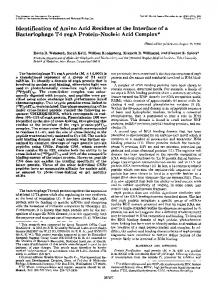
makes specific contacts with the neighboring subunit and, in Assembly of R e dsubunits into long, helical oligomers is required for ita roles in recombinational DNA repair some cases, also with positionsin the same subunit. andhomologousgeneticrecombination.Thecrystal We have introduceda large set of amino acid substitutions at structure of R e d reveals an extensive network of amino the five positions described above in order to address the folacid residues that lie at the subunit boundaries. We have lowing questions. 1) Which of the predicted intersubunit, as introduced a large set of substitutions at 5 clustered well as intrasubunit, interactions are actually important for residues, which are shown in the crystal structure to RecA function, i.e. which positions tolerate only very conservmake specific contacts with positions in the neighboring ative or no substitutions?2) What canbe determined about the monomer. We find that3 of the 5 residues are important chemical and steric constraints at each position based on the for RecA function (L~S~'~, Phez", and Ar822), whereas the other 2 (AsnZ1*and Wl8)are not. The patterns of observed pattern of functional substitutions? MATERIALS AND METHODS functionally allowed substitutions provide insight into the chemical and steric constraints required at these Analysis of the R e d Crystal Structure-Residues predicted to participate in monomer-monomer interactions are defined as those for positions.
which solvent-accessible surface area decreases by more than 15 Az upon oligomer formation (6).Fifty-five of the 303 amino acids in the RecA structure fit this criterion and can be grouped into six regions of The RecA protein is a central component of the processes of the structure. We chose the area defined by residues 213-222 because, of the six regions, this showed the largest decrease in solvent accessirecombinational DNA repair, homologous genetic recombinability following the monomer to oligomer transition. tion, and the cellular SOS response to DNA damage(1-4). The We performed a nearest neighbor analysis for all atoms in the side functional oligomeric form of RecA is a helical filament com- chains of residues 213,216, 217,218, and 222 using Turbo-Frodo verposed of hundreds to thousands of identical monomers(5).Elec- sion 3.0with a maximum interaction distance of 4.0A. The surface of tron microscopic studies of RecA have provided clear evidence the neighboring subunit that is contacted by these five side chains as to the general structure of this protein filament(9,and the comprises three regions of protein sequence: residues 94-98, 118-123, and 14@-156 (Fig. 1, B and C). Specific contacts that are seen in the x-ray crystal structure now givesa more detailed view of areas crystal structure are as follows. 1)For AsnZlS,the amide - N H z is within within the RecA monomer that are likely to participate in the H-bonding distance of the Thr160 -OH group of the neighboring monoformation of the intersubunit surfaces (6). Studies of other mer. In addition some of the side chain a t o m are within van der Waals DNA repair and recombination proteins (including DMC-1, distance of those in Lys216and Phe217within the same monomer. 2)For Rad51 and Rad57fromSaccharomycescerevisiae, and the Lys216,the +amino group interacts with the main-chain carbonyl oxyof Alaes on the neighboring monomer (Fig. 2 A ) . Although the nonUvsX protein from bacteriophage T4 (7, 8)) and the recent gen polar component of the Lys216side chain is within van der Waals disidentification of a h u m a n RecA-like gene (9) support the idea tance of Hise7in the neighboring monomer, no H-bonding interactions that such an oligomeric filament is a common structure found are seen between these 2 residues (Fig. 2 A ) . Additional van der Waals in RecA homologs among a wide variety of prokaryotic and interactions are observed between the Lys216 side chain and those of AsnZl3and Phez17 within the same monomer. 3) For Phe217,the side eukaryotic organisms. In this study we have used the x-ray crystal structure of chain is within van der Waals distance of ThrlS0and Ilel= on the as well asAsn21Sand Lys216within the RecA ( 6 )as a predictive tool for the identification of functionally neighboring monomer (Fig. B), same subunit. 4)For Ty1318, the side chain hydroxyl is within H-bondimportant interactions between neighboring monomers. The ing distance of the GlulS6side chain on the neighboring monomer. 5)For locations of those amino acidresidues whose solvent accessible the guanidinium group is positioned such that it can form an surface area decreases dramatically upon polymer formation ionic interaction with the side chain of Glum within the same subunit, as well as H-bonding interactions with the side chain of Hiss7 on the suggest that six areas of the protein structure play critical roles in subunit interactions (6). This study focuses on oneof these neighboring subunit. In addition the non-polar portion of the kg- side areas, which is located at residues 213-222 (Fig. L4).Analysis chain is within van der Waals distance of two side chains in the same monomer, Val247and Lys248(Fig. 2C). of the RecA crystal structure shows that 5 amino acid side Mutagenesis-A pBR322 derivative plasmid carrying the red gene chains in this region (Asn213, Lys216, Phe2I7,TyPs, a n d A r 8 2 2 ) under control of P,, (pTRecA220)was constructed such that unique are directed outward towardthe neighboring subunit and each restriction sites (BspEI and SnaBI) flank the coding region forresidues 213-222 (10). Mutations were introduced at codon 213,216,217,218, or 222 of a plasmid-borne red gene using cassette mutagenesis such that * This work was supported in part by National Institutes of Health each mutant carries a single amino acidsubstitution (11).Both strands Grant GM 44772 (to K. L. K.). The costa of publication of this article of the five oligonucleotidecassettes used in this study were synthesized were defrayed in part by the payment of page charges. This article must using an Applied Biosystems model 392 DNA/RNA synthesizer. Each therefore be hereby marked %duertisement" in accordance with 18 cassette contained the wild type r e d sequence except forthe codon to be mutated, which read NNGIC. Cassettes were ligated into the BspEY U.S.C. Section 1734 solely to indicate this fact. $ Postdoctoral fellow of the American Cancer Society. SnaBI backbone of pTRecA220, the resulting DNA transformed into 0 Cancer Research Scholar of the American Cancer Society, Massa- host cells carrying a chromosomal red deletion (MV1190;see Ref. 10) chusetta Division. and transformants were selected on LB-ampicillin-tetracycline.Amino
3823
Mutagenesis of R e d Protein Subunit Interface
3824
acid substitutions were determined by DNA sequence analysis of the cassette region plus approximately 25 flanking bases of the plasmidborne mutant r e d genes. Activity of Mutant R e d Proteins in Viv-RecA function was assessed as described previously(10) using three genetic screens: 1)cell growth in the presence of 4nitroquinoline 1-oxide (NQO),' 2) cell survival after exposure to W light, and 3)plaque formationby a red-gumChi+ A phage. Thefirst two assays measure M - m e d i a t e d rewmbination required for recovery from DNA damage, while the third measures the recombinational activity of RecA independent of DNA damage. The resulting scores of these testa allowed us to place mutants i n h one of four categories: 1) red+(wild type), 2) strong red+" (strong partial activity), 3) weak red'"(weak partial activiw), and 4) red-(no deteetable activity). These categories were d e k e d using the U V survival data as follows: red',fractional survival at 30 s 2 0.95; strong red+", fractional survival