Journal of the International Neuropsychological Society (2011), 17, 759–765. Copyright E INS. Published by Cambridge University Press, 2011. doi:10.1017/S1355617711000695
SHORT REVIEW
Functions of the Frontal Lobes: Relation to Executive Functions
Donald T. Stuss Ontario Brain Institute, Rotman Research Institute of Baycrest, University of Toronto, Toronto, Ontario, Canada (RECEIVED December 15, 2010; FINAL REVISION April 15, 2011; ACCEPTED April 15, 2011)
Abstract Proceeding from the assumptions that specific frontal regions control discrete functions and that very basic cognitive processes can be systematically manipulated to reveal those functions, recent reports have demonstrated consistent anatomical/functional relationships: dorsomedial for energization, left dorsolateral for task setting, and right dorsolateral for monitoring. There is no central executive. There are, instead, numerous domain general processes discretely distributed across several frontal regions that act in concert to accomplish control. Beyond these functions, there are two additional ‘‘frontal’’ anatomical/functional relationships: ventral–medial/orbital for emotional and behavioral regulation, and frontopolar for integrative—even meta-cognitive—functions. (JINS, 2011, 17, 759–765) Keywords: Task setting, Monitoring, Energization, Meta-cognition, Emotion regulation, Behavior regulation
25–33% of the entire cortex) with over 15 Brodmann areas, each with architectonic specificity and many having specific connectivity with non-frontal regions (e.g., Alexander, Delong, & Strick, 1986; Petrides & Pandya, 1994). Second, much of the classic literature on anatomical/functional correlations comes from clinicopathological studies of patients with significant cognitive deficits beyond just executive or with poorly localized lesions often extending beyond the frontal lobes. Third, many illnesses or injuries produce executive impairments with little to no demonstrable frontal injury—diffuse trauma, multiple sclerosis, vascular cognitive impairment, schizophrenia, even depression. In neither pairing—frontal lobe functions and the central executive nor frontal lobe damage and executive dysfunction—are the terms truly interchangeable.
INTRODUCTION Recent studies of the frontal lobes and their relation to executive functions have led to a revamping of theoretical constructs and to a change in experimental approaches. This review summarizes the lessons learned, and the results obtained. Because these are ‘‘lessons,’’ and the review is ‘‘short,’’ the examples provided will primarily come from our own research with supportive evidence from other laboratories as appropriate.
Terms: Executive or Frontal Lobe Functions Lezak defined executive functions as ‘‘those capacities that enable a person to engage successfully in independent, purposive, self-serving behavior’’ (Lezak, 1995, p. 42), and the index (p. 1006) identifies as specific executive functions initiation, planning, purposive action, self-monitoring, selfregulation, and volition. Other terms commonly used include inhibition and flexibility (shifting). Impairments in these functions have been most commonly observed after frontal lobe damage, and the terms ‘‘executive dysfunction’’ and ‘‘frontal lobe dysfunction’’ have been often used interchangeably. There are problems with such a rigid equation. First, the frontal lobes are very large (estimated at
A SHIFT IN MENTAL SET Clarifying the brain–behavior correlates between ‘‘executive functions’’ and the frontal lobes in adults therefore demands, at least in the initial stages of understanding brain–behavior relations, tethering of the psychological process to some defined anatomical region. Functional imaging and behavioral studies in non-focal lesion patients, and research in other populations such as patients with Alzheimer’s disease, do not inform us of the necessary relation of an anatomical region with a cognitive function. To determine if the relationship is primary and
Correspondence and reprint requests to: Donald T. Stuss, 3560 Bathurst Street, Toronto, Ontario, M6A 2E1. E-mail:
[email protected] 759
760 necessary, studies must be based on patients with very carefully defined and limited focal frontal lobe lesions (see Stuss et al., 2005 for definitions).
Statistical Approaches to Patient Subclassification A priori anatomical classifications such as frontal versus posterior, or unilateral frontal versus bi-frontal, have not been very successful in revealing specific frontal-behavioral relationships. One approach is ‘‘reverse engineering,’’ that is, discovering the principles of the functions of specific frontal regions through analysis of their structure and operations (for review of methods, see Stuss, Alexander et al., 2002). Splithalf division of all frontal patients based on their performance, for example, was successful in identifying that some but not all frontal patients had impaired recognition memory. Those who were impaired could in addition be divided into two subgroups: those with posterior inferior medial (e.g., septal region) damage who might be considered to have deficient limbic memory functioning affecting retrieval, and left lateral frontal patients whose impairment was more related to residual language deficits and impaired encoding (Stuss et al., 1994). There were two important lessons from this realization: tests (e.g., recognition memory) do not necessarily measure processes, and impairments in different processes can lead to similar test findings. The location of the lesions provides the clues to dissociating processes. More sophisticated methods, such as the Classification and Regression Tree (CART) analysis (Breiman, Friedman, Olshen, & Stone, 1984), can provide finer anatomical group classifications. For example, using the number of words generated in a verbal fluency task as an independent measure, CART identified four separate groups of performance patterns. When the subjects in the groups were assessed, they fell into four different regional injury patterns: left lateral, right lateral, superior medial, and inferior medial (Stuss et al., 1998; see also Stuss, Alexander et al., 2002, for further description of the method). The refinement of architectonic division within the frontal lobes provided by Petrides and Pandya (1994), the development of more process specific measurements (see below), and the availability of larger number of patients with well-defined circumscribed lesions led to development of an architectonic ‘‘hot-spotting’’ procedure (Stuss et al., 2005). For each functional outcome measure, the performance of all individuals with at least 25% involvement of a specific architectonic region was compared to that of all individuals who did not have damage to that region. All regions that led to significant impairment on that function were then considered to be areas necessary for the successful performance of that function.
Developing New Models of Frontal Lobe Functioning There are influential models of the functions of the frontal lobe (e.g., Fuster, 2008; Godefroy, Cabaret, Petit-Chenal, Pruvo, & Rousseaux, 1999; Grafman, 2002; Heilman & Watson, 1977; Knight, 1991; Luria, 1973; Mesulam, 1985; Mirsky, Anthony,
Stuss Duncan, Ahearn, & Kellam, 1991; Norman and Shallice, 1986; Paus et al., 1997; Posner and Petersen, 1990; Shallice, 1982; Sturm & Willmes, 2001; see Stuss & Knight, 2002, for details of other models). Some specifically emphasize a role in attention, with an anterior attentional system in the frontal lobe concerned with the ‘‘executive control’’ of attention, and a posterior system responsible for the spatial allocation of attention. We selected one as our starting point—the Supervisory Attentional System (SAS) model of Norman and Shallice (1986). One assumption guided our approach—there was no single basic frontal process. We searched for and reviewed all published papers up to 1994 that reported attentional impairments attributed to focal frontal lesions (see Stuss, Shallice, Alexander, & Picton, 1995). Allowing for different operational definitions from different researchers, we identified seven basic task types: sustaining, concentrating, sharing, suppressing, switching, preparing, and setting. Tasks, however, are not processes. Analysis of the demands of each task suggested that each might rely on one or some combination of the processes defined in our adaptation of the SAS model: energizing, monitoring, inhibiting, adjustment of contention scheduling, and logical analysis. Our hypotheses were (1) these processes could be experimentally defined, and (2) they would have different and specific correlations with regional frontal injuries. We spent the next 10 years examining these hypotheses.
A Different Approach to Assessment of Frontal Lobe Functions It has been commonly held that the frontal lobes are necessary when tasks are complex, have novel demands or require considerable attention (Norman & Shallice, 1986; Stuss et al., 1995). The very inherent complexity, novelty, or effort required for a complex task may mean that different processes instantiated in different frontal and non-frontal regions may be involved. If the processes themselves are straightforward but supraordinate and domain general, they would operate on or modulate or control the execution of many other functions regardless of task difficulty. If this hypothesis is correct, then a more appropriate assessment of impairments would assess more basic processes. ‘‘Complexity’’ could be built into tests by requiring more integration of multiple processes or placing more time or context constraints on use of a process. We created such tests to assess these processes—a Feature Integration Task (Stuss, Binns, Murphy & Alexander, 2002) and the ROtman-Baycrest Battery for the Investigation of Attention (ROBBIA) (Stuss et al., 2005); we also analyzed traditional multidimensional neuropsychological tasks—word list learning, Stroop, and Wisconsin Card Sorting Test—to examine if the same processes are required (Stuss & Alexander, 2007).
EVIDENCE FOR FRACTIONATION OF FRONTAL LOBE FUNCTIONING The conceptual heuristic guiding our research program is anatomically and functionally reductionist as a means of
Frontal lobe functions understanding the component processes associated with the frontal regions of the brain. Success has been partial. For some frontal regions, there is sufficient confidence to state that we are at least approximating the level of component processes. For other regions of the frontal lobes, however, research is still in the initial stages, and more general terms such as ‘‘functions’’ are more appropriate.
‘‘Supervisory’’ Attention as a Framework: A Revamped Attentional Model Three of the proposed processes (Energization, Monitoring, and Task Setting) and their correlations with regionally specific frontal injuries were readily identified in our studies on the role of the frontal lobes in attention. These results have been replicated across different patient groups, and different tasks (Stuss & Alexander, 2007 for review).
i) Energization Patients with superior medial (dorsomedial—primarily in areas 24, 9, and 6) damage had a unique cluster of deficits. They were significantly slower on all tasks that required speeded responses or time constrained suppression of responses. They could not sustain the beneficial effects of a warning signal over a 3-s period. They had a uniquely disproportionate decline in words during the last 45 s of a letter fluency task compared with the first 15 s. They underestimated a count of stimuli under both speeded and vigilant conditions, a deficit that worsened with task progression. Performance on all of these apparently disparate tasks is due to a failure of ‘‘energization,’’ that is, the process of initiation and sustaining any response (Alexander, Stuss, Picton, Shallice, & Gillingham, 2007; Alexander, Stuss, Shallice, Picton, & Gillingham, 2005; Picton et al., 2007; Shallice, Stuss, Alexander, Picton, & Derkzen, 2008; Shallice, Stuss, Picton, Alexander, & Gillingham, 2008; Stuss et al., 1998, 2005; Stuss, Binns, et al., 2002).
ii) Executive functions Two processes do fit a definition of executive functions. iia) Monitoring. Patients with right lateral damage, primarily in areas 44, 45, 46, 9, 9/46, and 47/12, had increased individual variability, impaired variable foreperiod effect, and an increase of all types of errors, including false negatives. They also had difficulty keeping track of the count of stimuli under speeded conditions only. This combination suggested poor monitoring of ongoing performance on very different tasks (Picton, Stuss, Shallice, Alexander, & Gillingham, 2006; Shallice, Stuss, Alexander, et al., 2008; Stuss et al., 2005; Stuss, Binns, et al., 2002). iib) Task setting. Patients with comparable left lateral damage had increased false positives (poor criterion setting) in any task (e.g., Stroop, word list learning, etc.) usually most prominent in the initial stages of learning (ROBBIA concentrate and ROBBIA suppress) (Alexander, Stuss, & Gillingham, 2009;
761 Alexander et al., 2005, 2007; Floden, Vallesi, & Stuss, 2011; Shallice, Stuss, Picton et al., 2008). Task setting requires both the processes of ‘‘if-then’’ logic and ‘‘adjustment of contention scheduling.’’
Other Functions of the Frontal Lobes Other research has suggested association of other functions with different regions of the frontal lobes. Whether there are component processes underlying these functions, and what they might be, remain to be determined.
Behavioral/emotional self-regulation Damage to the ventromedial cortex (VMPFC—areas 32, 25, 24, 14, 13, 12, 11) results in difficulty with integrating the motivational, reward/risk, emotional, and social aspects of behaviors more than with the executive functions required to implement a behavior. Performance on commonly used neuropsychological tests of executive functioning is normal. The tasks required to demonstrate these difficulties are experimental and often unstructured—deception, empathy, and gambling tasks (Bechara, Damasio, Damasio, & Lee, 1999). All involve reward/risk processing of sorts, for the individual or for others. The tasks are complex and await identification of the fundamental processes, some perhaps ‘‘executive’’ (Manes et al., 2002).
Metacognition/integration There is a final category of function—higher-order processing— that is much harder to define and to measure but which seems exquisitely related to frontal lobe integrity. This function is integrative and coordinating—orchestrating the energization, motivation, emotional perspective, and executive capacities that are necessary to accomplish complex, novel tasks. Damage to polar regions (10s and 10i) impairs these integrative/gateway functions (Burgess, Gilbert, & Dumontheil, 2007), although how to dissemble this putative function from the effects of VMPFC lesions is not certain. The tests for this category are also experimental and somewhat indirect— understanding humor, behaving from the perspective of another, recognizing the differences between what one knows from what one believes or remembers, amongst others. They are, thus, metacognitive.
Four Frontal Categories: Relation to Development and Connectivity In summary, there are at least four categories of frontal lobe functioning (Energization, Executive, Emotion/Behavioral Regulation, Metacognition), each related to a different region within the frontal lobes. An early and simplified version of this model was suggested by Stuss and Benson (1986). Somewhat different proposals for a fractionated frontal system also exist (e.g., Godefroy et al., 1999; Koechlin, Basso, Pietrini, Panzer, & Grafman, 1999; Shallice & Burgess, 1996).
762
Stuss
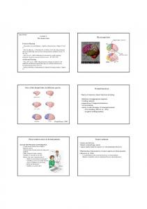
Fig. 1. This figure illustrates the frontal cortical—basal ganglia—thalamic circuits, supporting the fractionation of the frontal functional regions. Area 10 is not part of this circuitry, and is schematically presented in its polar location to suggest its integrative functions. For an expanded explanation of the anatomical and functional connections of Area 10 with other brain regions, see Gilbert, Gonen-Yaacovi, Benoit, Volle, & Burgess (2010) and Petrides and Pandya (2007). The figure also serves as a summary of the findings. STG 5 superior temporal gyrus; Right/Left 5 cerebral hemispheres.
There is other support for this model of discrete functional categories within the frontal lobes. Comparative anatomical studies and mapping of human brain development have identified two main frontal systems—a lateral one with primarily bidirectional connections to and from posterior cortices (executive) and an inferior/medial one with prominent limbic connections (emotional) (Pandya & Yeterian, 1996). These two systems are ‘‘energized’’ by the superior medial region. The frontopolar region—both phylogenetically and ontogenetically late developing—integrates the executive and the emotional processes. Of the exquisitely mapped, vertically segregated frontal–subcortical circuits (Alexander et al., 1986), three align with our categories of energization, executive, and emotional. The frontopolar region (integrative function) does not have major frontal–subcortical connections precisely because its role is integrating processes within the frontal lobes and with other regions (Petrides & Pandya, 2007). See Figure 1.
Brain Systems and Networks Our goal was to understand and fractionate the functions of the frontal lobes. For each frontal cortical functional region, there is a connection with a specific basal ganglia area, continuing to a defined-thalamic region. Are the functions of the connected regions the same? This question needs to be pursued
using similar operational definitions of processes as those outlined in the frontal patients. However, the demonstration of a parallel functional separation within the subcortical regions will be difficult, because of the smaller size of these areas. For example, in several of our studies (Stuss et al., 1998, 2000), patterns of performance after basal ganglia damage were similar to frontal patterns but, other than left–right differences, further distinctions could not be isolated. One interesting approach has been the use of deep brain stimulation in the subthalamic nucleus to demonstrate an alteration in a frontotemporal network related to the performance of a verbal fluency task (Schroeder et al., 2003). Similar questions could—and should—be raised about the functional similarities and dissimilarities in other frontal networks. We have pursued the question related to frontocerebellar connectivity. If characterization of patients is strict, and patients are studied in a chronic stage of recovery with lesions limited to the cerebellum, the functional similarity is quite limited and specific (Alexander, Gillingham, Schweizer, & Stuss, in press; Schweizer, Alexander, Gillingham, Cusimano, & Stuss, 2010). The potential specific role of white matter pathways also needs to be investigated. Understanding the role of specific brain regions within the frontal lobes is not phrenology; analysis of the simple tasks and how different regions, frontal and non-frontal, are required depending on task demands and difficulty, identifies
Frontal lobe functions these nodes as parts of flexible and dynamic networks (Stuss, 2006). More importantly, it provides a foundation for investigating and understanding the role of separate circuits, and the integration between and among circuits. The frontal lobes may play a key role in such integration but there is suggestion that integration and restructuring of neural assemblies can occur in different regions (Haber & Calzavara, 2009).
CONCLUSION Moving from multidimensional, clinical tasks to controlled experimental processes to reliably correlated brain regions has provided replicable evidence of fractionated frontal lobe functioning. Within the SAS model of attention, the processes of energization, monitoring, and task-setting (if-then logic and contingent responding) have been consistently identified and correlated with specific brain regions. It is highly likely that the appropriate experimental paradigm will reveal more frontal lobe processes, likely associated with other frontal brain regions. Our summary paragraph in the 1995 study, rephrased as follows, remains relevant today. The frontal lobes do not equal a central executive. Executive functions represent only one functional category within the frontal lobes. These frontal functions are domain general, possibly because of the extensive reciprocal connections with virtually all other brain regions, integrating information from these regions. Further integration of these processes with emotional and motivational processes allows the most complex behaviors (Alexander, 2006; Grafman, 2002).
FUTURE HORIZONS There are three: (1) Can experimental neuropsychology identify and extract additional basic processes—executive, emotional, others—as the raw material for understanding integrated brain functioning in the most complex tasks, including ‘‘consciousness’’? (Stuss & Benson, 1986; Stuss, Picton, & Alexander, 2001); (2) Can experimental neuroscience create methodologies for observing these processes in their normal operation through imaging? (a journey already started; e.g., Brass & von Cramon, 2004; Floden et al., 2011; Sturm & Willmes, 2001; Vallesi, McIntosh, Alexander, & Stuss, 2009; Vallesi, McIntosh, Shallice, & Stuss, 2009); (3) Can understanding these fundamental processes provide a framework for pharmacotherapeutic or behavioral treatments and assessments when the processes are impaired? (Cicerone, Levin, Malec, Stuss, & Whyte, 2006; Levine, Turner, & Stuss, 2008; Wang et al., 2007).
ACKNOWLEDGMENTS The following are gratefully acknowledged: all the co-authors on the various publications for their essential contributions to all aspects of the research program, especially M.P. Alexander, T. Shallice, T. Picton; funding agencies, in particular Canadian Institutes of
763 Health Research, the McDonnell Foundation, the Centre for Stroke Recovery; my research lab, in particular lab manager S. Gillingham; M.P. Alexander and D.P. Stuss for comments on an earlier draft, and S. Gillingham for figure preparation. There are no conflicts of interest to report.
REFERENCES Alexander, M.P. (2006). Impairments of procedures for implementing complex language are due to disruption of frontal attention processes. Journal of the International Neuropsychological Society, 12, 236–247. Alexander, G.E., Delong, M.R., & Strick, P.I. (1986). Parallel organization of functionally segregated circuits linking basal ganglia and cortex. Annual Review of Neuroscience, 9, 357–381. Alexander, M.P., Gillingham, S., Schweizer, T.A., & Stuss, D.T. (in press). Adults with chronic cerebellar lesions have only modest cognitive impairments. Cortex. Alexander, M.P., Stuss, D.T., & Gillingham, S. (2009). Impaired list learning is not a general property of frontal lesions. Journal of Cognitive Neuroscience, 21, 1422–1434. Alexander, M.P., Stuss, D.T., Picton, T., Shallice, T., & Gillingham, S. (2007). Regional frontal injuries cause distinct impairments in cognitive control. Neurology, 68, 1515–1523. Alexander, M.P., Stuss, D.T., Shallice, T., Picton, T.W., & Gillingham, S. (2005). Impaired concentration due to frontal lobe damage from two distinct lesion sites. Neurology, 65, 572–579. Bechara, A., Damasio, H., Damasio, A.R., & Lee, G.P. (1999). Different contributions of the human amygdale and ventromedial prefrontal cortex to decision-making. The Journal of Neuroscience, 19, 5473–5481. Brass, M., & von Cramon, D.Y. (2004). Selection for cognitive control: A functional magnetic resonance imaging study on the selection of task-relevant information. Journal of Neuroscience, 24, 8847–8852. Breiman, L., Friedman, J.H., Olshen, R.A., & Stone, C.J. (1984). Classification and Regression Trees (CART). Belmont, CA: Wadsworth. Burgess, P.W., Gilbert, S.J., & Dumontheil, I. (2007). Function and localisation within rostral prefrontal cortex (area 10). Philosophical Transactions of the Royal Society B: Biological Sciences, 362, 887–899. Cicerone, K., Levin, H., Malec, J., Stuss, D., & Whyte, J. (2006). Cognitive rehabilitation interventions for executive function: Moving from bench to bedside in patients with traumatic brain injury. Journal of Cognitive Neuroscience, 18, 1212–1222. Floden, D., Vallesi, A., & Stuss, D.T. (2011). Task Context and Frontal Lobe Activation in the Stroop Task. Journal of Cognitive Neuroscience, 23, 867–879. Fuster, J.M. (2008). The prefrontal cortex. London: Academic Press. Gilbert, S.J., Gonen-Yaacovi, G., Benoit, R.G., Volle, E., & Burgess, P.W. (2010). Distinct functional connectivity associated with lateral versus medical rostral prefrontal cortex: A metaanalysis. Neuroimage, 53, 1359–1367. Godefroy, O., Cabaret, M., Petit-Chenal, V., Pruvo, J.-P., & Rousseaux, M. (1999). Control functions of the frontal lobes. Modularity of the central-supervisory system? Cortex, 35, 1–20. Grafman, J. (2002). The structured event complex and the human prefrontal cortex. In D.T. Stuss & R.T. Knight (Eds.), Principles
764 of frontal lobe function (pp. 292–310). New York: Oxford University Press. Haber, S.N., & Calzavara, R. (2009). The cortico-basal ganglia integrative network: The role of the thalamus. Brain Research Bulletin, 78, 69–74. Heilman, K.M., & Watson, R.T. (1977). The neglect syndrome – A unilateral defect of the orienting response. In S. Harnad, R.W. Doty, J. Jaynes, L. Goldstein, & G. Krauthamer, (Eds.), Lateralization in the nervous system (pp. 285–302). New York: Academic Press. Knight, R.T. (1991). Evoked potential studies of attention capacity in human frontal lobe lesions. In H. Levin, H. Eisenberg, & F. Benton (Eds.), Frontal lobe functions and dysfunction (pp. 139–153). Oxford: Oxford University Press. Koechlin, E., Basso, G., Pietrini, P., Panzer, S., & Grafman, J. (1999). The role of the anterior prefrontal cortex in human cognition. Nature, 399, 148–151. Lezak, M.D. (1995). Neuropsychological assessment (3rd ed.). New York: Oxford University Press. Levine, B., Turner, G.R., & Stuss, D.T. (2008). Rehabilitation of frontal lobe functions. In D.T. Stuss, G. Winocur, & I.H. Robertson (Eds.), Cognitive neurorehabilitation, 2nd edition: Evidence and application (pp. 464–486). Cambridge: Cambridge University Press. Luria, A.R. (1973). The working brain: An introduction to neuropsychology (B. Haigh, Trans). New York: Basic Books. Manes, F., Sahakian, B., Clark, L., Rogers, R., Antoun, N., Aitken, M., & Robbins, T. (2002). Decision-making processes following damage to the prefrontal cortex. Brain, 125, 624–639. Mesulam, M.-M. (1985). Principles of behavioral neurology. Philadelphia: Davis. Mirsky, A.F., Anthony, B.J., Duncan, C.C., Ahearn, M.B., & Kellam, S.G. (1991). Analysis of the elements of attention: A neuropsychological approach. Neuropsychology Review, 2, 109–145. Norman, D.A., & Shallice, T. (1986). Attention to action: Willed and automatic control of behaviour. In R.J. Davidson, G.E. Shwartz, & D. Shapiro (Eds.), Attention to action: Willed and automatic control of behaviour (pp. 1–18). New York: Plenum. Pandya, D.N., & Yeterian, E.H. (1996). Comparison of prefrontal architecture and connections. Philosophical Transactions of the Royal Society of London, B: Biological Sciences, 351, 1423–1432. Paus, T., Zatorre, R.J., Hofle, N., Caramanos, J.G., Petrides, M., & Evans, A.C. (1997). Time-related changes in neural systems underlying attention and arousal during the performance of an auditory vigilance task. Journal of Cognitive Neuroscience, 9, 392–408. Petrides, M., & Pandya, D.M. (1994). Comparative architectonic analysis of the human and macaque frontal cortex. In F. Boller & J. Grafman (Eds.), Comparative architectonic analysis of the human and macaque frontal cortex (pp. 17–57). Amsterdam: Elsevier. Petrides, M., & Pandya, D.M. (2007). Efferent association pathways from the rostral prefrontal cortex in the macaque monkey. The Journal of Neuroscience, 27, 11573–11586. Picton, T.W., Stuss, D.T., Alexander, M.P., Shallice, T., Binns, M.A., & Gillingham, S. (2007). Effects of focal frontal lesions on response inhibition. Cerebral Cortex, 17, 826–838. Picton, T.W., Stuss, D.T., Shallice, T., Alexander, M.P., & Gillingham, S. (2006). Keeping time: Effects of focal frontal lesions. Neuropsychologia, 44, 1195–1209. Posner, M.I., & Petersen, S.E. (1990). The attention system of the human brain. Annual Review of Neuroscience, 13, 25–42.
Stuss Schroeder, U., Kuehler, A., Lange, K.W., Haslinger, B., Tronnier, V.M., Krause, M., y Ceballos-Baumann, A.O. (2003). Subthalamic nucleus stimulation affects a frontotemporal network: A PET study. Annals of Neurology, 54, 445–450. Schweizer, T.A., Alexander, M.P., Gillingham, S., Cusimano, M., & Stuss, D.T. (2010). Lateralized cerebellar contributions to word generations: A verbal and semantic fluency study. Behavioural Neurology, 23, 31–37. Shallice, T. (1982). Specific impairments of planning. Philosophical Transactions of the Royal Society of London, Series B: Biological Sciences, 298, 199–209. Shallice, T., & Burgess, P.W. (1996). Domains of supervisory control and the temporal organisation of behaviour. Philosophical Transactions of the Royal Society of London, B: Biological Sciences, 351, 1405–1412. Shallice, T., Stuss, D.T., Alexander, M.P., Picton, T.W., & Derkzen, D. (2008). The multiple dimensions of sustained attention. Cortex, 44, 794–805. Shallice, T., Stuss, D.T., Picton, T.W., Alexander, M.P., & Gillingham, S. (2008). Multiple effects of prefrontal lesions on task-switching. Frontiers in Human Neuroscience, 1, 1–12. Sturm, W., & Willmes, K. (2001). On the functional neuroanatomy of intrinsic and phasic alertness. Neuroimage, 14, S76–S84. Stuss, D.T. (2006). Frontal lobes and attention: Processes and networks, fractionation and integration. Journal of the International Neuropsychological Society, 12, 261–271. Stuss, D.T., & Alexander, M.P. (2007). Is there a dysexecutive syndrome? Philosophical Transactions of the Royal Society of London. Series B: Biological Sciences, 362, 901–915. Stuss, D.T., Alexander, M.P., Floden, D., Binns, M.A., Levine, B., McIntosh, A.R., y Hevenor, S.J. (2002). Fractionation and localization of distinct frontal lobe processes: Evidence from focal lesions in humans. In D.T. Stuss & R.T. Knight (Eds.), Principles of frontal lobe function (pp. 392–407). New York: Oxford University Press. Stuss, D.T., Alexander, M.P., Hamer, L., Palumbo, C., Dempster, R., Binns, M., y Izukawa, D. (1998). The effects of focal anterior and posterior brain lesions on verbal fluency. Journal of the International Neuropsychological Society, 4, 265–278. Stuss, D.T., Alexander, M.P., Palumbo, C.L., Buckle, L., Sayer, L., & Pogue, J. (1994). Organizational strategies of patients with unilateral or bilateral frontal lobe injury in word list learning tasks. Neuropsychology, 8, 355–373. Stuss, D.T., Alexander, M.P., Shallice, T., Picton, T.W., Binns, M.A., MacDonald, R., y Katz, D.I. (2005) Multiple frontal systems controlling response speed. Neuropsychologia, 43, 396–417. Stuss, D.T., & Benson, D.F. (1986). The frontal lobes. New York: Raven Press. Stuss, D.T., Binns, M.A., Murphy, K.J., & Alexander, M.P. (2002). Dissociations within the anterior attentional system: Effects of task complexity and irrelevant information on reaction time speed and accuracy. Neuropsychology, 16, 500–513. Stuss, D.T., & Knight, R.T. (Eds.). (2002). Principles of frontal lobe function. New York: Oxford University Press. Stuss, D.T., Levine, B., Alexander, M.P., Hong, J., Palumbo, C., Hamer, L., y Izukawa, D. (2000). Wisconsin Card Sorting Test performance in patients with focal frontal and posterior brain damage: Effects of lesion location and test structure on separable cognitive processes. Neuropsychologia, 38, 388–402. Stuss, D.T., Picton, T.W., & Alexander, M.P. (2001). Consciousness, self-awareness, and the frontal lobes. In S.P. Salloway, P.F. Malloy, & J.D. Duffy (Eds.), The frontal lobes and neuropsychiatric
Frontal lobe functions illness (pp. 101–109). Washington: American Psychiatric Publishing, Inc. Stuss, D.T., Shallice, T., Alexander, M.P., & Picton, T.W. (1995). A multidisciplinary approach to anterior attentional functions. Annals of the New York Academy of Sciences, 769, 191–212. Vallesi, A., McIntosh, A.R., Alexander, M.P., & Stuss, D.T. (2009). fMRI evidence of a functional network setting the criteria for withholding a response. Neuroimage, 45, 537–548.
765 Vallesi, A., McIntosh, A.R., Shallice, T., & Stuss, D.T. (2009). When time shapes behavior: fMRI evidence of brain correlates of temporal monitoring. Journal of Cognitive Neuroscience, 21, 1116–1126. Wang, M., Ramos, B.P., Paspalas, C.D., Shu, Y., Simen, A., Dogue, A., y Arnsten, A.F. (2007) A2A-Adrenoceptors strengthen working memory networks by inhibiting cAMP-HCN channel signaling in prefrontal cortex. Cell, 129, 397–410.