Commentary
Gene function: Getting specific, generally speaking S. W. Michnick* and F.-X. Campbell Valois
M
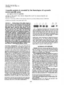
uch of modern biological research is of studying gene function and to another concerned with identifying genes class of problems: designing proteins that and the protein products of genes involved embody desired characteristics or function in cellular processes: determining how, of potential industrial, therapeutic, or funwhen, and where they are involved in damental interest. specific biochemical processes. The tools The essential elements of the strategy by which these aims are achieved can be Baker et al. (10) describe are variations on roughly divided into two types: those that two successful and popular technologies are specific to the study of individual or that have emerged in the last 10 years, classes of proteins versus those with more namely the yeast three-hybrid assay and general and broad utility. General tools protein ‘‘dimerizer’’ technology (11–20). are methods that allow for rapid inference The basis of their approach is described in of function of a gene Fig. 1. In the genproduct, either from eral concept, a chithe mRNA or the meric molecule (the An assay for detection of an active proteins it codes for, dimerizer) that confor any particular tains three compoform of a cephalosporinase of molecule or class of nents is synthesized: Enterobacter cloacae called P99 molecules. These are one moiety that is a is presented. increasingly in dehigh-affinity ligand for one protein, one mand, particularly that is a ligand for those that can be applied to entire genomes or large subsets of another protein, and a linker that contains the genes contained therein. At the same a substrate for some specific or general time, generality comes at a cost. By their class of enzymes. The dimer is introduced nature, general tools do not usually pro- into yeast containing a two-hybrid tranvide high-quality information about the scriptional reporter assay system confunction of a gene and may even mislead, sisting of the two proteins to which the particularly when applied across large dimerizer binds, fused to complementary numbers of genes. Examples include DNA DNA binding and RNA polymerase activatmicroarrays and multidimensional separa- ing domains, respectively. The dimerizer tion-MS and yeast two-hybrid strategies binds to the two proteins simultaneously, that detect protein–protein interactions or allowing for transcription of a reporter complexes (1–9). Although these ap- gene whose presence can be detected by proaches can, respectively, provide infor- enzymatic assays. The two dimerizing proteins are dihydrofolate reductase and glumation on whether and to what extent a cocortocoid receptor ligand-binding dogiven gene is being transcribed in a demains. These are fused to LexA DNA fined condition and with which proteins binding and B42 RNA polymerase activathe protein gene product is interacting tion domains, respectively. In the specific with, they cannot provide any insight into case presented by Baker et al. (10), an other crucial questions. For instance, assay for detection of an active form of a many proteins are enzymes that catalyze cephalosporinase of Enterobacter cloacae biochemical reactions. In the case of novel called P99 is presented. Thus, the dimergenes of unknown function it is not easy to izer consists of methotrexate linked via a definitively determine whether they are thioether to the -lactam cephalosporin enzymes nor what reactions they may cat- and in turn, to dexamethasone by a pepalyze. In this issue of PNAS Baker et al. tide bond (Mtx-cephem-Dex). When these (10) present a proof-of-principle study on proteins are expressed in the budding another approach that might fulfill this yeast Saccharomyces cerevisiae grown on need, but unlike the approaches described medium containing an appropriate conpreviously, the strategy is applied in in- centration of the dimerizer, simultaneous tact, living cells. Here we discuss the sig- binding of dihydrofolate reductase-LexA nificance of this distinction, the historical to the Mtx moiety and of glucocortocoid context of the development of the method, receptor-B42 to the Dex moiety of Mtxand potential applications to the problems cephem-Dex, results in reconstitution of www.pnas.org兾cgi兾doi兾10.1073兾pnas.012697899
an active LexA promoter and transcription of the -galactosidase reporter gene. The activity of the -galactosidase gene product is detected by using substrates (5-bromo-4-chloro-3-indolyl -D-galactoside and o-nitrophenyl-D--galactoside) that are converted to colored or fluorescent products by the -galactosidase. Detection of -lactamase activity is based on a loss of expression of -galactosidase if the -lactone ring is cleaved by a lactamase, resulting in expulsion of the leaving group at the C3⬘ position of the cephalosporin and therefore disintegration of the dimerizer induced dihydrofolate reductase-LexA-glucocortocoid receptor-B42 complex. Thus, a loss of activity indicates that a lactamase activity is present in the cell. Baker et al. tested the system by screening an artificial library of P99 the cephalosporinase containing either active WT or inactive mutant forms and demonstrate that selection for active enzyme can be reliably made. The Baker technology represents a first step in creating generalized detectors of enzyme activities in cells. It is a potentially valuable tool in proteomic and protein engineering for the discovery of novel enzymatic activities. There are, as the authors point out, many variations on this theme that could be devised. The dimerizer could contain general or specific substrates for many cleavage or ligation reactions, aimed at different classes of enzymes. Previously, a system in bacteria had been described based on AraC chimera dimerizer-regulated transcription to screen for dehydratase activity (21). The different basis of the dimerizer chemistry make these approaches complementary and might allow the screening of a larger set of enzymatic reactions. Second, reporter assays that are not limited to a cellular compartment or specific cell type could be used. For instance, a recent example of a fluorescence resonance energy transfer (FRET) assay for detecting specific protein kinase activities has been described, and such a FRET-based assay See companion article on page 16537. *To whom correspondence should be addressed. E-mail:
[email protected].
PNAS 兩 December 24, 2002 兩 vol. 99 兩 no. 26 兩 16513–16515
COMMENTARY
De´partements de Biochimie et Biologie Mole´culaire, Universite´ de Montre´al, C.P. 6128, Succursale Centre-ville, Montre´al, QC, Canada H3C 3J7
Fig. 1. A reaction-independent complementation assay for detecting enzyme activities of genes or artificial libraries of genes. (Upper) A specific ‘‘three-hybrid’’ assay for detecting lactamase activity. The two dimerized proteins consist of dihydrofolate reductase (rose) and glucocortocoid receptor ligand-binding domains (blue). These are fused to LexA DNA binding and B42 RNA polymerase activation domains and will reconstitute active RNA polymerase complex to transcribe the reporter gene -galactosidase when the two proteins are noncovalently ligated via the dimerizer dexamethasone-cephem-methotrexate molecule. (Lower Left) Examples of ligation or cleavage reactions that could be used in the assay. (Lower Right) Application of the assay to protein engineering. Figure was provided by Debleena Sengupta and Virginia Cornish (Columbia University, New York).
could also be used with a dimerizer system (22, 23). Equally, several protein fragment complementation assays could be used. These are designed to detect protein– protein interactions but small moleculeinduced dimerization of proteins have been demonstrated with these techniques, and in addition to detecting enzyme activities, some of these assays could be used to sublocalize these precisely inside the cells (24–28). However, one important limitation of enzyme activity assays for proteomic or drug discovery is that the enzymatic activity has to be studied in a cell model devoid or weakly displaying similar enzymatic activity for the screen to be easily tractable. The combination of these technologies in different cell types could expand the nature of enzymatic reactions assayable. Elegant strategies to allow for in vitro covalent tagging of specific enzymes with, for examples, biotin and fluorescent dyes have been developed. Ben Cravatt and coworkers at Scripps (29–34) have devel-
oped a number of experimental strategies that allow for identification of protease and other enzyme activities in different cell types under different circumstances such as in different types of cancer cells. As demonstrated, it allowed for phenotyping of these cells for invasiveness. With increased knowledge of the chemistry of diverse enzymatic activities, one could conceivably develop compounds for every biologically catalyzed reaction. Thus, the strategy described by Baker et al. (10) would be complementary to these other approaches in a proteomic perspective. How can these new methods aid in large-scale gene discovery? We think that the process of ontologically defining each gene of an organism could be envisioned in a three-step strategy going from general to more specific considerations. The first step consists of linking the gene of interest to all of the cellular processes it might be involved in, as inferred, for example, from the complete set of physical interactions map obtained from a large-scale genomic
16514 兩 www.pnas.org兾cgi兾doi兾10.1073兾pnas.012697899
screen with methods such as yeast twohybrid assays and MS or combinations of DNA and protein microarray data (2, 4–6, 35–38). The next step consists of the validation of the functional inference made with the results of the large-scale experiments. Generally speaking these can be attained by comparing the phenotype observed, whether through classical genetic approaches, RNAi, or newly emerging technologies, between the gene products thought to be involved in interactions network with the gene under study and this gene. This kind of comparison could then be expanded to the pattern observed in subcellular localizations and expression profiles and to genes involved in the inferred functional class, but that have not been shown to interact with the gene of interest. However, and, this is the last step, the addition of more specific assays for assigning function to genes would be a huge step toward completion of this goal. The new enzyme discovery tool described by Baker et al. would add an extra and Michnick and Campbell Valois
an enzyme activity is to express the library in a cell in which expression of library members with the desired characteristics confers growth capabilities in a given condition, in a specific medium or at nonpermissive growth temperature, for example. In the case where there is no such assay, the methods described by Baker et al. would be perfectly suited for screening novel enzymatic activities, but of course, with the same inherent limitations that we discussed for functional inference of gene products.
One way or another and for whatever purpose, we can hope to see the development of biological research tools that at once can be applied generally while at the same time provide specific information. But all of these technologies need to be challenged to check whether they can be useful for addressing real scientific questions. We are seeing that the development of novel strategies or combinations of smart technologies that already exist are creating new opportunities to explore the details of gene function in finer detail.
1. Fields, S. & Song, O. (1989) Nature 340, 245–246. 2. Ito, T., Chiba, T., Ozawa, R., Yoshida, M., Hattori, M., Sakaki, Y., Zhang, J., Ma, Y., Taylor, S. S. & Tsien, R. Y. (2001) Proc. Natl. Acad. Sci. USA 98, 4569–4574. 3. Lashkari, D. A., DeRisi, J. L., McCusker, J. H., Namath, A. F., Gentile, C., Hwang, S. Y., Brown, P. O. & Davis, R. W. (1997) Proc. Natl. Acad. Sci. USA 94, 13057–13062. 4. Uetz, P., Giot, L., Cagney, G., Mansfield, T. A., Judson, R. S., Knight, J. R., Lockshon, D., Narayan, V., Srinivasan, M., Pochart, P., et al. (2000) Nature 403, 623–627. 5. Ho, Y., Gruhler, A., Heilbut, A., Bader, G. D., Moore, L., Adams, S. L., Millar, A., Taylor, P., Bennett, K., Boutilier, K., et al. (2002) Nature 415, 180–183. 6. Gavin, A. C., Bosche, M., Krause, R., Grandi, P., Marzioch, M., Bauer, A., Schultz, J., Rick, J. M., Michon, A. M., Cruciat, C. M., et al. (2002) Nature 415, 141–147. 7. Boulton, S., Gartner, A., Reboul, J., Vaglio, P., Dyson, N., Hill, D. E. & Vidal, M. (2002) Science 295, 127–131. 8. Roberts, C. J., Nelson, B., Marton, M. J., Stoughton, R., Meyer, M. R., Bennett, H. A., He, Y. D., Dai, H., Walker, W. L., Hughes, T. R., et al. (2000) Science 287, 873–880. 9. Chu, S., DeRisi, J., Eisen, M., Mulholland, J., Botstein, D., Brown, P. O. & Herskowitz, I. (1998) Science 282, 699–705. 10. Baker, K., Bleczinski, C., Lin, H., Salazar-Jimenez, G., Sengupta, D., Krane, S. & Cornish, V. W. (2002) Proc. Natl. Acad. Sci. USA 99, 16537– 16542. 11. Belshaw, P. J., Spencer, D. M., Crabtree, G. R. & Schreiber, S. L. (1996) Chem. Biol. 3, 731–738. 12. Briesewitz, R., Ray, G. T., Wandless, T. J. & Crabtree, G. R. (1999) Proc. Natl. Acad. Sci. USA 96, 1953–1958.
13. Clackson, T., Yang, W., Rozamus, L. W., Hatada, M., Amara, J. F., Rollins, C. T., Stevenson, L. F., Magari, S. R., Wood, S. A., Courage, N. L., et al. (1998) Proc. Natl. Acad. Sci. USA 95, 10437– 10442. 14. Licitra, E. J. & Liu, J. O. (1996) Proc. Natl. Acad. Sci. USA 93, 12817–12821. 15. Rivera, V. M., Wang, X., Wardwell, S., Courage, N. L., Volchuk, A., Keenan, T., Holt, D. A., Gilman, M., Orci, L., Cerasoli, F., Jr., et al. (2000) Science 287, 826–830. 16. Rollins, C. T., Rivera, V. M., Woolfson, D. N., Keenan, T., Hatada, M., Adams, S. E., Andrade, L. J., Yaeger, D., van Schravendijk, M. R., Holt, D. A., et al. (2000) Proc. Natl. Acad. Sci. USA 97, 7096–7101. 17. Rosen, M. K., Amos, C. D. & Wandless, T. J. (2000) J. Am. Chem. Soc. 122, 11979–11982. 18. Sengupta, D. J., Zhang, B. L., Kraemer, B., Pochart, P., Fields, S. & Wickens, M. (1996) Proc. Natl. Acad. Sci. USA 93, 8496–8501. 19. Spencer, D. M., Wandless, T. J., Schreiber, S. L. & Crabtree, G. R. (1993) Science 262, 1019–1024. 20. Vogel, K. W., Briesewitz, R., Wandless, T. J. & Crabtree, G. R. (2001) Adv. Protein Chem. 56, 253–291. 21. Firestine, S. M., Salinas, F., Nixon, A. E., Baker, S. J. & Benkovic, S. J. (2000) Nat. Biotechnol. 18, 544–547. 22. Ting, A. Y., Kain, K. H., Klemke, R. L. & Tsien, R. Y. (2001) Proc. Natl. Acad. Sci. USA 98, 15003–15008. 23. Zhang, J., Ma, Y., Taylor, S. S. & Tsien, R. Y. (2001) Proc. Natl. Acad. Sci. USA 98, 14997–5002. 24. Galarneau, A., Primeau, M., Trudeau, L. E. & Michnick, S. W. (2002) Nat. Biotechnol. 20, 619– 622. 25. Michnick, S. W., Remy, I., Campbell Valois, F.-X., Vale´e-Belisle, A. & Pelletier, J. N. (2000) Methods Enzymol. 328, 208–230.
26. Pelletier, J. N., Campbell Valois, F. & Michnick, S. W. (1998) Proc. Natl. Acad. Sci. USA 95, 12141–12146. 27. Remy, I., Wilson, I. A. & Michnick, S. W. (1999) Science 283, 990–993. 28. Remy, I., Pelletier, J. N., Galarneau, A. & Michnick, S. W. (2001) in Protein–Protein Interactions: A Molecular Cloning Manual, ed. Golemis, E. A. (Cold Spring Harbor Lab. Press, Plainview, NY), pp. 449–475. 29. Adam, G. C., Cravatt, B. F. & Sorensen, E. J. (2001) Chem. Biol. 8, 81–95. 30. Adam, G. C., Sorensen, E. J. & Cravatt, B. F. (2002) Nat. Biotechnol. 20, 805–809. 31. Cravatt, B. F. & Sorensen, E. J. (2000) Curr. Opin. Chem. Biol. 4, 663–668. 32. Jessani, N., Liu, Y., Humphrey, M. & Cravatt, B. F. (2002) Proc. Natl. Acad. Sci. USA 99, 10335– 10340. 33. Kidd, D., Liu, Y. & Cravatt, B. F. (2001) Biochemistry 40, 4005–4015. 34. Liu, Y., Patricelli, M. P., Cravatt, B. F., West, M. W. & Hecht, M. H. (1999) Proc. Natl. Acad. Sci. USA 96, 14694–14699. 35. Zhu, H., Bilgin, M., Bangham, R., Hall, D., Casamayor, A., Bertone, P., Lan, N., Jansen, R., Bidlingmaier, S., Houfek, T., et al. (2001) Science 293, 2101–2105. 36. MacBeath, G. & Schreiber, S. L. (2000) Science 289, 1760–1763. 37. Ideker, T., Thorsson, V., Ranish, J. A., Christmas, R., Buhler, J., Eng, J. K., Bumgarner, R., Goodlett, D. R., Aebersold, R. & Hood, L. (2001) Science 292, 929–934. 38. Lee, T. I., Rinaldi, N. J., Robert, F., Odom, D. T., Bar-Joseph, Z., Gerber, G. K., Hannett, N. M., Harbison, C. T., Thompson, C. M., Simon, I., et al. (2002) Science 298, 799–804.
Michnick and Campbell Valois
PNAS 兩 December 24, 2002 兩 vol. 99 兩 no. 26 兩 16515
COMMENTARY
more specific functional inference in an obvious way to this scheme. We think the potential use of the method described by Baker et al. (10) to protein engineering is very promising. Effectively, in the search for novel enzymatic activity, the general strategy includes two distinct methods: one that can generate a diverse and large set of sequences and the other that allows for isolation of the sequences producing polypeptides displaying the sought-after characteristics. Obviously, an ideal way to screen for