THE JOURNAL OF BIOLOGICAL CHEMISTRY © 2000 by The American Society for Biochemistry and Molecular Biology, Inc.
Vol. 275, No. 34, Issue of August 25, pp. 26523–26529, 2000 Printed in U.S.A.
Gene-specific Silencing by Expression of Parallel Complementary RNA in Escherichia coli* Received for publication, April 4, 2000, and in revised form, June 6, 2000 Published, JBC Papers in Press, June 9, 2000, DOI 10.1074/jbc.M002833200
Nickolai A. Tchurikov‡§, Ludmila G. Chistyakova‡, Genadii B. Zavilgelsky¶, Iliya V. Manukhov¶, Boris K. Chernov储, and Yulia B. Golova储 From the ‡Department of Genome Organization and 储Group of Genes Chemical Synthesis, Engelhardt Institute of Molecular Biology Russian Academy of Sciences, Vavilov str. 32, Moscow 117984, Russia and the ¶Laboratory of Bacterial Genetics, State Scientific Center of Russian Federation GNIIGENETICA, 1th Dorozhnii pr. 1, Box 825, Moscow 113545, Russia
Gene-specific silencing refers to a phenomenon in which expression of an individual gene can be specifically repressed by different mechanisms on the levels of transcription, RNA splicing, transport, degradation in nuclei or cytoplasm, or blocking of translation. In different species gene-specific silencing was observed by expression or injections of antiparallel double-stranded RNA formed by a fragment of mRNA and antisense RNA. Here we show a potent and specific gene silencing in bacteria by expression of RNA, that is complementary in a parallel orientation to Escherichia coli lon mRNA. Moreover, the expression of parallel RNA is more effective at producing interference than expression of antisense RNA corresponding to the same mRNA region. Both effects of interference mediated either by parallel RNA or antiparallel RNA gradually decrease up to the 40th generation. Together with in vitro nuclease protection studies these results indicate that a parallel RNA duplex might be formed in vivo and both types of duplexes, antiparallel or parallel, can induce gene-specific silencing by similar mechanisms.
There has been dramatic recent progress in uncovering the gene-specific silencing in a number of organisms (1–3). Several lines of evidence suggest that dsRNA1 is the effector molecule responsible for RNA-mediated silencing or co-suppression. dsRNA is formed by mRNA and antisense RNA, that corresponds to the non-coding strand of the same gene. However, in experiments on Caenorhabditis elegans it was demonstrated that injections of gel-purified antisense RNA corresponding to an abundant transcript is less active at producing interference than in vitro annealed dsRNA samples (1). The purification
* This work was supported by Russian State Program “Frontiers in Genetics” Grant 99-1-085 and Russian Foundation for Basic Research Grants 97-04-49897 and 99-04-48065). The costs of publication of this article were defrayed in part by the payment of page charges. This article must therefore be hereby marked “advertisement” in accordance with 18 U.S.C. Section 1734 solely to indicate this fact. The nucleotide sequence(s) reported in this paper has been submitted to the GenBankTM/EBI Data Bank with accession number(s) J03896. § To whom all correspondence should be addressed. Tel.: 7-0951359753; Fax: 7-095-1351405; E-mail:
[email protected]. 1 The abbreviations used are: dsRNA, antiparallel double-stranded RNA; pRNA, RNA possessing complementary bases in the same polarity to mRNA; parlon, construct or its transcript possessing complementary bases in parallel orientation to a fragment of the lon mRNA; mirlon, construct or its transcript possessing mirror ordering of bases in respect to a fragment of the lon mRNA; antilon, construct or its transcript comprising the lon antisense RNA; RNAi, RNA interference; bp, base pair(s). This paper is available on line at http://www.jbc.org
was performed to remove the traces of dsRNA from in vitro synthesized RNA preparations because of the nonspecific activity of RNA polymerases. It was concluded that the observation of co-suppression and RNA interference uncovered the existence of a novel cellular mechanisms for regulation of gene expression (2, 4). The phenomenon has been described in fungi, protozoa, plants, invertebrates, and vertebrates (5–10). This suggests a evolutionary conservation of the physiological mechanisms involved. The mechanisms of RNAi remain largely unknown. It was concluded that RNAi and co-suppression work by an equivalent core mechanism produces decrease or elimination of a target mRNA transcript (2, 3, 4, 6). In experiments on gene silencing (“quelling”) in Neurospora crassa it was shown that in the mutant defective in quelling the gene specifying RNA-dependent RNA polymerase was affected (5). The genes involved in RNA degradation also could be connected with RNAi (12). It was considered that RNAi mechanisms might operate at the level of transcription and involve proteins of the polycomb complex (13). But recently it was clearly demonstrated that this transcriptional cosuppression mechanism is distinct from RNAi and involve homology recognition at the DNA level (14). The fact demonstrates that although the phenomenology of gene-specific silencing is similar in different taxa, the underlying molecular mechanisms are not yet understood. The physiological role of mechanisms leading to gene-specific silencing remains mysterious. Genes regulated by antisense transcripts were described in Eubacteria, Archaebacteria, and some primitive eukaryotes (15). RNAi was not described in prokaryotes. However, antisense control is bacteria is now recognized as an efficient and specific means of gene (16). This control occurs at many levels, including premature transcription termination, facilitated RNA decay, and direct or indirect blocking of translation. We had previously reported that RNA molecules are capable of forming parallel RNA-RNA duplexes in vitro (17). To address the capacity of parallel double-stranded RNA to induce genespecific silencing, we have used the construct expressing small RNA that is complementary in a parallel orientation (pRNA) to the endogenous lon mRNA in Escherichia coli cells. To our surprise, we found a potent and specific effect of interference on the expression of the gene. In vitro experiments with pRNA and corresponding sense RNA of the gene have revealed nuclease protection bands after annealing. Our results indicate that parallel double-stranded RNA might be formed in vivo and serve as effector for some mechanisms of gene regulation similarly to regular dsRNA.
26523
26524
Gene Silencing by Expression of Parallel Complementary RNA EXPERIMENTAL PROCEDURES
Construction of the Plasmids for in Vivo Experiments—For construction of parlon and mirlon plasmids the chemically synthesized 95-bp DNA (see Fig. 4A) was inserted into the SmaI site of the pUC12 vector in opposite orientations. For preparation of the antilon construct, the EcoRI-HindIII fragment from the pUC19AL1 construct, containing 341-bp HpaI-HindIII fragment of the lon gene (18), was subcloned in the pUC12 vector. All the constructs are expressed under control of the lac promoter of the vector and the in vivo transcripts possess identical stretches of 229 and 300 bases in length at their 5⬘ and 3⬘ ends, respectively, coming from the lac gene, as well as the different sequences in the middle, corresponding to the DNA inserts. The transcription termination signals are provided by pBR322 origin flanking the lac operon fragment in the pUC12 vector (19). All inserts interrupt the open reading frame of the lacZ gene and only the N-part of the gene could be translated. Degradation of Puromycin Peptides—HB101 transformants were grown for 14 h at 37 °C on plates. Separate colonies were inoculated separately in M9 medium containing 80 g/ml DL proline and 50 g/ml ampicillin and grown at 37 °C to A550 ⫽ 0.25– 0.35. Isopropyl-1-thio-D-galactopyranoside and puromycin were added to 0.5 mM and 100 g/ml, respectively, and after incubation for 20 min [3H]leucine (120 – 190 Ci/mmol) was added to 1 Ci/ml for 5 min. Then cells were centrifuged, washed, and suspended in the same medium but containing 300 g/ml unlabeled leucine and a number of 0.5-ml aliquots were taken immediately (zero point) and after 15, 30, 45, and 60 min of incubation. The samples were precipitated by 7% trichloroacetic acid with 10% bovine serum albumin for 30 min to determine the label of acid-soluble fraction. Protein degradation was calculated as a percentage of acidsoluble fraction to the total label incorporated in cells. Direct isolation and in vitro testing of the Lon preparations was performed. Fresh transformed HB101 cells were grown in L broth with 100 g/ml ampicillin at 37 °C to A550 ⫽ 0.3. Then isopropyl-1-thio--Dgalactopyranoside was added to 1 mM and incubation was continued to A550 ⫽ 1.0. Then suspensions of cells were diluted to the equal A550 and equal amounts of cells were peletted, and isolation of the Lon preparations on PC columns was performed as described earlier (18). Protein fractions enriched with the Lon protease were used for testing of ATPdependent proteolysis and results were calculated as % of hydrolysis of [14C]acetylcasein for 1 h at 37 °C with 1 mg of the Lon preparations in the presence of 3 mM ATP (18). To test the effect of KCN administration on degradation of puromycin peptides the HB101 transformants were grown in the presence of puromycin as described above in two tubes and KCN was added in one tube to 5 mM 1 h before cells were precipitated and degradation of 3 H-peptides was tested. To test the degradation of normal 3H-proteins the HB101 transformants were grown and analyzed as described above, but no puromycin was added. RNA Isolation and Northern Blot Analysis—After transformation the portions of HB101 cell suspension in L medium, containing up to 100 transformed cells were incubated in 20 ml of L medium containing 200 g/ml ampicillin for 14 h at 37 °C. Up to 105 of nontransformed cells of different strains were grown in L medium during the same time without ampicillin. For RNA isolation cells were precipitated, suspended in at 0 °C in solution containing 25% sucrose, 50 mM Tris-HCl, pH 8, and treated with 0.5 mg/ml lysozyme for 5 min and then 1 volume of the lysing buffer containing 2% SDS, 1% Brij 58, 0.4% deoxycholate, 0.1 M EDTA, 20 mM Tris-HCl, pH 8, was added on ice for 15 min. Then one-half volume of saturated NaCl solution was added. After precipitation at 16,000 rpm at 4 °C the supernatant was treated 5 times with phenol/chloroform mixture, pH 5, and ethanol precipitated. To remove DNA the pellet was dissolved in 100 l of water and RNA was precipitated with 300 l of 4 M sodium acetate, pH 4.8, after incubation for 1 h at ⫺20 °C. About 20 g of RNA was run in 1.2% agarose gel. Hybridization experiments were performed on identical blots separately with different 32P-labeled RNA probes (specific activities about 109 cpm/g): with antisense RNA synthesized with T7 RNA polymerase in vitro on the lon gene PstI-HindIII fragment (583–1036 bp, GenBank accession number J03896) cloned in the pGEM-1 vector; with probe, prepared by extension of the primer (5⬘-CCGGGTCGACTTAACGCGTTAGCTCC-3⬘) by avian myeloblastosis virus reverse transcriptase on 16 S ribosomal RNA; with RNAs complementary to antilon, parlon, or mirlon transcripts synthesized with T7 RNA polymerase in vitro on the inserts cloned in the pGEM-1 or pGEM-2 vectors. Hybridizations were performed in 5 ml of solution containing 50% formamide, 5 ⫻ SSC, Ficoll, polyvinylpyrolidone, bovine serum albumin (0.1% each) 0.1% SDS, denatured salmon DNA (50 g/ml), denatured pUC12 DNA (20 g/ml),
tRNA (50 g/ml), and 106 cpm of a probe. After hybridization for 24 h at 43 °C the filter was washed 2 times (20 min each) in 2 ⫻ SSC, 0.1% SDS solution at 43 °C, then 3 times at 65 °C in the same solution and finally 2 times at 65 °C in 0.2 ⫻ SSC, 0.1% SDS. In Vivo Measurements of the Luciferase Activity—The plasmid pAC16, containing the lux regulon (16 kilobase BamHI DNA segment from Vibrio fischeri) in pACYC184 vector was introduced in lon⫹ AB1157 cells and lon⫺ AB1899 cells. The obtained lon⫹ cells were transformed by the parlon or antilon expressing constructs or by pUC12 vector and immediately grown in L medium with 200 g/ml ampicillin (100 transformed cells/ml) at 28 °C. 200 l of suspension was mixed with 4 l of 0.001% n-decanal in ethanol for 0.5–1 min before measuring bioluminescence. Then the samples were transferred to the luminometer (consisting of a FEU-85 photomultiplier and a V2–15 microvoltmeter) in the special cells and the luminescence was measured in v (1 v ⫽ 107 quants/s). To study the effect of the lon gene inhibition up to 50 generations, the cells after transformation with constructs or pUC12 vector (100 transformants/ml) were grown as described above to A550 ⫽ 2.0 (4 ⫻ 108 cells/ml), which corresponds to 20 generations (n ⫽ 20), and the first measurement of the luminescence was performed. Then cells were 1000 times diluted in L medium and the growth was proceeded to A550 ⫽ 2.0 (n ⫽ 30) and the second measurement of the luminescence was performed. Similarly the measurements were performed until the 50th generation. Nuclease Protection Assay—The pGEM constructs were linearized completely. RNA transcription was performed in 20 l of solution containing 2–5 g of DNA, 40 mM Tris-HCl, pH 7.5, 6 mM MgCl, 2 mM spermidine, 10 mM NaCl, 10 mM dithiothreitol, 1 units/l of RNasin, ATP, GTP, CTP, UTP (500 M each), 2.5 M ␣-33P-labeled UTP (4000 Ci/mmol), or 50 M 3H-labeled ATP (27 Ci/mmol), and 20 –30 units of T7 RNA polymerase. Specific activities of 33P- and 3H-labeled RNAs were about 4.2 ⫻ l07/g and 1.6 ⫻ l06 cpm/g, respectively. For purification of [33P]RNA the electrophoresis in low melting temperature agarose gels containing methylmercuric hydroxide was performed followed by dithiothreitol treatment, short exposition, and isolation of RNA from a gel slice by heating on 0.5 M ammonium acetate, phenol/chloroform extraction, and ethanol precipitation. About 4.5 ng of [33P]RNA parlon was annealed alone or separately with 45 ng each of 3H-labeled RNAs in 6 l of solution containing 0.1 M NaCl, 10 mM Tris-HCl, pH 7.5, 10 mM MgCl2, and 0.1% SDS under oil in a wet chamber that was cooled from 37 to 0 °C or from 65 to 10 °C for 16 h. The parlon and mirlon RNAs were annealed at 53 °C for 16 h. Then to some samples 10 l of S1 nuclease in concentration from 0.1 to 5 units/l were added in a solution containing 0.3 M NaCl, 0.03 M Na acetate, pH 4.5, 3 mM ZnSO4, 0.5% glycerol, and 25 mg/ml bovine serum albumin and digestion was performed for 40 min at 10 °C. To the samples not treated with S1 nuclease 10 l of the same solution were added. Incubation was performed at 10 °C for 40 min. The antiparallel parlon/mirlon probe was incubated at 37 °C for 40 min. Then 25 l of 90% formamide containing 50 mM EDTA and dyes was added. 1-mm thick denaturing 4% polyacrylamide gels were run in the “Macrophor LKB System” at 65 °C. The samples before loading were heated at 100 °C for 2–3 min. After fractionation the gels were dried and autoradiographed to observe the fractionation of [33P]RNA. Software Used—RNA secondary structures constructions (Fig. 4C) were performed on the Genebee server. The longest antiparallel stretches formed by the lon mRNA and different transcript were selected. The Tm values of the stretches were determined on the Virtual Genome Center server. RESULTS
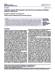
Relations between the lon mRNA and Two RNAs Expressed on Symmetrical DNA—To study the potential of pRNA in gene-specific silencing we selected the lon gene from E. coli. The gene specifies the ATP-dependent Lon protease that plays the important role in the selective degradation of abnormal proteins and limits the time of availability of critical regulatory proteins (21). Fig. 1 shows the relationships between the lon mRNA and short RNAs expressed under control of the lac promoter on three constructs in the pUC12 vector. One RNA, antilon, corresponds to the 341-nucleotide antisense RNA synthesized on the fragment of the lon gene (bases 247–587, numberings here and then as indicated in GenBank sequence accession number J03896). Earlier this fragment was successfully used for potent and selective inhibition of the lon
Gene Silencing by Expression of Parallel Complementary RNA
FIG. 1. The relations between the lon mRNA and RNAs expressed by the constructs. A, the relations between the lon gene and synthesized DNA, inserted in both orientations into the pUC12 vector under control of the lac promoter. B, the sizes of RNA molecules are not shown to scale. The sequences with gaps around triplets refer to complete texts of the parlon and antilon RNAs corresponding to the mRNA. In the lon mRNA the corresponding region possesses the 5⬘ non-coding sequence of 44 bases in length, initiation codon (shown as triplet with the gaps around) and the coding region specifying the following 16 amino acids. Texts under the mRNA and antilon RNA are restricted only to sequences corresponding to the parlon and mirlon RNAs. The axis shows the symmetry of nucleotide ordering between the fragment of mRNA and mirlon RNA.
gene expression by production of antisense RNA (18). That is why in our experiments we used the same region in the antilon construct as a positive control of inhibition by antisense RNA. The antilon RNA can form antiparallel duplex with the mRNA. The second RNA, parlon, has 95 nucleotides and is complementary in the same polarity to the lon mRNA fragment from 369 to 463 nucleotides, and potentially might bind in parallel orientation with the mRNA. The third RNA, mirlon, has inverted sequence corresponding to the same 95-nucleotide lon mRNA region and can form neither antiparallel nor parallel duplexes with the lon mRNA. The parlon and mirlon RNAs could be synthesized only on the heterologous DNA sequence possessing the mirror inversion of nucleotide sequence corresponding to the selected region of the gene. Such chemically synthesized DNA was inserted separately in different orientations in the pUC12 vector (Fig. 1A). All constructs were checked by sequencing. The constructs were introduced by transformation in E. coli cells. Gene-specific Silencing Tested Biochemically as Inhibition of Selective Energy-dependent Degradation of Puromycin Peptides—The expression of the lon gene in the transformants is tested by different approaches. The first one was based on the measurements of the degradation of abnormal proteins, synthesized in the presence of puromycin (20). The drug acts as an analog of aminoacyl-tRNA and terminates the polypeptide
26525
FIG. 2. Analysis of the lon gene expression in HB101 transformants. A, degradation of puromycin 3H-peptides. The cells were treated with [3H]leucine for 5 min in the presence of puromycin. The label was removed and protein degradation was measured in aliquots as percent of the acid-soluble fraction to the total label incorporated in cells as described earlier (18, 20). B, analysis of the lon mRNA content in HB101 transformants and in some nontransformed cells by Northern blotting and hybridization with the lon antisense [32P]RNA probe. Relative levels of the 16 S RNA, antilon, parlon, and mirlon transcripts were determined by hybridization with different probes (see “Experimental Procedures”). The numbers on the left indicate the lengths in kilobase.
chain. HB101 transformants containing the pUC12 plasmid or the mirlon construct released up to 9% of the labeled peptides for 1 h (Fig. 2A). In the presence of antilon or parlon RNA the peptides were degraded appreciably slower. The lon⫺ cells served as a negative control, demonstrating the effect of other proteases. The relative % of the Lon activity in different transformants were calculated. Up to 28 and 10% of the Lon activity were observed upon expression of the antilon and parlon RNAs, respectively (Fig. 2A). Surprisingly, parlon RNA was more effective at producing the interference that antilon RNA. In the first experiments we used the chemically synthesized DNA (synlon), corresponding to the 95-bp region of the lon gene, for the in vivo production of regular antisense RNA (not shown). The letter exactly corresponds to the same region of the mRNA as parlon RNA does (see Fig. 1B). As far as we observed that parlon reveals better inhibition of the lon gene than synlon, we decided to use the 341-bp region of the lon gene from the pUC19AL-1 construct as a positive control of inhibition (18).
26526
Gene Silencing by Expression of Parallel Complementary RNA TABLE I Testing of the energy dependence of proteolysis 14
Transformant
C/Acetylcaseina
ATPdependent proteolysis
Puromicin 3H-peptidesb
% of inhibition
Without KCN
67 71
100 29 11
%
pUC12, mirlon Antilon Parlon
41.7 18 12
Normal 3H-proteinsc
With KCN
Without KCN
11 12 10.3
100 84.3 83.3
%
With KCN %
87 80.5 81
a
Direct isolation and testing in vitro of the Lon preparations was performed. HB101 transformants were grown in the presence of puromycin in two tubes, but KCN was added in one tube to 5 mM 1 h before cells were precipitated and degradation of 3H-peptides was tested. The relative % values of intracellular proteolytic activity are indicated. The level of proteolysis in the pUC12 and mirlon containing cells in the absence of KCN are taken as 100%. c HB101 transformants were grown and analyzed as described in Footnote b, but no puromycin was added. The values of proteolysis in relative % are indicated as described in Footnote b. b
This region, including the 95-bp fragment, was described as the most effective in the selective inhibition of the gene by expression of antisense RNA (18). We found that the antilon construct, possessing this 341-bp fragment, and the synlon plasmid demonstrate the same inhibition of the lon gene activity and that both are less effective than the parlon construct. The higher effectiveness of the parlon transcript was independently confirmed by direct isolation of the Lon protease that could be readily isolated on the phosphocellulose (18) from equal amounts of cells (Table I, Footnote a). In these experiments the inhibition of the expression of the ATP-dependent Lon protease by antilon or parlon RNAs was observed. To inhibit the ATP-dependent proteolysis the administration of KCN to the cell suspension was performed as described earlier (18, 20). It was found that parlon and antilon RNAs affect only an energy-dependent proteolysis of abnormal 3Hpeptides and practically have no effect on the degradation of normal 3H-proteins (Table I, Footnotes b and c). These findings strongly suggest that both parlon and antilon RNAs are mediators of a potent and specific repression of the target gene expression, and parlon RNA is more efficient in this process. Gene-specific Silencing in E. coli HB101 Cells Does Not Reduce the mRNA Content—It was demonstrated earlier, that RNAi establishes an intracellular state leading to a specific decrease or elimination of the corresponding eukaryotic mRNA (4, 6). To elucidate a mechanism of the detected inhibition of the lon gene expression, we performed Northern blot analysis. The same amounts of mRNA were observed in all HB101 transformants (Fig. 2B). The method clearly can check the physiological decrease of the lon mRNA to 16 S RNA. In different nontransformed cells, incubated during the same time but grown to stationary state in the absence of ampicillin, an appreciable decrease of the lon transcripts was observed. The mRNA was absent only in the lon-146 mutant (22) that was used as a negative control. Thus, in our experiments the genespecific silencing does not induce an evident mRNA degradation. The Northern blot analysis also demonstrates that parlon, mirlon, and antilon constructs are expressed at the same level in the transformed cells (Fig. 2B). Thus, the differences in the effects of parlon, mirlon, and antilon constructs on the lon gene expression are not due to differences in amounts of the corresponding RNAs in the transformed cells. pRNA Action Tested Genetically as Inhibition of the Effect of the Gene on the lux Regulon—The last method that we have used for the study of gene-specific silencing established by expression of pRNA was based on an genetic approach allowing the observation of the lon⫺ phenotype easily. It was found earlier that the Lon protease is a negative regulator of the lux regulon, introduced in E. coli cells from Vibrio fischeri (23, 24). Inhibition of the lon gene expression considerably increases the bioluminescence provided by luciferase since the regulon activator, LuxR, is not attacked by the protease (Fig. 3A).
FIG. 3. Effect of the constructs on the lon expression in E. coli cells possessing the lux regulon. A, scheme showing the regulon genes (not in scale). Promoters pr and pl are located on different strands. Genes I, C, D, A, B, and E specify proteins essential for the luminescence. B, activation of luminescence in the lon⫹ AB1157 cells by constructs expressing the parlon or antilon RNAs (presented as luminometer data in v against A550, see “Experimental Procedures”); the lon⫺ AB1899 strain was used as a control. C, fading of the luminescence in the lon⫹ AB1157 cells up to 40 generations.
Plasmid pAC16, containing the lux regulon was introduced in both lon⫹ and lon⫺ cells. The resultant lon⫹ cells were transformed separately by the constructs expressing parlon RNA, antilon RNA, and pUC12 vector. The biochemical approach for testing the lon expression by the degradation of puromycin peptides allows one to measure the Lon activity only after 20 –23 generations after transformation. The sensitive bioluminescence method allows one to check the inhibition of the lon activity much earlier, after 12–15 generations. We
Gene Silencing by Expression of Parallel Complementary RNA
26527
FIG. 4. Texts of artificial sequences corresponding to the fragment of the lon gene and the pGEM-constructs used for in vitro experiments. A, relations between the fragment of the lon gene, parlon, and mirlon sequences used in the constructs. The numbers above the fragment of the gene refer to numbers in GeneBank accession number J03896. B, the schematic presentation of the constructs used for in vitro synthesis of the 33 P-labeled parlon transcript or the 3Hlabeled mirlon and sense RNAs. The constructs were linearized by restriction enzymes indicated on the scheme and used for RNA synthesis by T7 RNA polymerase. The parlon and mirlon RNAs possess 95-nucleotide sequences symmetric to the lon gene sequence and the attached sequences coming from the vectors. C, the longest antiparallel stretches that could be formed between the lon RNA and the transcripts derived from the constructs in vivo or in vitro.
observed that the luminescence of the lon⫹ cells expressing the parlon or antilon RNAs was increased 20 and 350 times, respectively, in comparison with the luminescence of the pUC12 containing lon⫹ cells (Fig. 3B). The luminescence of the lon⫺ cells was much higher. Nevertheless, the data strongly suggest the repression of the lon gene triggered by the parlon or antilon RNAs. The AB1157 lon⫹ transformants expressing the parlon RNA after growth on plates at 26 °C reveal the characteristic property of the lon⫺ cells and form rather mucous colonies. Thus, parlon RNA expression specifically induces changes in the phenotype of the lon⫹ host cells. These results are in good agreement with the data obtained by the biochemical approaches. Fig. 3C shows that activation of the luminescence in this system gradually decreases to zero up to the 40 generations. In separate experiments we found that this is not due to changes in the constructs. The constructs isolated from cells after 50 generations are capable of activating the luminescence after a new transformation (data not shown). Partial Nuclease Protection after Annealing of Parlon RNA and Sense RNA in Vitro—The observation of the readily reproducible inhibition of the lon gene induced by expression of pRNA encouraged us to study the possibility of formation of a parallel RNA duplex between the parlon RNA and sense RNA in vitro by nuclease protection assay. The 95-bp sequence of the lon gene and the corresponding stretches in the parlon and mirlon constructs are shown on Fig. 4A. The inserts from the parlon and antilon constructs in the pUC12 vector were subcloned into the pGEM vectors in the orientation allowing synthesization in vitro with T7 RNA polymerase, the parlon, mir-
lon, or sense RNA products (Fig. 4B). The correctness of the constructs was confirmed by sequencing. To purify the 33Plabeled parlon transcript from contamination with products of unintended synthesis on the opposite strand due to ectopic transcription, the electrophoresis in low gelling temperature agarose was performed. The gel-purified 33P-parlon RNA was annealed alone or with a 10-fold excess of 3H-labeled sense RNA in a wet chamber by the temperature shift from 37 to 0 °C or from 65 to 10 °C for 16 h. In separate experiments we selected the condition for the complete digestion of the 33P-parlon RNA preparation by adding S1 nuclease containing solution (0.5 units/l) to the substrate in the annealing buffer (Fig. 5, lane 2). Annealing with the sense RNA protected 33P-parlon RNA because the 70-bp band and two smaller bands appeared after both temperature shifts and the same digestion conditions (lanes 7 and 10). This was not simply due to an increase of RNA in the probes and as a result, partial digestion of single stranded 33P-parlon RNA. After adding the same amount of Drosophila Kruppel transcript, as was prepared and annealed in the same condition, no protection was observed (lanes 8 and 11). Similarly, adding of lon sense RNA without annealing also did not interfere with the complete digestion of 33P-parlon RNA (lane 13). It should be stressed that no protection after annealing of 33P-parlon RNA with the lon sense RNA was observed with 2 units/l of enzyme concentration (lane 5), while at 0.1 unit/l concentration the 134-bp full-length 33P-parlon RNA still remained (lane 3). The duplex formed does not survive adding of 2 units/l of S1 nuclease and incubation at 10 °C for 40 min, while regular antiparallel duplex, formed by the parlon and mirlon tran-
26528
Gene Silencing by Expression of Parallel Complementary RNA DISCUSSION
FIG. 5. Nuclease protection assay. 33P-Parlon RNA was annealed alone (lanes 1–3) or after adding of the sense RNA (lanes 4 –7, 9, and 10) in 6 l of the solution by temperature shifts (37– 0 °C or 65–10 °C) for 16 h. Then 10 l of the S1 solution were added in the concentrations as indicated. After incubation the probes were denatured and run on denaturing 4% polyacrylamide gel. In the probes shown on lanes 8 and 11, the sense RNA corresponding to the fragment of Kruppel cDNA was added instead of the lon RNA. Lane 12 presents the result of annealing antiparallel parlon and mirlon transcripts. Lane 13 shows the digestion of the mixed parlon and sense lon transcripts at 0 °C without incubation followed by S1 digestion. The numbers show marker (M) positions of pBR322-HinfI DNA fragments (left) or protected RNA bands (right). The 124-bp band corresponds to the full-length antiparallel region formed by parlon and mirlon RNAs containing the 95-bp stretch of the construct and the adjacent regions from the pGEM vectors.
scrips, are stable after adding of 5 units/l of the enzyme and incubation at 37 °C (lane 12). In the latter case the full-length 124-bp antiparallel duplex, including the attached polylinker sequence, was observed. The full-length complementary region between the sense RNA and parlon transcript is 95 bp long, but we have reproducibly observed only smaller protected bands. Nevertheless, the observation of partial protection after annealing of RNA molecules possessing complementary bases in the same polarity, suggests both the formation of non-canonical parallel duplex and its lesser stability compared with the regular antiparallel duplex. Fig. 4C presents the longest possible antiparallel stretches between the lon mRNA and the parlon or mirlon transcripts. The intermolecular duplex formed by the lon mRNA with itself is longer and more stable. Even -lactamase mRNA, that confers the resistance to ampicillin to the vector, could form the 9-bp antiparallel stretch with the lon mRNA. It follows that short antiparallel duplexes should not interfere in both the in vivo and in vitro studies described in this work.
The only possible explanation of the data on the gene-specific silencing observed in this study is the assumption that a parallel duplex could be formed between the lon mRNA and the parlon transcript. The transcripts coming from mirlon construct or pUC12 vector have two characteristics in common. They do not possess complementary nucleotide texts neither in antiparallel nor in parallel orientation with respect to the mRNA. Also, they do not induce gene-specific silencing. Although both parlon and mirlon RNAs potentially could form 8or 9-bp long antiparallel stretches with the mRNA, only the parlon transcript demonstrates a potent silencing ability, while mirlon RNA produces no effect at all. Moreover, the antilon transcript can form much longer antiparallel duplex with the lon mRNA (about 340 bp), but reproducibly induces less prominent effect than parlon RNA. The assumption concerning a possibility that the parallel RNA duplex formation was independently confirmed by in vitro studies. Our data show that only after annealing the parlon transcript with the sense lon RNA could partial protection be observed. It was difficult to select the condition for S1 analysis, because in this particular case the interval in which the complete digestion of single-stranded parlon RNA on one hand, and incomplete digestion of protected regions on the other, is narrow. We believe, that this is a case when testing aimed to reveal an object could disrupt it at the same time. Obviously, this is due to a low stability of the duplex formed. However, using a similar in vitro approach recently, we observed clearly more stable and longer duplex formed between a pRNA and a corresponding Drosophila sense RNA.2 There is one general argument in favor of the capability of RNA to shape a parallel duplex. It is known from the in vitro experiments that DNA molecules are physically capable of forming such structures (25, 26). There is evidence suggesting that versatile RNA molecules possess a higher plasticity (27). This view on RNA is clearly consistent with our data obtained in vivo and in vitro. The first experimental data demonstrating the possibility of parallel RNA-RNA duplex formation at acidic pH were provided about 40 years ago (28). It was concluded that the poly(A) duplex is stabilized by protonated symmetrical self-pairs formed by hydrogen bonds between N6 and N7. One more independent argument in favor of formation of parallel RNA duplexes came from EM observations of different viral molecules forming dimers in the physiological condition by joining their non-poly(A) sequences close to the 5⬘ ends (29). Recently the formation of parallel RNA duplexes between the molecules possessing alternating stretches of A and U bases was shown in vitro at neutral pH (17). The data suggest that these duplexes could be stabilized either by A-U bonds or A-A and U-U selfpairs, and that the symmetric A-A pair is formed by two hydrogen bonds between N1 and N6. The gene-specific silencing produced by expression of pRNA, described in this paper, strongly suggests that parallel RNARNA duplex also could serve as a potent and specific signal for similar factors and mechanisms as the regular dsRNA. It follows that exclusively the involvement of mRNA in any kind of duplex, antiparallel or parallel, is the effector of the interference. Independently the capability of pRNA to serve as a trigger for gene-specific silencing was observed in experiments on Drosophila Kruppel gene.2 It was convincingly demonstrated in C. elegans and Drosophila that single stranded molecules of aRNA or sense RNA are substantially less effective at producing interference than in
2 N. A. Tchurikov, N. G. Shostak, O. V. Okladnova, and B. K. Chernov, manuscript in preparation.
Gene Silencing by Expression of Parallel Complementary RNA vitro annealed and injected dsRNA. Clearly, sense RNA could induce the interference only if a preparation is contaminated with dsRNA (1). Interestingly, after injections of equal masses of dsRNA or purified aRNA corresponding to abundant mRNA, aRNA exhibits much lower interference than dsRNA. These data could be interpreted as a strong demonstration of the fact that the amount of dsRNA formed in vivo after injection of purified aRNA is low, and that annealing of 0.5–1 kilobase long RNA molecules in vivo is really hampered. Parallel duplexes are less stable and energetically more favorable for apparent regulatory cellular mechanisms then antiparallel ones (30). Thereafter, parallel RNA duplexes could be considered as more probable candidates for physiological interference mechanisms. The data presented here suggest at least two reasons in favor of this conclusion. First, parallel RNA could be more potent in inducing gene-specific silencing. Also, unstable parallel RNA duplex could be readily formed under in vivo conditions. It cannot be excluded that the observed higher activity of parallel RNA at producing interference is due to a higher sensitivity of some cellular factors to parallel RNA duplex than to antiparallel one. Whatever mechanism of gene-specific silencing in E. coli is operating, it does not give rise to degradation of mRNA. This result was not expected essentially because mRNAs in prokaryotes exhibit short half-lives when compared with eukaryotic mRNAs (31). The results presented here indicate that the block of translation plays a role in the interference described here. Our data are clearly consistent with the view of the important role of RNA duplexes for regulation of gene activity in prokaryotes (16). The data on the gradual fading of both types of gene-specific silencing observed by expression of the parlon or antilon RNAs suggest that common physiological mechanisms might be involved. We speculate that fading might reflect the existence of some mechanism of physiological adaptation that gradually switches off a gene silencing. The search of mutants resistant to the detected effect of pRNA and aRNA on the lon expression or to the fading effect, that we perform now, may answer some questions arisen by this study. The data presented in this study raise a question about the possible role of symmetrical sequences in the modulation of gene expression. The analysis of the nucleotide data bases revealed that in different genomes there are a lot of diverged sequences possessing this kind of symmetry (11). The sequences are about 50 bp in length and possess up to 80% symmetric positions of the same bases. These data may indi-
26529
cate that transcripts from symmetric sequences (mirror inversions of nucleotide sequences) could potentially serve as molecules for regulation of gene expression by physiological mechanisms. In any case, the data presented here suggest that mirror inversions of nucleotide sequences do possess biological activity. Acknowlegments—We thank Prof. A. A. Krayevsky for discussions; Prof. G. P. Georgiev and Prof. V. A. Gvozdev for support and encouragement; N. A. Ponomarenko for help in preliminary experiments and K. S. Pustovoit for technical assistance. REFERENCES 1. 2. 3. 4. 5. 6. 7. 8. 9. 10. 11. 12. 13. 14. 15. 16. 17. 18. 19. 20. 21. 22. 23. 24. 25. 26. 27. 28. 29. 30. 31.
Montgomery, M. K., and Fire, A. (1998) Trends Genet. 14, 255–258 Sharp, P. A. (1999) Genes Dev. 13, 139 –141 Bosher, J. M., and Labouesse, M. (2000) Nat. Cell Biol. 2, E31–E35 Fire, A., Xu, S., Montgomery, M. K., Kotas, S. A., Driver, S. E., and Mello, C. C. (1998) Nature 391, 806 – 811 Cogoni, C., and Macino, G. (1997) Proc. Natl. Acad. Sci. U. S. A. 94, 10233–10238 Ngo, H., Tschudi, C., Gull, K., and Ullu, E. (1998) Proc. Natl. Acad. Sci. U. S. A. 95, 14687–14692 Waterhouse, P. M., Graham, M. W., and Wang, M. B. (1998) Proc. Natl. Acad. Sci. U. S. A. 95, 13959 –13964 Kennerdell, J. R., and Carthew, R. W. (1998) Cell 95, 1017–1026 Sanchez-Alvorado, A., and Newmark, P. (1999) Proc. Natl. Acad. Sci. U. S. A. 96, 5049 –5054 Wianny, F., and Zernicka-Goetz, M. (2000) Nat. Cell Biol. 2, 70 –75 Tchurikov, N. A. (1992) Genetica 87, 113–117 Ketting, R. F., Haverkamp, T. H. A., van Luenen, H. G. A. M., and Plasterk, R. H. A. (1999) Cell 99, 133–141 Pal-Bhadra, M. U., Bhadra, U., and Birchler, J. A. (1997) Cell 90, 479 – 490 Pal-Bhadra, M. U., Bhadra, U., and Birchler, J. A. (1999) Cell 99, 35– 46 Nellen, W., and Lichtenstein, C. (1993) Trends Biochem. Sci. 18, 419 – 423 Wagner, E. G., and Simons, R. W. (1994) Annu. Rev. Microbiol. 48, 714 –742 Tchurikov, N. A., Ponomarenko, N. A., Golova, Yu. B., and Chernov, B. K. (1995) J. Biomol. Struct. & Dynam. 13, 507–513 Chistyakova, L. G., and Antonov, V. K. (1990) Biomedical Sci. 1, 359 –365 Vieira, J., and Messing, J. (1982) Gene (Amst.) 19, 259 –268 Goldberg, A. L. (1972) Proc. Natl. Acad. Sci. U. S. A. 69, 422– 426 Gottesman, S. (1996) Annu. Rev. Genet. 30, 465–506 Maurizi, M. R., Trisler, P., and Gottesman, S. (1985) J. Bacteriol. 164, 1124 –1135 Meighen, E. A. (1991) Microbiol. Rev. 55, 123–142 Zavil’gel’skii, G. B., and Manukhov, I. V. (1994) Genetika 30, 337–341 Van de Sande, J. H., Ramsing, N. B., German, M. W., Elhorst, B. M., Kitzing, E., Pon, R. T., Cleg, T. M., and Jovin, T. M. (1988) Science 241, 551–557 Tchurikov, N. A., Chernov, B. K., Golova, Yu. B., Nechipurenko, Yu. D. (1988) Proc. Russian Acad. Sci. 303, 1254 –1258 Cech, T. R. (1993) Gene (Amst.) 135, 33–36 Rich, A., Davies, D. R., Crick, F. H. C., and Watson, J. D. (1961) J. Mol. Biol. 3, 71– 86 Kung, H. K., Hu, S., Bender, W., Baily, J. M., Davidson, N., Nicolson, M. O., and McAllister, R. M. (1976) Cell 7, 609 – 620 Rippe, K., Ramsing, N. B., Klement R., and Jovin, T. M. (1990) J. Biomol. Struct. & Dynam. 7, 1199 –1209 Rauhat, R., and Klug, G. (1999) FEMS Microbiol. Rev. 23, 353–370