modify the nanoscale pore surfaces with DPC probe enables efficient sensing ... fabricated nanosensors led to high flux of the Cr(VI) ions to DPC probe without ...
Page 1 of 12
Materials Science and Technology (MS&T) 2007 September 16-20, 2007, Detroit, Michigan · Copyright © 2007 MS&T'07® NANOTECHNOLOGY: Innovative 3D Nanoparticulate Material Processing Organized by Kathy Lu, Alex Aning, and Ming-Jen Pan 3D Nanoparticulate Material Characterization
General and Simple Approach for Visual Detection of Chromium (VI) Ions Using ThreeDimensional Nanoscale Structures S. A. El-Safty*, A. A. Ismail, H. Matsunaga, Y. Kiyozumi, F. Mizukami Research Center for Compact Chemical Process, National Institute of Advanced Industrial Science and Technology (AIST), 4-2-1 Nigatake, Miyagino-ku, Sendai, 983-8551, JAPAN. Abstract The design of optical chemical nanosensors were successfully developed for simple and high-speed detection of toxic Cr(VI) ions. The systematic design of the optical nanosensors was based on densely patterned a chromogenic receptor, namely, diphenylcarbazide (DPC), as a selective binding site onto three-dimensional (3D) nanoscale structures. The ability to precisely modify the nanoscale pore surfaces with DPC probe enables efficient sensing responses as a result of Cr(VI)-DPC binding events. In this approach, a dense dispersion of cationic surfactant such as DDAB enabled of the surface-enhanced polarity of the 3D carriers, which led to successful design of DPC-based nanosensors. In addition, the use of DDAB as modifier enhanced the accessibility and adsorptive characteristics of the DPC probe onto the 3D carriers with retaining the intrinsic mobility of the fabricated nanosensors. Such flexibility in the fabricated nanosensors led to high flux of the Cr(VI) ions to DPC probe without significant kinetic hindrance. The synthetic nanosensor can be used for an effective recognition of Cr(VI) ions to low level of concentrations in the range of 10-10-10-6 M (i.e. 0.07 ppb-200 ppb) with rapid response time (in the order of seconds). Moreover, these new classes of the design-made hybrid nanosensors exhibited long-term stability of signaling and recognition functionalities that in general provided extraordinary sensitivity, selectivity, reusability, and fast kinetic detection and quantification of various deleterious metal ions in our environment. Introduction The sensitive detection of pollutants is of particular significance in environmental science and technologies [1-4]. Attractive means of improving monitoring of metal ion concentrations would be the use of simple, inexpensive, rapid responsive and portable sensors [5]. Sensors have the advantage of possessing high sensitivity and selectivity, as well as providing on-line and realtime analysis that has revolutionized the field of chemical analysis, particularly in critical care analysis of blood and serum samples [6]. Chemical sensors are molecular receptors that transform their chemical information into analytically useful signals upon binding to specific guests. These sensors are attracting attention owing to their potential for easy detection and quantification of the pollutant species in many fields of application, such as waste management, environmental chemistry, clinical toxicology, and bioremediation of radio-nuclides [7]. This need has been recognized, and a number of potential means of measuring specific metal ion concentrations using on-site, inexpensive facilities have been investigated [8]. These methods have often employed electrochemical ion-selective electrodes [5], luminescence [9], and colorimetric measurements [10]; however, they suffer either from high detection limits or from the need for preconcentration procedures, besides generation of organic wastes. Also, extensive
9
Page 2 of 12
efforts have been made to prepare and characterize chemo-sensors for specific metal ions with the intent of incorporating these molecules in sensors [11]. Fabrication of solid state colorimetric sensors for visual detection is much less advanced, even though colorimetric sensors can allow on-site, real time qualitative/semi-quantitative detection, without complicated analytical instruments. Also, molecular interactions with metal ions in solid phase medium often offer interesting results that are in conspicuous divergence from solution chemistry. However, such developments in the form of compact instrumental free ion-sensors are still under research level and currently being investigated using different techniques like, modified sol-gel membranes, molecular imprinted polymers, nano-thin film techniques, etc [12-14]. New technologies can offer totally new approaches to chemical analysis thus; pushing the limits can transform technologies into real methods and often leads to newer technologies. The extensive use of chromium in the metallurgic, leather tanning, electroplating, lumber, electricity generating, and other industries have promoted enormous ecological impact in numerous sites that are being contaminated by chromium [15]. Nowadays, it is well known that the toxicological and biological properties of many elements depend upon their chemical forms. Chromium (III) is considered an essential element controlling glucose lipid and protein metabolism in mammals, while chromium (VI) is definitely highly toxic for biological systems, with mutagenic and potential carcinogenic properties [16-26]. In addition, there is an increasing interest in determination hazardous substances at trace level, such as heavy metals, because of persistent and bio-accumulative effect in the environment and living organisms. Determination of trace amount of Pb(II) and Cr(VI) ions in natural water samples plays an important role in the environmental pollution monitoring the environmental pollution. Due to the constant interest for monitoring of toxic elements such as Cr(VI) and lead, extended reviews about the analytical methodologies for their determination are recently published [17]. Several methods have been reported for the successful determination and quantification of Cr(VI) in solution [16-20]. Some of these techniques rely on spectroscopic methods [16,17] while others depend on mass-sensitive devices [21,22] and electrochemical detection [23,24]. Of the spectroscopic techniques, many make use of diphenylcarbazide for the colorimetric determination of Cr(VI) and and and adsorption of Cr(VI) at low concentration10 ppm [16,17]. Recent efforts to improve the visual detection of Cr (VI) using luminescence quenching or bacterial sensors or even chromophore (diphenylcarbazide, DPC) in both homogenous and heterogeneous systems have significantly led to accurately sensitive of Cr (VI) target with detection limits as low as 1 µg L-1 [25]. Some challenges that remain for these broad ranges of visual sensing systems are the high-power of non-flexible and intensive designs, relatively high-cost, and long real-time quantitative determination, making them less likely candidates for large-scale domains of Cr (VI) detections [26]. The ability of the optically defined sensors to display control recognition and signaling of toxic species with a simple and rapid assessment process is still a major challenge of these applicable sensing systems [1-4, 11]. Manipulation of matter at the nanoscale level has enabled the development of optochemical sensors that exhibit high strength, as well as recognition and signaling of a broad range of chemical species [27-32]. The advanced functionality of manipulating chromophore probes into nanomaterials as sensing receptors has received attention in the design of chemical sensor arrays for responsive recognition of several species such as metal cations[38-40], and charged and neutral organic molecules [28,29]. Recently, we recently constructed chromophore probes onto large cage mesostructures whose surfaces have been modified by the functionalizing agents [11]. Although these optical cage sensors exhibit improved detection of trace amounts of
10
Page 3 of 12
specific metal ions up to sub-nanomolar concentrations [11], the field of optical sensor technology still demands a simple yet general approach to the reliable design of optical chemical sensors-based nanostructure materials for fast, sensitive, selective, inexpensive, and specific recognition of a broad range of toxic metal ions. A rational strategy is needed to develop optical nanosensor arrays that can be used to control the stable immobilization process of the molecular indicator probes and to control accurate recognition and signaling abilities of chemical species for ion-sensing purposes. The most common method used for design of optical sensor arrays is the grafting technique [1-4]. However, in grafting strategy, the solid carriers are modified first to enhance the polarity of the pore surfaces by using silanting coupling agents and then to strongly bind the probe molecules. Although this technique can control the potential leaching of the incorporated probes into the sample solution, limitations include limited range of probe molecules that can be incorporated, time-consuming syntheses, and high capital and operating costs [28-37]. To the best of our knowledge, no general design for optical chemical nanosensor has been reported that incorporates simplicity in terms of fabrication time and low-cost materials but still retains sensitivity and fast-response detection of multiple metal ions [18, 35-37]. Here, we developed the design of optical chemical nanosensors based on densely patterned a chromogenic receptor (DPC), as a selective binding site onto three-dimensional (3D) nanoscale structures. The synthetic nanosensor can be used for an effective recognition of Cr(VI) ions to low level of concentrations in the range of 10-10-10-6 M (i.e. 0.07 ppb-200 ppb). Optimization of control sensing conditions was established for achieving enhanced signal response and color intensities of Cr(III)–diphenylcarbazone complex up to sub-picomolar detection limits ( 10-11 mol/dm3) of Cr(VI) ions, for the first time, with rapid response time (in the order of seconds). The nanosensors exhibited long-term stability of signaling and recognition functionalities that in general provided extraordinary sensitivity, selectivity, reusability of Cr(VI) ions. Experimental Section All materials were used as produced without further purification. The dilauryl dimethyl ammonium bromide (DDAB), sodium dodecyle sulfate (SDS) and Brij 56 (C16EO10) surfactants, pentadecane (C15-alkane), and tetramethylorthosilicate (TMOS), which was used as the silica source, were obtained from Sigma-Aldrich Company Ltd. USA. The Cr (VI) standard solution was obtained from Wako Company Ltd. (Osaka, Japan). The commercially available chromophores diphenylcarbazide (DPC) were purchased from Gelest, Japan. Buffer solutions of 0.2 M KCl-HCl, and CH3COOH-CH3-COONa were used to adjust the pH in the 1 to 6 range. A mixture of 2-(cyclohexylamino) ethane sulfonic acid (CHES), 3-morpholinopropane sulfonic acid (MOPS), and N-cyclohexyl-3-aminopropane sulfonic acid (CAPS) was used to adjust the pH in the 7 to 11 range by using 0.2 M NaOH. MOPS and CAPS were purchased from Dojindo Chemicals, Kumamoto, Japan. Chemicals and reagents like, KCl & CH3COONa and HCl & CH3COOH were purchased from Wako Pure Chemicals, Osaka, Japan. Synthesis of 3D Optical Nanosensors 3D cubic Fm3m HOM-10 nanostructure carriers were firstly fabricated using microemulsion phases formed by addition of C15- alkane to Brij 56 (C16EO10)/ (TMOS) mixture domains at Brij 56/TMOS ratio of 70 wt% as templates [38]. Second, a combinatorial
11
Page 4 of 12
immobilization of 0.1 M ethanol solution of DDAB into 0.5 g of HOM monoliths was used to fabricate positively charged carriers. Third, DPC hydrophobic probe was successfully incorporated to the solid HOM-DDAB monoliths (charge carrier surfaces). The removal of the ethanol was necessary by gentle vacuum at room temperature. Note that the immobilization of hydrophobic was repeated several times until the equilibrium adsorption capacity ‘saturation’ of probe molecules was detected spectrophotometrically. Fourth, the HOM-DDAB-DPC monoliths were thoroughly washed by deionized water until no elution was observed. The resulting HOM sensors was dried at 65 oC for 2 hours and then grinded to fine powder of 100 µm size before used for analyte detection. In general, the adsorption capacity of the probe molecules at saturation step was determined by monitoring the absorbance of the probe with the adsorption time in this synthesis design. However, the adsorption amounts of the DPC probes into cylindrically cubic Fm3m (HOM-10), was estimated to be 0.073 mmol/g respectively. Colorimetric Determination of Cr(VI) Ions The colorimetric detetermination of Cr(VI) analyte ions by using hydrophobic DPC and DZ probe-doped building-blocks was carried out by adding a mixture containing specific concentrations of Cr (VI) ions adjusted at pH ca. 2.2 (by using 0.2 M of KCl/HCl/H2SO4) and containing 1 ml (0.01 M) SDS to 4-5 mg of the solid sensors at constant volume (20 cm3) with shaking. After an interval of time, the solid material was collected by using suction and 25-mmdiameter cellulose acetate filter paper (Sibata filter holder). The color of the collected sample was estimated qualitatively by using the naked-eye, and quantitatively by using UV-Vis spectrometry. In general, the concentration of the metal ions was analyzed by using ICP-AES before and after recognition by the nanosensors. To ensure both accuracy and precision of the metal ions sensing systems, successive measurements were carried out using wide-range concentrations of the standard “well-known” solutions of metal ions. The calculated standard deviation of all metal ions was in the range of 0.01-0.05%. However, The UV-Vis absorption intensities of the [Cr-DPC]n+ complex formed by using both the standard solutions and target samples were compared to estimate the [Cr6+] analyte ions. Analyses The metal ion concentration was determined using a Seiko SPS-1500 Inductively Coupled Plasma Atomic Emission Spectrometer (ICP-AES). The absorbance spectrum of the solid material was recorded using UV-Visible spectrometer (Shimadzu 3150, Japan). Smallangle powder X-ray diffraction (XRD) patterns were measured by using an MXP 18 diffractometer (Mac Science Co. Ltd.) with monochromated CuK radiation. The textural surface properties of the HOM-solid carriers and nanosensors, including the specific surface area (SBET) and pore volumes (Vp), were determined by measuring the N2 adsorption-desorption isotherms at 77K by using a BELSORP36 analyzer (JP. BEL Co. Ltd). The pore size distribution ® was then determined from the adsorption curve of the isotherms by using nonlocal density functional theory (NLDFT). Transmission electron microscopy (TEM) images were obtained by using a JEOL TEM (JEM-2000EXII) operated at 200 kV with a side-mounted CCD Camera (Mega View III from Soft Imaging System Co.). Energy Dispersive X-ray microanalysis (DS130S) was used to determine the elemental compositions of the functionalized HOM carriers and sensors. Fourier transform infrared spectra (FTIR) were recorded by using an FTIR Prestige-21
12
Page 5 of 12
(Shimadzu, Japan). Thermogravimetric and differential thermal analyses (TG and DTA, respectively) were done using a Thermo Plus TG8120 (Rigaku, Japan). Results & Discussions Systematic Design of Chemical Nanonsensors The design of nanosensors was based on the formation of robust chromophore-modified carriers. For instance, the pore surfaces of the HOM monoliths were firstly tuned by a net positive charge through the dense dispersion of cationic surfactant (DDAB). However, the physisorption of DDAB on the silica monolithic surfaces took place mainly through dispersive interactions (i.e. van der Waals interactions), forming partially positive surfaces. The formation of positive carriers led to higher accessible and functional DPC chromophore (Scheme 1). Despite this entangled construction of HOM-DDAB-DPC nanosensors, elution was appeared when the Cr (VI) analyte solution were added to the solid nanosensor. To this sensing systems, the addition of anionic surfactants (SDS), which carry a net negative charge, led to stabilize this [Cr-DPC]n+ complexes. There is no elution was evident, indicating the formation of stable [CrDPC]n+ complex (violet) onto nanosensor monoliths. The significant binding between the oppositely charged surfactants (SDS & DDAB) and between SDS and the formed [Cr-DPC]n+ complex provided a degree of stability in this chemical sensing system [39,40]. In addition, the electrostatic equilibration in the sensing system might also induce an adequate potential distribution on the HOM surface charge islands (Scheme 1).
Scheme 1 Schematic model of the design of DPC nanosensor through the high dispersion of DDAB modifier and the sensing recognition of Cr(VI) ions by the fabricated nanosensors.
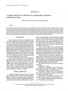
Characterization of HOM Carriers and Nanosensors A major advantage of the development nanosensor-based monoliths here is the flexibility to access the nanosacle geometries and ordered structures that have 3D arrays, uniform cylindrical channels, large pore sizes and volumes, high surface areas, and macroscopic particle size-like monoliths, when combined with inexpensive DPC probe molecules. Furthermore, the functional use of the optically transparent silica monoliths with cubic Fm3m (HOM-C10) structures enhanced their potential technologies as low processing costs. TEM image (Fig.1) revealed that the long-range ordered pores in large-scale domains were characteristics the optically cylindrical sensors, despite the dense construction of the building blocks. Furthermore, 3D TEM surface pattern exhibited a uniform arrangement with continuous order of nanosensors. This finding indicated the fabrication of ordered nanosensors for high accessibility and retainability of Cr(VI) ions during the sensing responses.
13
Page 6 of 12
Fig. 1 Representative TEM (a) and 3D surface (b) images of cubic Fm3m HOM-DPC nanosensor recorded along the [100] direction. The 3D surfaces of the TEM micrograph with tilt of 35o revealed the features of the network surfaces of the HOM-DPC sensors. Note the bar is equal to 20 nm.
The XRD pattern (Fig.2) shows finely resolved diffraction peaks, which are indicative of highly ordered cubic Fm3m structure. Based on the N2 isotherms, uniform and open pore cylindrical-like channels with large surface area and pore volumes were also characteristics the HOM-DPC sensor monoliths, leading to facile diffusion and accessibility of Cr (VI) analyte towards the DPC probe molecules. Despite the little decrease in the structure parameters of the HOM-DPC nanosensor (Fig. 2B, insert), the significant retention of ordering and uniformly cylindrical mesopores of the optical nanosensor. 111
(A)
600
2
V ml(STP). g-1
Intensity
0 20
310 222
220
(a) calcined HOM-10 400 440
d111= 6.9 nm
(a) 750 (b) 700 (c) 660
X
d111= 6.9 nm
1
2
3
4
5
1.09 0.99 0.97
X XX X
X XX XX X X XX XX X X XX
XXX
X XXX
X X X X X X X
X X X X X X X X X X X X X X
X X
XX X XX
X
X X
X X X X X X X X X X X X XX X
(B)
XX X X X X X X X X
(c) HOM-DDAB-DPC
2 /o
6.2 6.1 6.0
400
200
(b) HOM-DDAB
3
SBET(m /g) R(nm) VP (cm /g)
d111= 6.9 nm
6
0.0
0.2
0.4
0.6
0.8
1.0
P/Po
Fig. 2 XRD patterns (A) and N2 adsorption/desorption isotherms at 77 K (B) of highly ordered cubic Fm3m (HOM10) (a), HOM-DDAB charged carriers (b), and HOMDDAB-DPC nanosensor(c). Insert (B) is the physical characteristics of the solid materials.
The high loading capacity of the immobilized DDAB and probe moieties onto surface carriers was revealed based on thermal and spectroscopic techniques. The TG curves revealed that DDAB and DPC probe (Fig. 3) moieties decreased in mass at around 180-650 oC. This decrease accompanied exothermic peaks in the corresponding DTA curves, indicating decomposition of the organic components. The decrease in DDAB and DPC probe moieties, for example, was 26.6 and 36.8 mass%, respectively, coinciding with the adsorption capacity (Q) of the loaded organic moieties of these materials. These results from the TG analyses were consistent with elemental content results from EDS X-ray microanalysis of C, H, N, and O. Based on elemental analyses, the composition of CHNO with DDAB-modified carriers and probe sensors was 25 and 32 mass%, respectively [41].
14
Page 7 of 12
Fig. 3 TG and DTA analyses of HOM-DDAB silica carriers(A) and HOM-DDAB-DPC nanosensors (B).
The FTIR spectra (data not shown) revealed that the functionalized DDAB and probe moieties constructed using the building-blocks approach were the rigid “backbone-like” part of the surface matrices of the HOM carriers. With HOM, HOM-DDAB, and HOM-DDAB-probe samples, the appearance of a broad absorption band in the 3000-3500 cm-1 region indicated the presence of Si-OH asymmetric stretching. In addition, two strong bands at 1100 and 960 cm-1 were assigned to Si-O-Si and Si-OH stretching vibrations, respectively [41]. The silica-modified TMAC showed additional absorption bands at 2960 and 1400 cm-1, which were due to the aliphatic C-H and the stronger C-N bonds, respectively [41]. Control Recognitions of Cr(VI) Using Nanosensor High Performance Metal Ion Sensing Systems High performance of the DPC-based nanosensor in terms of sensitivity and kinetic reaction between the metal ions and chromophore-doped HOM surfaces (time-response) depended on key factors such as the amount of the HOM-based sensor and SDS and the pH value. Changes in these control experiments can significantly affect the redistribution of the charge polarity and the electron and energy transfer within the probe molecule into the pore surfaces, permitting a high threshold level of disturbance of the sensing system. In this study, we carried out a series of experiments to systematically define and evaluate the relative importance of these factors to a HOM-Probe sensor for analyte detection (Fig. 4). The influence of SDS concentration from 0 - 4 mM was studied, while the other factors such as amount of HOM sensors (4 mg) and pH value (at pH=2.2) remained constant. Our findings showed that the absorbance increased with increasing SDS concentration from 0 to 0.5 mM. With a further increase in SDS concentration ( 0.5 mM), the reflectance spectra drastically decreased, indicating that using the appropriate amount of SDS is critical in our sensing design. Results indicated that the addition of the appropriate amount of SDS surfactant was the key factor to enable the stable formation of the metal-ligand complexes. In addition, the sensitivity of nanosensors strongly depends on the adsorbed amount of DPC-probe onto HOM monolithas and pH values (Fig. 4).
15
Page 8 of 12
Fig 4 Effect of SDS concentration amount of HOM-DPC sensor material, and pH values on optical reflectance spectra of [Cr-DPC] complex during the detection of Cr6+ concentration of [100 ppb] at 550 nm.
Visual Detection of Ultra-Traces Cr(VI) Ions Although, the DPC receptor was used to sensitive determination of Cr(VI) analyte ions in solution up to 10-7 mol/dm3, as previously reported [42], the DPC-doped nanosensor led to simple separation and visual detection over a wide, adjustable range, and sensitive quantification of analyte ions at trace levels ( 10-10 mol/dm3). The rapid and flexible Cr(VI)-DPC binding events with the formed complexes onto nanosensors led to the separation and preconcentration of Cr(VI) ions even at a trace concentration level (response-time 60 sec). However, the color change provided a simple procedure for sensitive, selective detection of Cr(VI) ions without the need for sophisticated instruments (Fig. 5) [30-37]. In addition, visual detection of Cr(VI) ions at a wide detection range (DR) of 0.07 ppb- 200 ppb was clearly achieved by using 3D nanosensors (Fig. 5).
Fig. 5 Concentration-dependent changes in color developed sequence of DPC-doped building-blocks sensor after detection of Cd2+ analyte.
Colorimetric Determination of Cr(VI) ions The design-made nanosensors show that the colorimetric determination using UV-Vis reflectance spectroscopy could quantitatively validate the wider concentration range of the metal ions (13 x10-10 to 3 x 10⁻⁶ mol/dm3) compared with the metal ion recognitions in solution [42]. However, the signaling change in the reflectance spectra of chemosensors was monitored during the Cr(III)–diphenylcarbazone complex formation (Fig. 6). The reflectance spectra of the this complex was observed at max of 550 nm, as a result of the binding of Cr(VI) target ions with the DPC-probe ligand (Fig. 6 ). The signaling responses indicated the formation of charge-transfer complex. The color intensity of the Cr(III)– diphenylcarbazone complex increased in a liner correlation of the Cr(VI) concentrations up to the maximum plateau “saturation step”, as evidenced from the calibration curves of these sensing systems (Fig. 6, insert). Furthermore, results show that the design-made nanosensor enabled possible detection limit (DL) of Cr(VI) ions down to concentration of 81 x 10-11 mol/dm3 by using this simple sensing system [11].
16
Page 9 of 12
6+
at 550 nm [Cr ] ppb 200 100 50.0 30.0 10.0 5.00 2.50 1.00 0.50 0.10 0.07 0.00
Absorbance
0.6
0.6
Absorbance
0.8
0.5 0.4 0.3 0.2 0.1 0.0 0
1
2
10
3 4 [Cr6+ ] M
5
10
0.4
0.2 350
400
450
500
550
600
650
700
750
Wavelength (nm)
Fig. 6 Concentration-dependent changes in UV-Vis reflectance spectra and the calibration curve (inset) of HOMDPC sensors based on the DPC- doped nanosensor after detection of Cr6+ analyte ions.
The selectivity of the DPC-doped nanosensor for Cr(VI) ions was checked. Results show that the nanosensor did not have potentially interfering components such as anions and cations (Fig. 7). No changes in color developed or reflectance intensities of the Cr(III)– diphenylcarbazone complex formation with addition of the interfered species. For example, the addition of various types of anion species, even at concentrations up to 500 times higher than that of analyte concentrations, did not affect the qualitative and quantitative analyses of Cr(VI) sensing systems (Fig. 7). Among all possible interfering cation species, the Pb2+ and Fe3+ ions at high concentration showed significant change in the reflectance spectra and color intensities of complex, indicating a disturbance in the Cr(VI) ion sensing systems. To counteract such disturbance, the addition of 0.005 mM citrate or phathalate to the specific sensing conditions can enhance the tolerable concentration of the active the Pb2+ and Fe3+ ions, respectively (Fig. 7).
0.70
Absrobance
0.65 0.60 0.55 0.50 0.45 0.40
Anions 1 2 3 4 5 6 Catio 7 ns an d anio 8 9 10 11 ns in 12 terfe rence 13 14 15 s
Cations
Fig. 7 Effect of the anions and cations as interference ions on the absorbance spectra of Cr-DPC complex formed during the detection of Cr (VI) ion at [100 ppb]. Note, interfered cations from 1-15 are as follows; Mo, Ni, Co Al, Fe, Mg, Ca, Cu, Zn, Bi, Hg, Pb, Sb, Cd, Cr. Interfered Anions from 1-14 are as follows; CTAB, Triton X, C8H4O4- , C6H5O73-, C2O42-, Cl-, CH3COO- , NO2- , NO3-, SO32- , SO42-, CO32-, and PO43-
17
Page 10 of 12
The achievement of the retention of high-sensitive character of DPC-doped nanosensor designs after a number of regeneration cycles is a unique and interesting challenge [43,44]. Simple treatment procedures, using specific concentration of acetic acid (0.2 M) as a stripping agent was possible for the removal of the Cr (VI) ion (i.e. decomplexation). The results revealed that the nanosensor remained its sensitivity even after extended regeneration and reuse cycle, despite 10 % decrease in the sensitive recognition of Cr(VI) ions after 3 cycles, indicating the reversible nanosensor. Conclusion We developed the design of optical chemical nanosensors for simple, high-speed detection of toxic Cr(VI) ions. The systematic design of the optical nanosensors was based on densely patterned a chromogenic receptor (DPC), as a selective binding site onto threedimensional (3D) nanoscale structures. The ability to precisely modify the nanoscale pore surfaces with DPC probe enables efficient sensing responses as a result of Cr(VI)-DPC binding events. A key feature of this design strategy is that the pore surface-enhanced polarity by dense dispersion of cationic surfactant such as DDAB led to increased accessibility and adsorptive characteristics of the DPC probe with high intrinsic mobility systems. Such flexibility in the fabricated nanosensors led to high flux of the metal analytes across the probe molecules without significant kinetic hindrance. The synthetic nanosensor can be used for an effective recognition of Cr(VI) ions to low level of concentrations in the range of 10-10-10-6 M (i.e. 0.07 ppb-200 ppb). Optimization of control sensing conditions was established for achieving enhanced signal response and color intensities of Cr(III)–diphenylcarbazone complex up to sub-picomolar detection limits ( 10-11 mol/dm3) of Cr(VI) ions, for the first time, with rapid response time (in the order of seconds). The optical chemical nanosensors are reversible and have the potential to serve effectively in on-site field analysis of environmental samples, eliminating the necessity for instrument-dependent analysis. Moreover, these new classes of the design-made hybrid nanosensors exhibited long-term stability of signaling and recognition functionalities that in general provided extraordinary sensitivity, selectivity, reusability, and fast kinetic detection and quantification of various deleterious metal ions in our environment. References [1] F. Turner, Biosensors-sense and sensitivity, Science 290, 2000, 1315. [2] P. Chen, C. He, A general strategy to convert the MerR family proteins into highly sensitive and selective fluorescent biosensors for metal ions . J. Am. Chem. Soc. ,126, 2004, 728. [3] L. Basabe-Desmonts, J. Beld, R. S. Zimmerman, J. Hernando, P. M. Mary, F. G. Parajo, N. F. van Hulst, A. v. den Berg, D. N. Reinhoudt, M. Crego-Calama, Simple approach to sensor discovery and fabrication on self-assembled monolayers on glass. J. Am. Chem. Soc, 126, 2004, 7293. [4] R. A. Potyrailo, Polymeric sensor materials: toward an alliance of combinatorial and rational design tools? Angew. Chem. Int. Ed., 45, 2006,702. [5] H. Zheng, Z. Yan, H. Dong, B Ye, Simultaneous determination of lead and cadmium at a glassy carbon electrode modified with Langmuir–Blodgett film of p-tert-butylthiacalix[4]arene Sens. Actuators B: Chemical, 120, 2007, 603.
18
Page 11 of 12
[6] U.S. Spichiger-Keller, Chemical Sensors and Biosensors for Medical and Biological Applications; Wiley-VCH: Weinheim, Germany, 1998. [7] A. Miyawaki, J. Lopis, R. Helm, J.M. McCaffery, J.A. Adams, M. Ikura, R.Y. Tsien, Fluorescent indicatorsfor Ca2+ based on green fluorescent proteins and calmodulin, Nature, 388, 1997, 882. [8] V.K. Gupta, A.K. Singh, B. Gupta, Schiff bases as cadmium(II) selective ionophores in polymeric membrane electrodes, Anal. Chim. Acta 583, 2007,340. [9] X. Peng, J. Du, J. Fan, J. Wang, Y. Wu, J. Zhao, S. Sun, T. Xu, J. Am. Chem. Soc. 6, 2007,1500. [10] C. Zhang, K.S. Suslick, A colorimetric sensor array for organics in water, J. Am. Chem. Soc. 127, 2005, 11548. [11] S.A. El-Safty, T. Balaji, H. Matsunaga, T. Hanaoka, F. Mizukami, Optical sensors based on nanostructured cage materials for detection of toxic metal ions, Angew. Chem. Int. Ed. , 45, 2006, 7202. [12] M.A. Kalinina, N.V. Golubev, O.A. Raitman, S.L. Selector, V.V. Arslanov, A novel ultrasensing composed Langmuir–Blodgett membrane for selective calcium determination in aqueous solutions, Sens. Actuators B, 114, 2006, 19. [13] K. Seiler, Ion-selective Optode Membrane, Fluka, Buchs, Switzerland, 1993. [14] U. Drechsler, B. Erdogan, V.M. Rotello, Nanoparticles: Scaffolds for Molecular Recognition, Chem. Eur. J., 10, 2004, 5570. [15] M. Miro, J.M. Estela, V. Cerda, Application of flowing stream techniques to water analysis Part III. Metal ions: alkaline and alkaline-earth metals, elemental and harmful transition metals, and multielemental analysis, Talanta 63 2004, 201. [16] M. Zevin, R. Reisfeld, I. Oehme, O.S. Wolfbeis, Sol-gel-derived optical coatings for determination of chromate, Sens. Actuators B 38-39, 1997, 235. [17] Y.M. Scindia, A.K. Pandey, A.V.R. Reddy, S.B. Manohar, Chemically selective membrane optode for Cr(VI) determination in aqueous samples, Anal. Chim. Acta 515 ,2004, 311. [18] N. A. Carrington , G. H. Thomasa, D. L. Rodman, David B. Beach , Z-L. Xue, Optical determination of Cr(VI) using regenerable, functionalized sol–gel monoliths Analytica Chimica Acta 581 ,2007, 232–240 [19] V.P. Dedkova, O.P. Shvoeva, S.B. Sawin, Test determination of Cu(II), Ni(II), and Cr(VI) in a single sample, J. Anal. Chem. 56, 2001, 758. [20] S. Tao, C.B. Winstead, H. Xian, K. Soni, A highly sensitive hexachromium monitor using water core optical fiber with UV LED, J. Environ. Monit. 4 (2002) 815. [21] M. F. Bergamini, D. P. dos Santos, M. V. B. Zanoni, Development of a voltammetric sensor for chromium(VI)determination in wastewater sample , Sens Actuators B 123, 2007, 902–908 [22] Y. Zhang, H.-F. Ji, G.M. Brown, T. Thundat, Detection of CrO42- using a hydrogel swelling microcantilever sensor , Anal. Chem. 75, 2003, 4773. [23] I. Svancara, P. Foret, K. Vytras, A study on the determination of chromium as chromate at a carbon paste electrode modified with surfactants , Talanta 64 , 2004, 844. [24] Y.-J. Yang, H.-J. Huang, A Polyaniline-modified electrode-based FIA system for sub-ppb-Level chromium(VI) Analysis, Anal. Chem. 73, 2001,1377. [25] N.A. Carrington, L. Yong, Z.-L. Xue, Electrochemical deposition of sol–gel films for enhanced chromium(VI) determination in aqueous solutions, Anal. Chim. Acta 572 , 2006, 17. [26] S. J. Lippard, J. M. Berg, Principles of Bioinorganic Chemistry, (University Science Books, Mill Valley, CA, 1994).
19
Page 12 of 12
[27] M. Comes, G. R. Lopez, M.D. Marcos, R. M. Manez, F. Sancenon, J. Soto, L. A. Villaescusa, P. Amoros, D. Beltran, Host solids containing nanoscale anion-binding pockets and their use in selective sensing displacement assays, Angew. Chem. Int. Ed. 44, 2005, 2918. [28] M. Comes, M.D. Marcos, F. Sancenon, J. Soto, L. A. Villaescusa, P. Amoros, D. Beltran, Chromogenic discrimination of primary aliphatic amines in water with functionalized mesoporous silica, Adv. Mater., 16, 2004, 1783. [29] A. B. Desacalzo, K.Rurack, H. Weisshoff, R. M. Manez, M.D. Marcos, P. Amoros, K. Hoffmann, J. Soto, Rational design of a chromo- and fluorogenic hybrid chemosensor material for the detection of long-chain carboxylates, J. Am. Chem. Soc.,127, 2005,184. [30] G. Wirnsberger, B. J. Scott, G. D. Stucky, pH sensing with mesoporous thin films Chem. Commun., 1, 2001, 119. [31] L. Nicole, C. Boissiere, D. Grosso, P. Hesemann, J. Moreau, C. Sanchez Advanced selective optical sensors based on periodically organized mesoporous hybrid silica thin films, Chem. Comm., 2004, 2312. [32] S. J. Lee, S. S. Lee, J. Y. Lee, J. H. Jung, A functionalized inorganic nanotube for the selective detection of Copper(II) ion, Chem. Mater., 18, 2006, 4713. [33] R. Metivier, I. Leray, B. D. Lebeau, B. Valeur, A mesoporous silica functionalized by a covalently bound calixarene-based fluoroionophore for selective optical sensing of mercury(II) in water, J. Mater. Chem., 15, 2005, 2965. [34] T. Balaji, M. Sasidharan, H. Matsunaga, Naked eye detection of cadmium using inorganic– organic hybrid mesoporous material,. Anal. Bioanal. Chem., 384, 2006, 488. [35] A. Safavi, M. Bagheri, Design and characteristics of a mercury (II) optode based on immobilization of dithizone on a triacetylcellulose membrane, Sens Actuators B , 99, 2004, 608. [36] A. A. Ensafi, M. Fouladgar, Development of a mercury optical sensor based on immobilization of 4-(2-pyridylazo)-resorcinol on a triacetylcellulose membrane, Sens Actuators B 113, 2006, 88. [37]Jeronimo, P.A., Araujo, A. N.; Montenegro, M. C., Satinsky, D., Solich, P. Colorimetric bismuth determination in pharmaceuticals using a xylenol orange sol–gel sensor coupled to a multicommutated flow system, Anal. Chim. Acta 504, 2004, 235–241. [38] S. A. El-Safty, T. Hanaoka, Fabrication of crystalline, highly order three- dimensional silica monoliths (HOM-n) with large, morphological mesopore structures. Adv. Mater. 15, 2003, 1893. [39] K. L. Herrington, E. W. Kaler, D. D. Miller, J. A. Zasadzinski, S. Chiruvolu, Phase behavior of aqueous mixtures of dodecyltrimethylammonium bromide (DTAB) and sodium dodecyl sulfate (SDS). J. Phys. Chem. 97, 1993,13792 . [40] J. W. Gilliland, K.Yokoyama, W. T. Yip, Effect of coulombic interactions on rotational mobility of guests in sol-gel silicate thin films. Chem. Mater.16, 2004, 3949. [41] X. Ji, Q. Hu, J. E. Hampsey, X. Qiu, L. Gao, J. He, Y. Lu, Synthesis and Characterization of Functionalized Mesoporous Silica by Aerosol-Assisted Self-Assembly , Chem. Mater., 18, 2006, 2265. [42] E. B. Sandell, Colorimetric Determination of Traces of Metals, 3th Edition, Interscience Publisher, INC., NY, P326, 1959. [43] M. K. Nazeeruddin, D. D. Censo, R. Humphry-Baker, M. Gratzel, Highly selective and reversible optical, colorimetric, and electrochemical detection of mercury(II) by amphiphilic Ruthenium complexes anchored onto mesoporous oxide films. Adv. Funct. Mater. 16, 2006,189 . [44] E. Palomares, R.Vilar, A. Green, J. R. Durrant, Alizarin complexone on nanocrystalline TiO2: A heterogeneous approach to anion sensing. Adv. Funct. Mater. 2, 2004,111.
20