Biologia, Bratislava, 57/Suppl. 11: 129—136, 2002
Glucan binding domain of streptococcal glucosyltransferases Deepan S. H. Shah & Roy R. B. Russell* Oral Biology, Dental School, University of Newcastle, Newcastle upon Tyne, NE2 4BW, UK; tel.: ++ 44 191 222 7859, fax: ++ 44 191 222 6137, e-mail:
[email protected]
SHAH, D. S. H. & RUSSELL, R. R. B., Glucan binding domain of streptococcal glucosyltransferases. Biologia, Bratislava, 57/Suppl. 11: 129—136, 2002; ISSN 0006-3088. Glucosyltransferase I (GTF-I) of Streptococcus downei synthesises an insoluble α-1,3-linked glucan from sucrose. It contains a catalytic domain that is a circularly-permuted homologue of the catalytic domain of Family 13 enzymes and a C-terminal glucan-binding domain (GBD) that consists of a series of tandem repeats, each ∼35 amino acids long. In order to examine the contribution of these repeats to binding, a nested series of plasmids was constructed that expressed truncated forms of GTF-I or GBD in Escherichia coli, with an N-terminal His6 tag. The tag facilitated rapid purification and also allowed development of a novel microtitre tray assay in which binding of biotin-labelled dextran to immobilised GTF-I or GBD could be quantitatively determined. The capacity to bind dextran was proportional to the number of repeats, with a minimum of four repeats necessary to demonstrate binding in this assay. Key words: Streptococcus, glucan binding, dextran, Family 70. Abbreviations: GBD, glucan binding domain; GBP, glucan binding protein; GTF, glucosyltransferase.
Introduction The link between dietary sugar and dental caries is well established. While any fermentable carbohydrate may be utilised by plaque bacteria to generate the acids that attack enamel substance, sucrose is recognised as being particularly important in the caries process. Not only can it be fermented to produce acid, but it also serves as substrate for extracellular enzymes of plaque bacteria that synthesise sucrose-derived polymers (COLBY & RUSSELL, 1997). These polymers are of central importance in adhesive interactions in plaque, where
they mediate attachment of bacteria to the tooth surface and to other bacteria thus stabilising the plaque biofilm, serve as energy stores aiding the survival of plaque bacteria, and modulate the permeability of plaque and hence the level of acid at the enamel surface. The enzymes forming polymers from sucrose, particularly the glucosyltransferases (GTFs) making water-soluble and insoluble glucans, as well as the glucan-binding proteins (GBPs), have thus attracted considerable interest as potential targets for inhibition of the processes which can lead to caries and as a means for directing therapeutic agents to plaque.
* Corresponding author
129
GTFs are extracellular enzymes produced by bacteria of the closely related genera Streptococcus, Leuconostoc and Lactobacillus that synthesise, from sucrose, polymers consisting solely of glucose (glucans). They form part of a larger group referred to as glucansucrases, that also includes amylosucrase of Neisseria spp. (VAN GEELSCHUTTEN et al., 1999; REMAUD-SIMEON et al., 2000; ALBENNE et al., 2002). The enzymes are capable of synthesising α1,6-, α1,2- or α1,3- linkages between the glucose units and are sometimes referred to according to their product e.g. dextransucrase, mutansucrase, alternansucrase. The relative proportions of the linkages and the degree of branching determine the ultimate properties of the glucan. The nucleotide sequences of 16 streptococcal GTFs are available, as well as sequences for dextransucrases and alternansucrase of Leuconostoc. All GTFs are of high molecular weight (∼160,000 kDa) and consist of a signal peptide, a highly variable stretch of approximately 200 amino acids, a conserved core region of about 900 amino acids including residues at the active site, and a C-terminal glucan-binding domain that contains about 400 amino acid residues (MONCHOIS et al., 1999b; ARGÜELLO-MORALES et al., 2000; REMAUD-SIMEON et al., 2000). The enzymes are all placed in the Family 70 of the glycoside hydrolase clan but sequence alignments and secondary structure predictions show that the GTF catalytic module is related to the α-amylase superfamily, glycoside hydrolase Family 13 (MOOSER, 1992; MACGREGOR et al, 1996). The distinctive feature of the alignment is that the first helix in the GTF sequence aligns with helix-3 (H3) of other Family 13 enzymes, while the β- and α- segments normally found at the start of proteins such as amylase (E1 H1 E2 H2 E3) come in the latter part of the GTF sequence. In other words, in the GTF the usual arrangement of alternating sheets and helices has undergone cyclic permutation in a novel construction. No three-dimensional structure has yet been reported for a GTF but the occurrence of a (β/α)8 barrel motif is supported experimentally by circular dichroism experiments (MONCHOIS et al., 1999b). The similarity in structure of GTF and amylases is further reflected in the mechanism; a ‘catalytic triad’ of carboxyl groups is found in all members of the glycoside hydrolase Family 13. These invariant Asp, Glu and Asp catalytic residues are located at the C-terminus of β strands 4, 5 and 7 respectively (MACGREGOR et al., 2001). Corresponding residues are conserved at the matching positions in GTF and their im-
130
portance in catalysis has been demonstrated by site-directed mutagenesis (DEVULAPALLE et al., 1997). This suggests that the mechanism of initial glycosidic cleavage of sucrose is the same in GTF as in other glycosidases. Other amino acid residues essential for the function of GTF occur in the first one-third of the core region, outside the predicted (β/α)8 barrel (MONCHOIS et al., 2000). It should be noted that amylosucrase does not fit this pattern, but has a non-permuted (β/α)8 barrel structure (SKOV et al., 2001). The carboxyl-terminal one-third of GTFs consists of a series of related but non-identical tandem amino acid repeats. This has been identified as a glucan binding domain (GBD) and has been shown to be responsible for the ability of GTF to bind dextran, both by experiments in which GTF was truncated by protein engineering (FERRETTI et al. 1987; ABO et al., 1991) and by proteolysis of GTF and capture of dextran-binding fragments (MOOSER & WONG, 1988). The strength of binding has been determined by a number of workers, and dissociation constants of the order of 10−5 to 10−7 M have been reported (LANDALE & MCCABE, 1987; HAAS & BANAS, 2000; KASEDA et al., 2000). This binding has allowed the purification of GTF by affinity chromatography on dextran, and the binding domain can be fused to other proteins to allow their purification, or exploited in targeting enzymes to dental plaque. However, the precise function of the binding domain remains unclear and may differ between different GTF enzymes. Amongst the proposed functions are: i) modulating the balance between glucan synthesis and sucrose hydrolysis (ABO et al., 1991; KONISHI et al., 1999); ii) aiding extension of the growing glucan chain (MONCHOIS et al., 1998); iii) binding dextran which enhances catalytic activity (ROBYT et al., 1995; MONCHOIS et al., 1999a; KONISHI et al., 1999); iv) modulating the types of glycosyl linkage introduced to the glucan (VICKERMAN et al., 1996); v) localising GTF at the bacterial surface (KATO & KURAMITSU, 1991); vi) binding to pre-formed or nascent glucan to mediate bacteriabacteria or bacteria-surface adhesion (FERRETTI et al., 1987). A number of different types of repeating units have been identified in the primary sequence of GTF, termed A, B, C, or D repeats. These vary in length from 20 to 48 amino acids long but in all cases certain residues are highly conserved, particularly aromatic amino acids and glycine, so it has been suggested that the A, B, C, D repeats are all derived from a basic 9-residue ‘YG’ repeat (GIFFARD & JACQUES, 1994). However, the only
Glucosyltransferase
Material and methods
Dextransucrase
Alternansucrase
Fig. 1. Schematic diagram of glucansucrases showing the N-terminal variable region, central catalytic core and C-terminal domain that contains tandem amino acid repeats. Repeats that fit the defintion of ‘A’ repeats are shown as black bars.
A A1
A2
A3
A4
B1 A5
B2 A6
B Repeat sequences A1 LYYFGQDGYMVTGAQNIKGSNYYFLANGAALRNTVY A2 WRYFK-NGVMALGLTTVDGHVQYFDKDGVQAKDKII A3 WYLGK-DGVAVTGAQTVGKQHLYFEANGQQVKGDFV A4 WFYLGKDGAAVTGAQTIKGQKLYFKANGQQVKGDIV A5 WVYVK-SGKVLTGAQTIGNQRVYFKDNGHQVKGQLV A6 WLYVK-DGKVLTGLQTVGNQKVYFDKNGIQAKGKAV B1 VNGKTYYFGSDGTAQTQANPKGQTFKDGSGVLRFYNLEGQYVSGSGWY B2 VNGKTYYFGSDGTAQTQANPKGQTFKDGSGVLRFYNLEGQYVSGSGWY
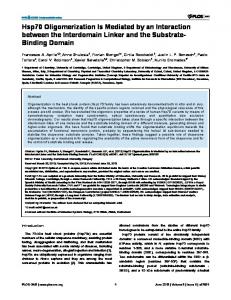
Fig. 2. The glucan binding domain of GTF-I. (A) Schematic diagram showing the C-terminal region with the locations of the A (open boxes) and B (filled boxes) repeats. (B) The amino acid sequences of the A and B repeats. Conserved residues in the A repeats are shown in bold. The B repeats are identical to each other (FERRETTI et al., 1987).
repeat found in all GTF is the ‘A’ repeat first described in GTF-I of Streptococcus downei (FERRETTI et al., 1987) and its occurrence in the different glucansucrases is illustrated in Figure 1. S. downei produces four different GTFs with distinct properties (COLBY & RUSSELL, 1997) and GTF-I, which produces a glucan with 93 % α-1,3 linkages, has been the subject of extensive study in our laboratories. This paper describes the construction of a series of mutants of GTF-I truncated at the C-terminus (Fig. 2), and characterisation of their ability to bind dextran.
General methods E. coli strains XL1-Blue or JM109 (pREP4) were grown on Luria-Bertani medium (LB) supplemented with 100 µg/mL ampicillin and/or 50 µg/mL kanamycin when necessary. S. downei MFe28 was grown on Brain Heart Infusion (BHI) or on Todd Hewitt medium supplemented with 0.5% (w/v) yeast extract (THYE). Standard DNA manipulations were carried out using protocols described in SAMBROOK & RUSSELL (2001). Restriction enzymes were obtained from commercial sources and used according to manufacturers’ instructions. Large-scale plasmid extractions for DNA sequencing were carried out using the Plasmid Midi-Kit (Qiagen). DNA sequencing was carried out at the Molecular Biology Unit, University of Newcastle, using Thermosequenase and dye terminator chemistry (Amersham) on an ABI377 sequencer. DNA sequence was assembled and analysed using OMIGA v2.0 software (Oxford Molecular). Polymerase Chain Reaction (PCR) experiments were carried out with the high fidelity, pre-mixed Extensor Long PCR Master Mix (ABgene) without oil overlays using cycling conditions recommended by the manufacturer, on a GeneAmp9700 thermal cycler (Applied Biosystems). All PCR primers were custom synthesised by Genset. PCR products for cloning were purified from the agarose electrophoresis gels using the Qiaex II kit (Qiagen). Over-expression and purification of GTF-I and its deleted forms E. coli overnight starter cultures in LB with antibiotics were diluted 1 in 10 in fresh medium and shaken for 1 h at 37 ◦C prior to the addition of IPTG at 0.1 mM final concentration and a further incubation of 6 h shaking at 37 ◦C. Bacterial cells were harvested by centrifugation and resuspended in B-PER lysis solution (Pierce) containing complete protease inhibitor cocktail (Boerhinger Mannheim). The suspensions were frozen at −20 ◦C overnight. Cleared lysates were obtained by centrifugation and then filtration through a 0.2 µm syringe filter. The lysates were diluted in 2× binding buffer to give a final binding buffer containing 20 mM sodium phosphate, 0.5 M NaCl, 20 mM imidazole, pH 7.4. An AKTA Prime chromatography system (Amersham Pharmacia) with the pre-programmed HisTag purification protocol was used for subsequent purification steps: the lysate was loaded onto a nickelcharged HiTrap chelating column (Amersham Pharmacia) equilibrated with binding buffer. After washing with 10 column volumes of binding buffer to remove unbound material, the His-tagged GTF-I derivatives were eluted with an imidazole gradient of 20 mM to 500 mM and collected in 1 mL fractions. In some cases, cleared lysates were obtained as described above and proteins purified using the Ni-NTA Spin Kit (Qiagen) according to manufacturer’s instructions. SDS PAGE Protein composition and GTF activity of samples were determined after gel electrophoresis as described by
131
FERRETTI et al. (1987). For detection of histidinetagged proteins on western blots, proteins were transferred from SDS gels onto Optitran BA-S 85 cellulose nitrate membranes (Schleicher & Schuell). After transfer, histidine-tagged proteins were detected by probing with a Ni-NTA alkaline phosphatase conjugate (Qiagen) as follows. Membranes were washed twice for 10 min in TBS (10 mM Tris HCl pH 7.5, 150 mM NaCl), blocked overnight in 3% (w/v) BSA in TBS, washed three times for 10 min with TBS and incubated for 1 h at RT with 1/1000 dilution of Ni-NTA AP conjugate in TBS-Tween (TBS containing 0.05% (v/v) Tween 20). The conjugate was removed by washing three times for 10 min in TBS-Tween. The colour reaction was carried out by incubation with BCIP/NBT stock solutions (Zymed) according to manufacturer’s instructions. For detection of dextran binding proteins on western blots, the membrane was washed three times for 15 min in PBS-T buffer (20 mM sodium phosphate pH 7.3, 150 mM NaCl, 0.05% (v/v) Tween-20) and blocked overnight in 3% (w/v) BSA in PBS-T. The membranes were washed as before, incubated with 100 µg/mL of biotin-dextran (Sigma) in PBS-B (20 mM sodium phosphate pH 7.3, 150 mM NaCl, 0.2% (w/v) BSA) for 1 h, washed as before and incubated with 1/20000 dilution of Extravidin-alkaline phosphatase conjugate (Sigma) in PBS-B for 1 h. The membrane was washed as before and the colour reaction was carried out as above. Glucan binding assay 20 pmol samples of purified protein were added to NiNTA coated 96-well HisSorb plates (Qiagen) in PBS-T to a final volume of 200 µL and incubated overnight at 4 ◦C. The protein solutions were removed and the wells washed four times for 1 min with PBS-T. 200 µL of a 100 µg/mL solution of biotin-dextran in PBSB were added and incubated for 10 min. After washing as before, wells were incubated with 200 µL of a 1/20000 dilution of Extravidin-alkaline phosphatase conjugate (Sigma) in PBS-B for 30 min and washed again. 100 µL of phosphatase substrate solution (Sigma 104 substrate) was added and colour change monitored by readings in a Titertek Multiskan MCC 340 Plate Reader.
S. downei chromosomal DNA was used as template. The forward primers incorporated a BamHI site at the 5’ end of the product and the reverse primers incorporated a HindIII site at the 3’ end. Gel-purified PCR fragments were digested, ligated into pQE30 cut with BamHI and HindIII and introduced into E. coli XL1-Blue. This resulted in constructs encoding GTF or GBD derivatives with an N-terminal fusion with six histidine residues under the control of the inducible Ptac . The constructs were checked by nucleotide sequencing. In the course of this work plasmid GTF-I1 was found to have arisen in a population originally thought to correspond to GTFI-0. GTFI-I has only one B repeat and no A5. Sequence analysis suggested that this can most likely be attributed to a spontaneous homologous recombination event between the two B repeats, which have 100% sequence homology. Crossover between the two B repeats results in an internal deletion removing one B repeat and A5. In order to maintain stability of plasmids, all the constructs were thereafter maintained in the recA strain JM109 containing the kanamycin-resistance plasmid pREP4 that provides constitutive expression of the lac repressor in trans. The full set of deletions obtained is illustrated schematically in Figure 3.
Results
Protein purification, detection and activity in gels The recombinant forms of GTF were purified by affinity chromatography on nickel chelating columns. The yield and purity of different factions was monitored by SDS PAGE and Coommassie Blue staining revealed the presence of a contaminating band in some of the preparations. In the case of the proteins containing the catalytic domain, this band was shown to possess a histidine-tag by western blotting with Ni-NTA alkaline phosphatase conjugate and is likely to be either a proteolytic fragment or the result of premature termination of translation. Contaminating bands could be removed by further chromatography steps.
Mutant construction In order to express truncated forms of GTF-I in E. coli, fragments of the gtfI gene were cloned into plasmid pQE30 (Qiagen) to yield fusion proteins with an N-terminal hexahistidine tag. A series of nested deletion mutants was constructed by PCR amplification using a forward primer targeted to the region encoding the start of the catalytic domain or the beginning of the region encoding the GBD. Reverse primers targeted different sites internal to the region encoding the GBD.
Glucan binding activity of the purified proteins The glucan binding assay was based upon a method using biotin-labelled dextran introduced by LIS et al. (1995) but exploiting the specific binding of the His-tagged proteins to Ni-NTA coated microtitre plates as described in Materials and methods. The data obtained for the mutants constructed in this study are shown in Figure 4. The decrease in size of the GBD resulted in a corresponding decrease in binding activity. GTF-I1A with repeats A1-A4 and B1 intact had some bind-
132
GTFI0
A1 A2 A3
Catalytic domain
A4 B1 A5
GTFI1
B1/2
GTFI1A
B1/2
GTFI2
B1
B2 A6
GTFI2A
GTFI3
GTFI4
B1
Catalytic D i
B2
GBD0 B1/2 GBD1 B1/2
Catalytic D i
GBD1A
Catalytic D i
GBD2
B1
Fig. 3. Deletion derivatives of GTF-I constructed for this study. In all cases the signal peptide and variable region have been replaced by an N-terminal His6 tag. GTF-I0 and GBD0 represent the full-length core + GBD and GBD alone wild-type proteins, respectively. The catalytic domain, A repeats and B repeats are indicated. Repeats B1/2 are believed to have formed by recombination of repeats B1 and B2.
ing activity but removal of repeat B1 from this abolished binding as shown by the data for GTFI2A. Thus, the minimal binding unit appears to consist of repeats A1-A4 and B1. The data from the GBD constructs demonstrates that the catalytic domain is dispensable for glucan binding. Thus, the GBD is an independent domain capable of binding glucan, the ability of the various truncated forms to bind dextran being unaffected by the presence or absence of the core domain. The glucan-binding properties of the deletion derivatives were also investigated by western blotting with biotin-dextran as probe. This method proved to be more sensitive for detecting weak binding as even GTF-I3 was shown to bind dextran, although to a considerably lesser degree than the longer constructs.
Discussion In this study we have constructed a series of nested deletions in the C-terminal, glucan-binding domain of GTF-I from S. downei. The proteins were purified and their glucan binding properties investigated by a new microtitre plate assay that yields quantitative data on the binding capacity and will be of great value in future studies involving the screening of large numbers of mutants. The results obtained confirm and extend earlier reports that C-terminal truncation of GTF-I results in loss of dextran binding activity. We have demonstrated a correlation between number of repeats and binding activity. Furthermore, it was found that the repeats may not contribute equally to the binding. GTF-I1, GTF-I2, GBD1 and GBD2 all have the
133
2.4 2.0 1.6 1.2 0.8
GBD 2
GBD 1A
GBD 1
GBD 0
GTFI c
GTFI 3
GTFI 4
GTFI 2A
GTFI 2
GTFI 1A
0.0
GTFI 1
0.4 GTFI 0
Phosphatase activity (arbitrary units)
2.8
Protein M u ta n t G T F I0 G T F I1 G T F I2 G T F I1 A G T F I2 A G T F I3 G T F I4 G T F Ic GBD0 GBD1 GBD2 GBD1A
R e p e a ts p re s e n t A1 A2 A3 A4 B1 A1 A2 A3 A4 B1 A1 A2 A3 A4 B1 A1 A2 A3 A4 B1 A1 A2 A3 A4 A1 A2 A3 A1 A2 None A1 A2 A3 A4 B1 A1 A2 A3 A4 B1 A1 A2 A3 A4 B1 A1 A2 A3 A4 B1
A5 A6 A5
B2
A6
A5 A6 A5
B2
A6
Fig. 4. Glucan binding activity. The bar chart shows the binding activity of the different deletion mutants as revealed by alkaline phosphatase activity measured 5 min after addition of substrate. The averaged results of three experiments are shown. The table shows the repeats possessed by each mutant.
same number of repeats in the C-terminus. GTFI1 and GBD1 are missing repeat A5 whereas GTFI2 and GBD2 are missing repeat A6. However, GTF-I1 and GBD1 have higher binding activity than GTF-I2 and GBD2 respectively indicating that A6 has more affinity than A5. There are no major differences in the amino acid sequences of these two repeats to explain this difference (Figure 2) so the interchangeability of repeats underscores the modular construction of the GBD. It should be noted however, that GTF-I1 and GBD1 have a tail of ∼30 amino acids after the last repeat whereas GTF-I2 and GBD2 have only three amino acids after the last repeat. It is possible that this, too, could influence binding affinity either by interacting with the dextran or affecting folding of the domain. During the course of this study, it was found that an internal deletion of the cloned gtfI gene had occurred due to recombination between the identical DNA sequences encoding the two B repeats. It is not known at what stage of the cloning
134
procedure this occurred and it is possible that there is some selective advantage for the host E. coli cell in carrying a reduced size GTF-I. The internal deletion event is similar to that found in a spontaneous mutation in the gtfG gene of Streptococcus gordonii, between homologous repeats (VICKERMAN et al., 1996). No systematic study of variation within a GTF has yet been described, though the phenomenon clearly provides a mechanism for generating diversity within a population of bacteria, and modulating the amount and nature of glucan produced. One intriguing question concerns the evolution and divergence of repeats in the various GTFs. It is probable that the tandem repeats have arisen by a process of genetic duplication (GIFFARD & JACQUES, 1994) although it is not apparent whether the evolutionary pressure to generate diversity has arisen to generate variability in binding specificity or affinity, as a mechanism for modulating glucan synthesis or (in the case of the oral streptococci) to generate antigenic variability as a form of immune evasion. We have previously reported that sequential truncation of the C-terminus resulted in a reduction both in the catalytic activity of GTF-I and in the extent to which activity is stimulated by added dextran (MONCHOIS et al., 1999a). However, the catalytic core retains the ability to synthesise an α-1,3 glucan indistinguishable from that synthesised by the intact enzyme and clearly can function as an autonomous domain. In the present study we have expressed the C-terminal glucan-binding region and show that it also functions as an independent domain, binding dextran as efficiently as when it is part of the intact GTF-I enzyme. Understanding of the way in which the two domains interact with each other, and with dextran, must await elucidation of the three-dimensional structure. This paper has focused upon the A repeats found in the C-terminal one third of GTF-I. Similar repeats, varying in number and spacing but with the same pattern of highly conserved residues, are found in all streptococcal GTFs and Leuconostoc dextransucrases. Alternansucrase however, has a series of short repeats that do not match this pattern (JANEČEK et al., 2000). We recently reported that a partial A repeat is found at the N-terminal end of the catalytic core of GTFs but there is a distinct difference between different glucansucrases because dextransucrases have two full A repeats whereas alternansucrase has three (Fig. 1; JANEČEK et al., 2000). It is not yet known whether these repeats have binding capacity, or what function they serve in glucan for-
mation. It is also important to appreciate that dextran may not be the only, or even the biologically most significant, ligand bound by repeat regions. A repeats are not necessarily associated with glucan synthesis and A repeats are found in the GbpA glucan binding protein of Streptococcus mutans, which appears to have no catalytic activity (BANAS et al., 1990). Furthermore, the first half of the A repeat of GTF enzymes matches closely the consensus sequence of a repeat unit that occurs in the choline-binding proteins of Streptococcus pneumoniae and the toxins of Clostridium difficile that bind to Galα1-3Galβ1-4GlcNAc (WREN, 1991; JANEČEK et al., 2000). Recently, analysis of the S. mutans genome sequence has revealed the presence of a previously-unrecognised protein containing 3 A repeats near its N-terminus, but no other similarity to any proteins in the public databases (R. R. B. RUSSELL, unpublished results). Domains containing A repeats and related sequences are thus carbohydrate-binding modules that occur in proteins with different functions and specificities. Many questions remain about what the roles of the repeats are in many of these proteins and what components of the repeats and surrounding primary sequence are determinants of binding specificity and affinity. Owing to the iterative nature of the sequence within these binding domains it is difficult to investigate the effects of single amino acid changes within the repeats since any change may be compensated for by the other repeats. The identification of a minimal-size binding unit for the GBD of GTF-I will greatly facilitate such studies in our laboratory, where we propose to use site-directed mutagenesis of specific amino acid residues within the minimal binding unit and measure the effects with the refined HisSorb glucan binding assay. In addition, the ability to express and purify the binding domain in large quantities has enabled us to initiate studies aimed at determining the structure of the GBD by X-ray crystallography. Such studies should lead to important insights into the biology of these fascinating proteins. References ABO, H., MATSUMURA, T., KODAMA, T., OHTA, H., FUKUI, K., KATO, K. & KAGAWA, H. 1991. Peptide sequences for sucrose splitting and glucan binding within Streptococcus sobrinus glucosyltransferase (water-insoluble glucan synthetase). J. Bacteriol. 173: 989–996. ALBENNE, C., POTOCKI DE MONTALK, G., MONSAN, P., SKOV, L., MIRZA, O., GAJHEDE, M. &
REMAUD-SIMEON, M. 2002. Site-directed mutagenesis of key amino acids in the active site of amylosucrase from Neisseria polysaccharea. Biologia, Bratislava 57 (Suppl. 11): 119–128. ARGÜELLO-MORALES, M. A., REMAUD-SIMEON, M., PIZZUT, S., SARC ¸ ABAL, P., WILLEMOT, R. M. & MONSAN, P. 2000. Sequence analysis of the gene encoding alternansucrase, a sucrose glucosyltransferase from Leuconostoc mesenteroides NRRL B1355. FEMS Microbiol. Lett. 182: 81–85. BANAS, J. A., RUSSELL, R. R. B. & FERRETTI, J. J. 1990. Sequence-analysis of the gene for the glucanbinding protein of Streptococcus mutans Ingbritt. Infect. Immun. 58: 667–673. COLBY, S. M. & RUSSELL, R. R. B. 1997. Sugar metabolism by mutans streptococci. J. Appl. Microbiol. 83: S80–S88. DEVULAPALLE, K. S., GOODMAN, S. D., GAO, Q., HEMSLEY, A. & MOOSER, G. 1997. Knowledgebased model of a glucosyltransferase from the oral bacterial group of mutans streptococci. Protein Sci. 6: 2489–2493. FERRETTI, J. J., GILPIN, M. L. & RUSSELL, R. R. B. 1987. Nucleotide sequence of a glucosyltransferase gene from Streptococcus sobrinus MFe28. J. Bacteriol. 169: 4271–4278. GIFFARD, P. M. & JACQUES, N. A. 1994. Definition of a fundamental repeating unit in streptococcal glucosyltransferase glucan-binding regions and related sequences. J. Dent. Res. 73: 1133–1141. HAAS, W. & BANAS, J. A. 2000. Ligand-binding properties of the carboxyl-terminal repeat domain of Streptococcus mutans glucan-binding protein A. J. Bacteriol. 182: 728–733. JANEČEK, Š., SVENSSON, B. & RUSSELL, R. R. B. 2000. Location of repeat elements in glucansucrases of Leuconostoc and Streptococcus species. FEMS Microbiol. Lett. 192: 53–57. KASEDA, K., YOKOTA, H., ISHII, Y., YANAGIDA, T., INOUE, T., FUKUI, K. & KODAMA, T. 2000. Singlemolecule imaging of interaction between dextran and glucosyltransferase from Streptococus sobrinus. J. Bacteriol. 182: 1162–1166. KATO, C. & KURAMITSU, H. K. 1991. Molecular-basis for the association of glucosyltransferases with the cell-surface of oral streptococci. FEMS Microbiol. Lett. 79: 153–158. KONISHI, N., TORII, Y., YAMAMOTO, T., MIYAGI, A., OHTA, H., FUKUI, K., HANAMOTO, H., MATSUO, H., KOMATSU, H., KODAMA, T. & KATAYAMA, E. 1999. Structure and enzymatic properties of genetically truncated forms of the water-insoluble glucan-synthesizing glucosyltransferase from Streptococcus sobrinus. J. Biochem. 126: 287–295. LANDALE, E. C. & MCCABE, M. M. 1987. Characterization by affinity electrophoresis of an α-1,6glucan-binding protein from Streptococcus sobrinus. Infect. Immun. 55: 3011–3016. LIS, M., SHIROZA, T. & KURAMITSU, H. K. 1995. Role of the C-terminal direct repeating units of
135
the Streptococcus mutans glucosyltransferase-S in glucan binding. Infect. Immun. 61: 2040–2042. MACGREGOR, E. A., JANEČEK, Š. & SVENSSON, B. 2001. Relationship of sequence and structure to specificity in the α-amylase family of enzymes. Biochim. Biophys. Acta 1546: 1–20. MACGREGOR, E. A., JESPERSEN, H. M. & SVENSSON, B. 1996. A circularly permuted α-amylasetype α/β-barrel structure in glucan-synthesizing glucosyltransferases. FEBS Lett. 378:263–266. MONCHOIS, V., ARGÜELLO-MORALES, M. A. & RUSSELL, R. R. B. 1999a. Isolation of an active catalytic core of Streptococcus downei MFe28 GTF-I glucosyltransferase. J. Bacteriol. 181: 2290–2292. MONCHOIS, V., LAKEY, J. H. & RUSSELL, R. R. B. 1999b. Secondary structure of Streptococcus downei GTF-I glucansucrase. FEMS Microbiol. Lett. 177: 243–248. MONCHOIS, V., REVERTE, A., REMAUD-SIMEON, M., MONSAN, P. & WILLEMOT, R. M. 1998. Effect of Leuconostoc mesenteroides NRRL B-512F dextransucrase carboxy-terminal deletions on dextran and oligosaccharide synthesis. Appl. Environ. Microbiol. 64: 1644–1649. MONCHOIS, V., VIGNON, M., ESCALIER, P. C., SVENSSON, B. & RUSSELL, R. R. B. 2000. Involvement of Gln937 of Streptococcus downei GTF-I glucansucrase in transition-state stabilization. Eur. J. Biochem. 267: 4127–4136. MOOSER, G. & WONG, C. 1988. Isolation of a glucan-binding domain of glucosyltransferase (1,6α-glucan synthase) from Streptococcus sobrinus. Infect. Immun. 56: 880–884. MOOSER, G. 1992. Glycosidases and glycosyltransferases. The Enzymes 20: 187–233.
REMAUD-SIMEON, M., WILLEMOT, R. M., SARC ¸ ABAL, P., POTOCKI DE MONTALK, G. & MONSAN, P. 2000. Glucansucrases: molecular engineering and oligosaccharide synthesis. J. Mol. Catal. B10: 117– 128. ROBYT, J. F., KIM, D. & YU, L. 1995. Mechanism of dextran activation of dextransucrase. Carbohyd. Res. 266: 293–299. SAMBROOK, J. & RUSSELL, D. W. 2001. Molecular Cloning. A laboratory manual. 3rd Edition. CSHL Press, Cold Spring Harbor, New York. SKOV, L. K., MIRZA, O., HENRIKSEN, A., DE MONTALK, G. P., REMAUD-SIMEON, M., SARC ¸ ABAL, P., WILLEMOT, R. M., MONSAN, P. & GAJHEDE, M. 2001. Amylosucrase, a glucan-synthesizing enzyme from the α-amylase family. J. Biol. Chem. 276: 25273–25278. VAN GEEL-SCHUTTEN, G. H., FABER, E. J., SMIT, E., BONTING, K., SMITH, M. R., TEN BRINK, B., KAMERLING, J. P., VLIEGENTHART, J. F. & DIJKHUIZEN, L. 1999. Biochemical and structural characterization of the glucan and fructan exopolysaccharides synthesized by the Lactobacillus reuteri wild-type strain and by mutant strains. Appl. Environ. Microbiol. 65: 3008–3014. VICKERMAN, M. M., SULAVIK, M. C., MINICK, P. E. & CLEWELL, D. B. 1996. Changes in the carboxyterminal repeat region affect extracellular activity and glucan products of Streptococcus gordonii glucosyltransferase gene. Infect. Immun. 64: 5117– 5128. WREN, B. W. 1991. A family of clostridial and streptococcal ligand-binding proteins with conserved Cterminal repeat sequences. Mol. Microbiol. 5: 797– 803. Received October 15, 2001 Accepted February 12, 2002
136