HIPPOCAMPUS 23:1103–1124 (2013)
COMMENTARY
Homeostatic Regulation of Memory Systems and Adaptive Decisions Sheri J.Y. Mizumori,* and Yong Sang Jo ABSTRACT: While it is clear that many brain areas process mnemonic information, understanding how their interactions result in continuously adaptive behaviors has been a challenge. A homeostatic-regulated prediction model of memory is presented that considers the existence of a single memory system that is based on a multilevel coordinated and integrated network (from cells to neural systems) that determines the extent to which events and outcomes occur as predicted. The “multiple memory systems of the brain” have in common output that signals errors in the prediction of events and=or their outcomes, although these signals differ in terms of what the error signal represents (e.g., hippocampus: context prediction errors vs. midbrain=striatum: reward prediction errors). The prefrontal cortex likely plays a pivotal role in the coordination of prediction analysis within and across prediction brain areas. By virtue of its widespread control and influence, and intrinsic working memory mechanisms. Thus, the prefrontal cortex supports the flexible processing needed to generate adaptive behaviors and predict future outcomes. It is proposed that prefrontal cortex continually and automatically produces adaptive responses according to homeostatic regulatory principles: prefrontal cortex may serve as a controller that is intrinsically driven to maintain in prediction areas an experience-dependent firing rate set point that ensures adaptive temporally and spatially resolved neural responses to future prediction errors. This same drive by prefrontal cortex may also restore set point firing rates after deviations (i.e. prediction errors) are detected. In this way, prefrontal cortex contributes to reducing uncertainty in prediction systems. An emergent outcome of this homeostatic view may be the flexible and adaptive control that prefrontal cortex is known to implement (i.e. working memory) in the most challenging of situations. Compromise to any of the prediction circuits should result in rigid and suboptimal decision making C 2013 The and memory as seen in addiction and neurological disease. V Authors. Hippocampus Published by Wiley Periodicals, Inc. KEY WORDS: hippocampus; striatum; prefrontal cortex; predictions; error correction
INTRODUCTION Different motivational systems can be simultaneously active, and as such, they must compete for control over decision processing and behav-
This is an open access article under the terms of the Creative Commons Attribution-Non-Commercial-NoDerivs License, which permits use and distribution in any medium, provided the original work is properly cited, the use is non-commercial and no modifications or adaptations are made. Psychology Department, University of Washington, Seattle, Washington Grant sponsor: National Institute of Mental Health; Grant number: MH 58755. *Correspondence to: Sheri J.Y. Mizumori, Psychology Department, Box 351525, University of Washington, Seattle, WA 98195-1525, USA. E-mail:
[email protected] Accepted for publication 17 July 2013. DOI 10.1002/hipo.22176 Published online 8 August 2013 in Wiley Online Library (wileyonlinelibrary.com).
ioral responses. For example, a hungry animal may explore a territory for food while “keeping an eye out” for potential mates and predators. In this case, a single behavioral sequence is engage but it may be driven from moment to moment by whichever motivation is the strongest. The current and strongest motivation should determine which cues draw one’s attention, the appropriate behavioral responses, and one’s expectations for particular outcomes of choices and actions. As such, motivation state likely influences the organization of currently active neural representations in memory-related brain structures. Indeed, it has been shown that different hippocampal place field “maps” (which presumably reflect different memories) are recalled depending on whether rats are searching the same space for food or water after being made hungry or thirsty, respectively, in a single environment (Kennedy and Shapiro, 2004). The ability to efficiently switch between different mnemonic influences on choices and actions is undoubtedly essential for the continuous execution of adaptive behaviors. A current challenge for the field of memory research is to understand the mechanisms by which different memory systems are dynamically and continuously coordinated according to one’s current goal. There are numerous excellent reviews of literatures that define the functionality of individual memory systems of the brains (e.g., Eichenbaum and Cohen, 2001; Tulving, 2002; Boyden et al., 2004; Colombo, 2004; Yin and Knowlton, 2006; Fuster, 2009; Kesner, 2009). To address the issue of how these memory systems work together to provide what appear to be unitary control over behaviors, the following first presents an organizational scheme for memory processing in the brain that includes decision neural circuitry (Schultz and Dickinson, 2000; Hikosaka et al., 2008). Then we discuss how the different mnemonic brain regions may be dynamically regulated via a homeostatic-like mechanism that insures the continuous generation of adaptive decisions.
MULTIPLE PROCESSORS OF MNEMONIC INFORMATION From the early reports of memory problems of brain damaged amnesic patients came the view that different brain areas are responsible for distinct types of memories.
C 2013 THE AUTHORS. HIPPOCAMPUS PUBLISHED BY WILEY PERIODICALS, INC. V
1104
MIZUMORI AND JO
Decades of experimental research supported this view since targeted lesions in animals produced memory dissociations that were comparable to those observed clinically. For example, hippocampal damage produced episodic (including event or spatial) memory deficits that were not shown by animals with striatal lesions; the latter instead showed habit learning deficits. The accumulation of similar anatomically defined distinctions led to the commonly held view that hippocampus mediates episodic or event-related memory and the striatum mediates habit or procedural memories (Sherry and Schachter, 1987; Packard et al., 1989; Knowlton et al., 1996; Squire and Zola, 1996; White and McDonald, 2002). Additional research identified roles for other brain structures in specialized memory functions: the amygdala is considered important for learning and remembering emotionally relevant information (Kim and Jung, 2006; Johansen et al., 2011), the prefrontal cortex preferentially mediates working memory (e.g. Jacobsen, 1936; Fuster, 2006, 2009), and sensory and motor memories are thought to involve parietal cortex, medial temporal cortex, and cerebellum (e.g. Duhamel et al., 1992; Bisley et al., 2004, Taylor and Ivry, 2012; Ito, 2013; Rochefort et al., 2013). Figure 1A provides a schematic of these memory systems, illustrating the parsimonious view that each separately contributes to a single decision or action selection process. It is often suggested that the differential strengths and efficiencies of the memory processing centers of the brain control behaviors according to task demands (Packard and McGaugh, 1996; Miziumori et al., 2004; Fig. 1B). In fact numerous physiological and theoretical models describe mechanisms, from broad neural systems to cellular perspectives, by which these brain structures enable efficient and accurate mnemonic functions. By comparison, little is known about how these multiple processors of mnemonic information work interactively to result in continuously adaptive, experiencedependent behaviors and choices in a complex natural world. By definition, all memory systems have a common purpose, and that is to remember information. However, all memory systems do not play equal roles in determining the impact of experience on particular decisions or actions. What accounts for this difference? Abundant evidence from primate and rodent studies show that (internal and external) sensory, behavioral, task rule and reward information are often represented experientially in brain areas thought to mediate different forms of memory (e.g., Wiener et al., 1993; Lavoie and Mizumori, 1994; Wiener, 1996; Pratt and Mizumori, 2001; Yeshenko et al., 2004, Barnes et al., 2005; Smith and Mizumori, 2006; Puryear et al., 2010). Thus, the nature of the represented information per se cannot explain the unique mnemonic contributions of different brain areas. Rather their unique mnemonic contributions are likely to be more strongly determined by brain area-specific computational network architectures, and their afferent and efferent connections.
HIPPOCAMPUS AND CONTEXT ANALYSIS There is substantial evidence to support the view that hippocampus is essential for the processing of context information (Hirsh, 1974; Myers and Gluck, 1994; Anagnostaras et al., 2001; Maren, 2001; Fanselow and Poulos, 2005; Bouton et al., Hippocampus
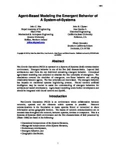
FIGURE 1. A: Different types of memory are shown to contribute to the determination of decisions and actions. The efficiencies of each of these memory systems can be regulated by neuromodulators such as dopamine (DA), serotonin, acetylcholine, norepinephrine, or a variety of stress or reproductive hormones. B: Memory systems are often considered to compete for the control of behaviors, depending on the situation. For simplicity, the relative contributions of only three memory systems are shown. (left) When context or reward prediction errors occur, hippocampal (HPC) output may exert greater control over (thick arrow) decisions and actions relative to output from the striatal (STR) habit system (thin arrow). Since the prefrontal cortex (PFC) receives the HPC error messages, it should also increase control over behaviors (moderate arrow) as it seeks to resolve the uncertainty caused by the prediction error in HPC (center). After the prediction errors have been resolved, behavior may be driven by learned sensorymotor associations (i.e., habits). In this case, behaviors are controlled primarily by the STR habit system. (right) If, on the other hand, no prediction errors are detected during context processing, the HPC likely maintains a moderate increase in activity (e.g. relative to the activity level during habit based behaviors) since it would be most adaptive if it remained keenly tuned to possible context prediction errors. Almost by definition, the monitoring of errors in context-based predictions involves more levels of comparisons than the habit system, and thus the PFC should become more engaged. [Color figure can be viewed in the online issue, which is available at wileyonlinelibrary.com.]
2006). For example conditioned fear responses to contextual stimuli are eliminated with hippocampal lesions even though responses to discrete conditional stimuli remain intact (Kim and Fanselow, 1992; Phillips and LeDoux, 1992).
MEMORY PROCESSING AND ADAPTIVE DECISION MAKING In these earlier experiments, the term “context” was used to refer to only the external sensory environment. However, more recently, it has become evident that the type of context processing carried out by hippocampus is more complex, and that it includes sensory (external and internal), behavioral, and motivational information that characterizes and defines a specific situation or event (Nadel and Wilner, 1980; Mizumori et al., 1999; Nadel and Payne, 2002; Jeffery et al., 2004; Smith and Mizumori, 2006; Mizumori, 2008a; Penner and Mizumori, 2012a,b). The firing patterns of individual hippocampal neurons change when any aspect of a familiar situation changes, including internal and external sensory information, the behavior and cognitive requirements to achieve a desired goal, one’s motivational state, and one’s expectations for a particular reward value (e.g., Markus et al 1994; Wood et al., 2000; Jeffery et al., 2004; Yeshenko et al., 2004; Ferbinteneau and Shapiro, 2006; Smith and Mizumori, 2006; Leutgeb et al., 2007). These place field changes have been described as being of one of two types: “global remapping” in which the location of the field is altered, and “rate remapping” in which the in-field firing rates is altered but not the absolute field location (e.g., Leutgeb et al., 2004). The former is thought to reflect switches between episodic memories or maps, while the latter is considered to reflect changes in a familiar memory, or map. In both cases place fields are thought to represent sensory, behavioral or cognitive features of a context, as well as the learned value (i.e., economic utility; Penner and Mizumori, 2012a,b), or salience, of each context defining feature. An example is the learned probability of obtaining a reward when a particular choice is made in a particular location. Evidence is indeed beginning to emerge to support this view as place fields have been observed to change according to the probability of expected reward values (Hollup et al., 2001; Lenck-Santini et al., 2001, 2002; Lee et al., 2012; Penner et al., 2012). Also, recent neuroimaging studies with humans show that HPC becomes selectively active during value-based decision making (Wimmer and Shohamy, 2012), perhaps to generate prediction error signals during learning (Kumaran and Maguire, 2007; Kuhl et al., 2010; Chen et al., 2011; Dickerson et al, 2011; Foerde and Shohamy, 2011; Duncan et al., 2012a,b). In freely-navigating animals, hippocampal context information is represented within a spatial framework (Nadel and Wilner, 1980; Mizumori et al., 1999, 2000; Nadel and Payne, 2002; Mizumori et al., 2007; Mizumori, 2008a,b). A spatial bias to hippocampal neural representations has been described as location- and=or directionally selective firing in mice, rats, bats, birds, and monkeys (O’Keefe, 1976; Rolls et al., 1989; Nakazawa et al., 2002; Hough and Bingman, 2004; Yartsev and Ulanovsky, 2013). Human hippocampal neurons also show spatially organized representations as subjects solve virtual spatial navigation tasks (e.g. Maguire et al., 1998; Burgess and O’Keefe, 2003; Etchamendy et al., 2012). It is possible, then, that the imposition of a spatial organization on context information is a fundamental process that facilitates hippocampal mnemonic operations, and as such, it
1105
is prominent and commonly found across species. In this case, hippocampus can be thought of as representing the value of sensory, behavioral, and cognitive information relative to one’s motivation and spatial experiences in a given environment. The multifaceted neural code that is associated with unique contexts provides hippocampus with appropriately detailed information to strategically guide future decisions, behaviors, and memories based on event-specific information. Events are distinguished not only by their sensory and behavioral significance, but also the variation of the significance across time. Thus an important function of hippocampus may be to identify when one significant event ends and the next begins (Smith and Mizumori, 2006). One way to make such identifications is to signal when expectations are, or are not, met. Of interest in this regard, there is growing evidence that an important hippocampal algorithm is to identify times when actual experiences vary from those expected based on past experiences. It is worth noting that the same basic algorithm can be applied in novel situations and when familiar conditions change (Fig. 2). Many laboratories have shown that hippocampal place fields change characteristics (i.e. remap) during initial exposures to novel situations (e.g. Frank et al., 2006; Roth et al., 2012) but that with continued exposure the same place fields stabilize (i.e. they do not continue to remap). The degree of stability is related to the amount of direct experience that an animal has with a particular environment (Rowland et al., 2011). In novel situations, any “expectation” would probably not match actual experiences since by definition there is no prior knowledge from which to form accurate expectations. Thus, all input should initially generate a mismatch signal, and these should subside with experience. After familiarity is established, hippocampal neural activity shows sensitivity to changes in familiar environments, or contexts: significant alterations in hippocampal neural representations are observed when subjects are exposed to unexpected (i.e. mismatch) conditions (e.g. Mizumori et al., 1999; 2000; Kumaran and Maguire, 2007; Mizumori, 2008a,b; Chen et al., 2011; Duncan et al., 2012b; Penner and Mizumori, 2012a). A general function of the hippocampus, then, may be to detect mismatches between the expected and actual contextual features of a task so that “context prediction error” (CPE, Fig. 2) signals can be forwarded to efferent systems for subsequent evaluation of the significance of the change. In this way CPEs can ultimately be used to define contexts associated with specific outcomes or intentions. This ability to discriminate contexts has been proposed to reflect a fundamental computation that defines and predicts events, or meaningful episodes (O’Keefe and Nadel, 1978; Cohen and Eichenbaum, 1993; Dusek and Eichenbaum, 1998; Redish, 1999; Smith and Mizumori, 2006; Lisman and Redish, 2009; Gill et al., 2011; Penner and Mizumori, 2012a), a function that makes hippocampus essential for normal episodic memory. Hippocampus
1106
MIZUMORI AND JO
FIGURE 2. A: Schematic illustration of the feedback loops involved in the analysis of context-based predictions. One’s expectations (yellow highlight) about the contextual features of a particular situation are based on past experiences. These expectations are compared against features that are actually experienced, and a match or mismatch is determined. Detected mismatches generate an error signal that is used to update ones expectations for the future. Match detections should reinforce or strengthen the memories that originally generated the most recent expectations. B: The same logic is applied to our understanding of how mismatches in expectations about the outcomes of choices (e.g., reward) can also update future reward expectations or strengthen the memories used to derive the initial expectations. [Color figure can be viewed in the online issue, which is available at wileyonlinelibrary.com.]
STRIATAL EVALUATION OF RESPONSE OUTCOMES Analogous to hippocampus, the midbrain dopaminergic system may also generate prediction error signals but in this case the focus is on whether the outcome of goal-directed behaviors occurs as predicted based on past experience (Hollerman and Schultz, 1998; Hollerman et al., 1998; Bayer and Glimcher, 2005; Mizumori et al., 2009; Stalnaker et al., 2012). In particular, it is thought that dopamine neurons transmit information about the subjective value of rewards in terms of reward prediction error signals (RPEs; Fig. 2). RPEs are thought to initiate three distinct and parallel loops of information processing between striatum and neocortex as new associations become learned sufficiently to habitually drive behaviors (e.g., Alexander et al., 1986; Alexander and Crutcher, 1990; Haber, 2003). Penner and Mizumori (2012b) recently summarized Hippocampus
this vast literature starting with the limbic loop through which information flows between ventromedial prefrontal cortex with the ventral striatum (Alexander and Crutcher,1990; Graybiel et al., 1994; Voorn et al., 2004; Yin and Knowlton, 2006; Graybiel, 2008; Pennartz et al., 2009) to mediate learning about the significance of previously neutral stimuli (i.e. as occurs in Pavlovian learning). The associative loop involves the medial prefrontal cortex and the dorsomedial striatum to support action-outcome learning. The sensorimotor loop involves transmission between somatosensory and motor cortical areas with the dorsolateral striatum. The latter loop is suited for incremental sensory-motor learning as happens when new procedural memories are formed. It is hypothesized that the transformation of newly learned behaviors to habits occurs as a result of multiple iterations of information flow through these three information loops starting with the limbic loop, the associative loop, and then finally the sensorimotor loop. Importantly, information flow through these systems is thought to be continually informed about the expected values of goals via dopamine signaling from the ventral tegmental area (VTA) and=or the substantia nigra (SN; Horvitz, 2002; Nicola et al., 2004; Schultz, 2010). Thus, when performing learned habits, the striatum is particularly suitable to rapidly control behavior or to provide feedback about behaviors that led to prediction errors (Stalnaker et al., 2012) because of its rather unique pattern of reciprocal connections with sensory and motor cortical regions (Alexander and Crutcher, 1990; Groenewegen et al., 1999; Haber, 2003), and because striatum can receive immediate feedback when goal outcomes are not what was expected. In this way, midbrain signals of errors in predicting rewards may initiate adjustments to future planned behaviors (Penner and Mizumori, 2012b). Sensory and motor predictions: In addition to hippocampus and striatum, various sensory and motor cortical and cerebellar areas have been reported to generate prediction errors when expected sensory or motor-related input does not match expectations (e.g. Tanaka et al., 2009; Scheidt et al., 2012). This sort of feedback permits temporally and spatially precise behavior adjustments based on past outcomes. Also, information about expected sensory and motor events can be used to plan future sensory expectations and specific anticipatory movements (e.g. Duhamel et al., 1992). Such prediction error mechanisms are thought to fine tune actions to optimize the chances of securing a desired goal.
ERROR SIGNALING IN THE BRAIN: IMPACT ON MEMORY PROCESSING Converging evidence indicates that (at least) hippocampal, striatal, neocortical, and cerebellar neural responses signal occasions when actual events or information do not match those expected based on past experiences. Such error signals allow organisms to appropriately refine movements and choices
MEMORY PROCESSING AND ADAPTIVE DECISION MAKING
1107
Mizumori, 2009, Penner and Mizumori, 2012a,b; Fig. 3). Midbrain-generated reward prediction error signals may destabilize cortical neural (memory) networks so that they become more readily updated with new information (Mizumori, 2008a; Penner and Mizumori 2012b). The updated memory information can then be passed on to hippocampus in the form of the most up-to-date context expectations (Fig 3). This view of how error signals can inform future processing in other prediction regions of the brain suggests a high level of interdependence across mnemonic structures regardless of the task (as suggested by Yeshenko et al., 2004; Mizumori et al., 2004). FIGURE 3. Building on the model shown in Figure 2, here it is shown that if hippocampal context prediction analysis concludes that there were NO changes in the context (i.e. a “match”), then the currently active memory network becomes strengthened to increase the likelihood that it will be activated the next time that animals are in the same context. If, on the other hand, the hippocampus concludes YES there was a context change (i.e. a “mismatch”), then the prediction error signal that is transmitted to the midbrain-striatal system begins an evaluation of the significance of the change. The midbrain-striatal system should then determine whether the value of the outcome occurred as expected originally. If the answer is NO, then the memory network that generated the initial expectation should be strengthened since it still produced the desired goal. If, on the other hand, it was determined that YES, the outcome is different from what was expected, then the memory network should be updated accordingly. At other times, there may be an unexpected change in reward outcome, and this should initiate a prediction error signal at the level of the midbrain-striatum. This YES conclusion should lead to a memory update that the expected context did not result in rewards as predicted, and this in turn would be reflected in the future expectation information provided to the hippocampus. From this point, a similar context comparison will be carried out as described above. [Color figure can be viewed in the online issue, which is available at wileyonlinelibrary.com.]
relative to their perceived utility or value, and thus ultimately determine future decisions and behavior (e.g. Schultz and Dickinson, 2000; Doll et al., 2012; Walsh and Anderson, 2012). In fact it has been suggested that the ability to predict behavioral outcomes has essentially driven the evolution of the entire brain (Llinas and Roy, 2009). If this is the case, the underlying organization and detailed neural mechanisms of predictions are likely foundational for complex cognitive functions like decision making, learning and memory, and thus likely to be highly conserved across species (Watson and Platt, 2008; Adams et al., 2012). At a cellular level, prediction error signals may elevate the level of excitability of efferent neurons such that they become more responsive to outcome signals. This greater neural responsiveness may enhance the temporal and spatial resolution of future neural responses, and this in turn should ultimately result in improved accuracy of future predictions. For example, if hippocampus detects a mismatch between expected and actual contextual features, it may generate an error signal that “alerts” striatal efferent structures so that they become more responsive to future rewards (Schultz and Dickinson, 2000; Mizumori et al., 2000, 2004; Lisman and Grace, 2005;
SETTING THE BASELINE FROM WHICH ERROR SIGNALS EMERGE Individual neurons face a continual barrage of excitatory inputs across tens of thousands of synaptic connections. Yet, neurons cannot maintain high levels of excitability and remain viable in the long term. Fortunately, individual neurons appear to be able to naturally and automatically engage mechanisms that control their level of excitability. This may occur by sensing and controlling the flow of various ions across cell membranes (e.g., Turrigiano, et al., 1998; Burrone et al., 2002; Turrigiano, 2008; see more detailed description below). Optimal levels of neuronal activity can be maintained also by achieving a relatively constant balance of excitatory and inhibitory synaptic input (e.g. Burrone et al., 2002). Together these factors define the baseline level of tonic activity against which phasic error signals are imposed. Interestingly, the tonic level of cell excitability can be set according to the motivational state of an animal (Fig. 4; Pecina et al., 2003; Cagniard et al., 2006; Puryear et al., 2010). In this way, ones motivational state may play a significant role in determining the threshold for phasic neural and behavioral responding. Motivational state information (e.g. signals of hunger or thirst) may arrive in prediction error structures such as the hippocampus or striatum via hypothalamic afferent systems. For example, lateral hypothalamus signals of hunger that reach brain areas that evaluate predictions may increase subsequent reward-responsiveness of efferent target neurons. Elevated responses to reward could presumably reflect higher subjective values of the reward, and this interpretation is consistent with the biological needs of an animal. The amygdala, on the other hand, is thought to mediate a different motivational variable, and that is the emotional state of animals (Johansen et al., 2011). A message reflecting the current emotional state may emerge from the amygdala’s role in associating cues with their aversive consequences (e.g. Chau and Galvez, 2012; Paz and Pare, 2013). One scenario is that the amygdala may alter its neural activity in response to fear (Ciocchi et al., 2010; Haubensak et al., 2010; Li et al., 2013). Since the amygdala has direct excitatory effects on substantia nigra or VTA neurons (Lee et al., 2005; Zahm et al., 2011), fear-induced amygdala activation may increase the likelihood that dopamine neurons Hippocampus
1108
MIZUMORI AND JO
FIGURE 4. (top) The prediction analysis described in Figures 2 and 3 is illustrated for times when animals perform a familiar task when prediction errors and associated stress and arousal should be minimal. (middle) One’s motivation can dramatically alter the profile of activity of the prediction error areas of the brain. In particular, when emotions are elevated, amygdala will increase activity, and this may in turn elevate the activity level more generally across prediction areas of the brain. Shown here is elevated activity (shown by the larger circles relative to top) in the midbrain-striatum (which generates reward prediction errors, or RPE, signals) and hippocampus (which generates context predic-
tion errors, or CPE, signals). This elevated neural activity should result in stronger input to action and decision areas of brain (thicker arrows). (bottom) When flexible processing of error signals is needed, the prefrontal cortex may elevate the activity generally across all prediction centers so that they can generate the most adaptive behavioral response to future detected mismatches. The need to resolve the uncertainty that results from prediction errors is postulated to engage working memory operations in the prefrontal cortex (see text for further discussion). [Color figure can be viewed in the online issue, which is available at wileyonlinelibrary.com.]
transition to a more excitable “up-state” (Wilson, 1993; Wilson and Kawaguchi, 1996) when hippocampal messages arrive (Fig. 4). In this way, in urgent situations, animals can more readily assess the value of a changed context since transitioning to an “up-state” could make the dopamine cells respond more quickly to an input. This could be adaptive since responses can be implemented more quickly,
In addition to generally biasing the levels of neural excitability (which may translate to biasing the threshold for prediction error signaling), the amygdala may modulate prediction errorbased learning efficiency on a trial by trial basis. For example, it is known that there is increased attention to cues or rewards that are unexpected or surprising based on past experiences (Rescorla and Wagner, 1972; Pearce and Hall, 1980). The
Hippocampus
MEMORY PROCESSING AND ADAPTIVE DECISION MAKING
1109
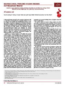
FIGURE 5. A homeostatic model of memory processing posits that neural systems interactions are driven internally to reduce uncertainty so that the most adaptive decisions can be made. According to this model, a key to successful outcomes is the presence of a number of brain systems that detect whether expected events (either input or outcomes) occur as predicted. Shown here are the striatum (reward prediction error, or RPE), hippocampus (context prediction error, or CPE), sensory cortex (sensory prediction error, or SPE), and the motor cortex (motor prediction errors, or MPE). There are others as well (area X. . .; X prediction error, or XPE). These prediction error areas connect with each other, and they have reciprocal connections to the prefrontal cortex. Messages from the prediction brain areas relay activity state information to the prefrontal cortex. It is proposed that in a familiar situation (i.e. when there are no prediction errors) the prefrontal cortex is continually driven to maintain afferent neural activity to a set point via a complex pattern of excitatory (1) and
inhibitory (-) efferent connections to the brain areas that sent state information to the prefrontal cortex previously. In this way, the prediction neuronal networks will be prepared to generate temporally and spatially accurate error signals when needed. After prediction errors are detected, error messages are sent to other prediction centers and to prefrontal cortex. The latter may coordinate the efficiencies of the different prediction centers based on recent outcomes of behaviors. This heightened coordinated activity could result in transient co-modulation across brain structures, and it could effectively extend working memory operations beyond the prefrontal cortex. Motivation may tune the efficiency of the prediction analysis system by raising or lowering prediction thresholds across multiple brain areas. Motivational influences likely arrive from structures such as the amygdala and hypothalamus. [Color figure can be viewed in the online issue, which is available at wileyonlinelibrary.com.]
dopamine system clearly plays a role in surprise-induced enhancement of learning (e.g. Schultz et al., 1997; Schultz and Dickinson, 2000), and this may relate to an influence of the amygdala on dopamine neurons since this prediction errorbased learning effect is abolished in rats with amygdala disruption (Holland and Gallagher, 2006) with no effects on the subsequent expression of surprise-induced enhanced learning (Lee et al., 2008). The amygdala and hypothalamus, then, may orchestrate information processing circuits=systems by ultimately setting the threshold for future error detection via direct connections to prediction error structures such as the hippocampus, striatum, sensory and motor cortex, and the cerebellum. The prefrontal cortex can also be thought of as playing a modulatory role in the processing of prediction errors but for reasons that are different than the amygdala. The prefrontal cortex is commonly thought to be important for holding information on-line in a working memory buffer (e.g., Fuster, 2008; Arnsten et al., 2012). This function is considered essential to be able to select appropriate responses and=or for switching behavioral strategies (Ragozzino et al., 1999a,b; Young and Shapiro, 2009), and this interpretation is consistent with findings that transient functional connections exist between the prefrontal cortex and the hippocampus or striatum especially when working memory is helpful for optimal behaviors. For example hippocampal and prefrontal theta become co-modulated at times when animals make choices (e.g.,
Hyman et al., 2005; Shirvalkar et al., 2010) but not at other times during task performance. Co-activation of striatal and prefrontal activity has also been observed when working memory is required for accurate response selection (Levy et al., 1997; Scimeca and Badre, 2012). Thus, the functional connections between striatum and prefrontal cortex, or between hippocampus and prefrontal cortex, can vary in strength and impact depending on the current task demands (Fig. 4). Presumably this variation reflects the phasic task-dependent coordination of patterns of excitation and inhibition between prefrontal cortex and its efferent targets. Since the prefrontal cortex is thought to play a role in prediction analysis (e.g. Holyroyd et al., 2002), we suggest the possibility that the coordination across prediction areas is at least in part regulated by prefrontal cortex via regulation of its inhibitory and excitatory projections to multiple types of neurons (i.e. both interneurons and projection neurons) in efferent prediction brain areas (as reviewed in Khan and Muly, 2011; Arnsten et al., 2011, 2012), neurons that then return information back to prefrontal cortex. Neocortex has indeed been shown to regulate the excitability states of subcortical neurons (e.g. Plenz and Arnsten, 1996; Plenz and Kitai, 1998; Calhoon and O’Donnell, 2013). Prefrontal cortex in particular may likely continually receive information about the current level of neural activity in target regions, and then use these afferent data to determine the extent and type of excitatory and inhibitory control needed to achieve optimal tonic activity within each of the multiple Hippocampus
1110
MIZUMORI AND JO
FIGURE 6. A: A homeostatic model of memory processing suggests that the primary goal of prefrontal cortex interactions with prediction centers of the brain (e.g., hippocampus and the midbrain-striatal area) is to maintain the baseline (tonic) firing rate of neurons within these centers at a set point level that is optimal for detecting future prediction errors. Modulating factors such as one’s motivation or emotional state can elevate or reduce the baseline firing rates. According to Figure 5, the prefrontal cortex continually receives information from the prediction areas regarding the current population firing rates. If the baseline rates become elevated (e.g., due to stress) the prefrontal cortex is equipped to anatomically and physiologically restore firing rates (red straight arrows) to their optimal (baseline) levels. If the rates become too low (e.g., in depression), the prefrontal cortex should engage mechanisms to elevate firing to optimal levels over time. When a prediction error signal arrives in, or is generated by, a given prediction structure, firing rates can increase (in cases when prediction errors are positive) or decrease (when prediction errors are negative). The degree of rate increases or decreases scales to the degree of mismatch that is detected, and the slope of the increase or decrease in firing may vary between individuals and=or as a function of experience (blue arrow). B. The prefrontal cortex is responsible for restoring the firing rates back to optimal levels and this reduces the uncertainty that was generated by the prediction error signals. [Color figure can be viewed in the online issue, which is available at wileyonlinelibrary.com.]
efferent prediction error systems (Fig. 5). When the tonic activity level is low, for example at times when there are no prediction error signals, prefrontal cortex may elevate the stae of neural excitability so that the afferent cells are more responsive to future error signals (Fig. 6), a feature that should increase the speed and accuracy of the error signaling. If, on the other hand, the baseline of a target region is higher than is optimal for the detection of prediction errors, further increasing the excitability of the cells may be detrimental for the cell’s health and ability to produce clear error signals. In this case, it would Hippocampus
be most adaptive if the prefrontal cortex lowered the level of excitability of its target cells so that optimal responsivity can be restored in the target region. Recurrent neurocircuitry within the prefrontal cortex is thought to contribute to its working memory capacity (e.g. Arnsten et al., 2012), and as such this circuit is a clear candidate system to not only integrate error signals arriving from the different prediction error brain regions, but to also bias the thresholds and strengths of subsequent error-related signals from the brain regions that originally produced the error signal. The particular constellation of excitatory and inhibitory biases presumably will result in the most desired behavioral outcome. In summary, at specific times when working memory is needed, the intrinsic recurrent neural cirtuits of the prefrontal cortex (Arnsten et al., 2012) may selectively and strategically exploit (differentially or in concert) its rich array of excitatory and inhibitory efferent connections to regulate firing rates in different prediction areas of the brain. In this way, the prefrontal cortex may regulate the relative responsiveness of different prediction brain regions in task-dependent ways. When prediction errors are detected and firing rates change, the prefrontal cortex may not only integrate the signal within its recurrent (working memory) intrinsic circuitry, but it may have a key restorative function in efferent structures such that the firing rates return to a baseline tonic level that optimizes subsequent responsiveness to input. Thus, the prefrontal cortex may bias efferent neurons’ ability to engage in, or efficiently use, prediction error analysis and hence their ability to adaptively guide future behaviors.
A PREDICTION MODEL OF MEMORY ORGANIZATION The brain contains a dynamic and interactive architecture by which memories of past experiences can continually and adaptively guide choices and behaviors. The fundamental elements of such an adaptive memory system necessarily include a) mechanisms for detecting errors in predictions about sensory, motor, decision and event outcomes (e.g. Yiv and Schoenbaum, 2008), b) the computational architecture to then coordinate different sorts of mismatches or errors between memory-based predictions and actual events with the goal of selecting adaptive decisions or actions, c) mechanisms for selecting and predicting the future outcomes of the chosen decision or action, and d) the ability to quickly and strategically restore baseline activity levels in such a way that the outcomes of future predictions can be rapidly and optimally assessed. This sort of adaptive memory system is made possible by natural neural regulatory systems that are driven to resolve uncertainties that result from failed predictions (Fig. 6; e.g. Dayan, 2012). Indeed, brain mechanisms have been identified that respond to outcomes only (or primarily) under uncertain conditions (Fiorillo et al., 2003, 2008). An inability to accurately resolve uncertainties will preclude the ability to correctly predict the outcomes of responses or significant events, and this in turn will result in maladaptive decisions and actions.
MEMORY PROCESSING AND ADAPTIVE DECISION MAKING The fundamental elements of the proposaed prediction model of memory include the following:
DETECTING ERRORS IN PREDICTIONS ABOUT SENSORY, MOTOR, DECISION, AND EVENT OUTCOMES It was emphasized above that most of the brain areas that traditionally have been ascribed to have specific memory functions (e.g. hippocampus, striatum, amygdala, sensory and motor cortex, cerebellum) engage in match-mismatch operations for the purpose of detecting prediction errors. The specific types of information that are evaluated vary across brain areas as this is determined by their different constellation of inputs. We suggest that these error detection systems have in common output that effectively alerts other prediction error systems (either directly or via prefrontal cortex regulation, Fig. 5) so that memories can be updated (and behaviors implemented) as quickly and accurately as possible.
COORDINATING DIFFERENT SORTS OF MISMATCHES OR ERRORS IN PREDICTION By virtue of its unique and sophisticated pattern of connections with prediction areas of the brain, as well as its capacity to retain information within an internal recurrent collateral system, the prefrontal cortex is strategically situated to receive and integrate prediction error messages simultaneously from the various memory regions of the brain via both inhibitory and excitatory mechanisms. It is proposed that much of the coordination is driven by natural and automatic tendencies to maintain neural networks in stable states. This also just happens to be the state where the neural sensitivity to prediction errors is maximal. That the brain is driven to resolve uncertainties that arise with prediction errors (Fiorillo et al., 2003; Knill and Pouget, 2004; Doya, 2008; Schultz et al., 2011; Bach and Dolan, 2012; Dayan, 2012) should ensure adaptive and predictable outcomes. Given the role for neighboring orbital frontal cortex in risky or probabilistic decision making (e.g. O’Neill and Schultz, 2010; Roitman and Roitman, 2010; Schultz et al., 2011), it is likely that the orbital frontal and prefrontal cortex normally work together to result in the most accurate decisions that reduce uncertainty (Rushworth and Behrens, 2008; O’Neill and Schultz, 2010). Future work aimed at understanding in more detail the relationship between the orbital and prefrontal cortices should shed new light on the mechanisms of the frontal cortical response to uncertainty and prediction errors. Also, since one’s past experiences can bias one’s interpretation of prediction errors and outcome values, prefrontal cortex coordination of prediction-based brain areas should also be impacted by expectancy information provided by existing longterm memories. When no prediction errors are present (i.e., when working memory is not needed for successful performance of a task),
1111
the prefrontal cortex may continue to automatically monitor (and then maintain) a level of neural activity that is optimal for detecting future errors of prediction. This ability to constantly check for possible errors in prediction seems necessary to optimally execute adaptive behaviors. If prefrontal cortex has an active role in coordinating activity within and across prediction centers of the brain, organisms would be endowed with a high degree of behavioral flexibility at precisely the times when multiple factors need to be considered simultaneously to make a choice or decision. This type of processing is consistent with a common definition of (prefrontal-mediated) working memory in which a working memory “buffer” allows information to be manipulated. However, analogous to an alternative view of prefrontal function proposed by Fuster (2006, 2009), it is suggested here that managing the network activity between the prefrontal cortex and connected prediction areas of the brain is what produces working memory capacity. If this is the case, the critical role of the prefrontal cortex in adaptive behaviors should be most obvious when multiple prediction errors need to be processed simultaneously. Indeed, combinations of prefrontal cortex and connected prediction areas appear co-activated during “working memory” function (e.g. Levy et al., 1997). Prefrontal coordination of prediction centers may be continuous even in situations that do not require working memory, thereby enabling organisms able to rapidly change behaviors as needed by changing environmental conditions. With prefrontal cortex damage, then, one may not observe behavioral deficits unless conditions change and prediction errors need to be detected in order to make accurate choices. The continuous and automatic functioning of memory regions of the brain has been postulated by a number of investigators (e.g. Mizumori et al., 2004; Tse et al., 2007). Prefrontal cortex damaged that results in inefficient behaviors could be due to an impaired ability of error detection systems to guide adaptive behaviors and=or relatively ineffective compensatory mechanisms that were engaged (Ragozzino et al., 1999a). In summary, working memory may be possible because it is involved in (or in this case controls) organized exchanges across widely distributed networks across the brain (Fuster, 2006, 2009). An important implication is that prefrontal cortex does not by itself make executive decisions but rather it has the highly influential role of biasing the impact of one or a combination of prediction error analyses depending on the successes and failures of recent predicted outcomes. In this way, prefrontal cortex ultimately determines which behavior should next be implemented.
SELECTING FUTURE BEHAVIORS AND PLANNED OUTCOMES The circuit described thus far accounts for animals’ ability to correct behaviors based on past outcomes. What happens when there is more than one option for a decision or action? In order to make that decision, one needs to look ahead in time Hippocampus
1112
MIZUMORI AND JO
to determine the expected outcomes one, two, or more choices down the road, then select the option that will eventually lead to the most desired outcome. If one can determine that the ultimate goal can be achieved by going down path A but not B, greater certainty could be associated with choice A. While it is not known how such prospective analysis takes place in the brain, recent data reveal that neurons in at least the hippocampus have the capacity to prospectively and retrospectively code spatial context information (e.g. Wilson and McNaughton, 1994; Buzsaki, 1989; Frank et al., 2000; Ferbinteanu and Shapiro, 2003; Gupta et al., 2012). It is possible, then that when animals are faced with choice points, the hippocampus guides the prefrontal cortex analysis into the future by providing it with a sequence of information that does not reflect the current location of the animals, but rather the expected locations and their significance. When prefrontal cortex receives “expected” location information, it may naturally retrieve the relevant information from long term memory about the associated expected outcomes. Indeed patients with hippocampal damage lose the ability to plan for the future (Hassabis et al., 2007; Schacter and Addis, 2007), a finding consistent with evidence that rodent hippocampus can construct experienced and never-before-experienced paths (Gupta et al., 2010).
RESTORING BASELINE ACTIVITY LEVELS As elaborated above, the prefrontal cortex is thought to continually monitor the tonic activity levels of connected prediction areas of the brain to ensure an optimal threshold for detection of prediction errors. This is the case regardless of whether prediction errors are detected or not. In this way prediction-relevant neural networks are driven to reduce uncertainty so that future predictions are accurate and so that desired goals are achieved in the most efficient way.
IMPLICATIONS FOR THEORIES ON THE ORGANIZATION OF MEMORY IN THE BRAIN The proposed prediction model of memory asserts that different brain areas to mediate different forms of memory because they support different type of prediction analysis. The hippocampus evaluates the extent to which contextual features have changed, while the midbrain-striatum evaluates whether responses resulted in the expected goal outcomes. Sensory regions of brain determine whether expected sensory events occurred as predicted, and motor cortex and cerebellum signal whether a specific behavioral act resulted in a specific outcome. These different prediction analyzers interact to inform each other if prediction errors occur, and this may have the effect of altering future prediction thresholds across the brain. As an example, errors of one type (e.g. episodic memory) may increase the bias to use other (e.g. response) strategies to solve Hippocampus
a task by lowering the threshold for detecting a prediction error. A coordinated and goal-directed set of responses to prediction errors is needed when multiple error messages are generated. The prefrontal cortex is suggested to coordinate the thresholds for prediction detection across many brain areas in order for the animal to produce the most adaptive responses to unexpected events. Similar to the suggestion by Fuster (2009), working memory reflects not just processing internal to prefrontal cortex, but this function emerges from the diverse and simultaneous interactions of the prefrontal cortex with prediction centers. In this case, prefrontal cortex’s control may be defined by the nature of its feedback messages in response to changes in the activity level in the connected prediction areas. The prefrontal cortex may seek to maintain optimal (stable) activity levels in the prediction centers so that they are maximally prepared for meaningful responses when needed. Prefrontal cortex, then, could be thought of as effectively regulating the balance between stable and flexible neural processing in connected brain areas. By this logic, the prediction model of memory views the vertebrate brain as having a single memory system that is continuously integrating different sorts of prediction analyses. The pattern of differential biases of controlled neural activity by the prefrontal cortex determines the selection of a particular adaptive behavior or choice. Decisions, then, reflect the relative weighting of signals across brain areas that generate prediction error signals. The clear benefit of this type of system is that when one type of prediction analysis fails, others may ramp up their influence over future decisions and actions without having to “learn from scratch”. The effectiveness of the regulation of prediction centers by prefrontal cortex likely reflects modulating influences from motivation and=or emotion processing areas such as the amygdala. Such modulation reflects the current (or expected) emotional or motivational state, and this regulation is manifested by changing baseline firing rates in prediction brain areas (Fig. 6). If the baseline remains too high (e.g., in states of high arousal) or low (e.g., in states of depression), prefrontal cortex should receive feedback about these changes in firing rates and then implement mechanisms to restore neural activity to normal levels via its bidirectional-regulatory system. If prefrontal cortex is damaged, then neural activity levels may remain exceptionally high or low, resulting in poor choices, decisions, and behavioral adaptation. If one of the prediction centers is damaged (and this results in abnormal activity levels) then it would be expected that prefrontal cortex will not be able to restore optimal activity levels. If baseline activity in an intact brain is restored after the onset of an arousing incident, one should become able to analyze a situation by coordinating the normal prediction-error based analysis that leads to adaptive decisions and behavior selections. Supporting this view are reports that altered emotional states (e.g., depression) are correlated with changes in prefrontal cortex function (Baxter et al., 1989; Mayberg et al., 1999). The connection between the prefrontal cortex and the amygdala provides reciprocal functional regulation since targeted disruption of prefrontal cortex results
MEMORY PROCESSING AND ADAPTIVE DECISION MAKING
1113
FIGURE 7. Schematic illustration of the activation patterns of amongst context prediction error (CPE) and reward prediction error (RPE) systems, and their coordination permitted by working memory (WM). Activated systems are circled in red, and thicker lines reflect increase information transfer. A: When there are no prediction errors, neural systems are stable and there is little or no uncertainty in terms of outcome expectations. Arrows reflect the baseline levels of reciprocal information exchange between RPE, CPE and WM systems. B: (1) An error signal is initially generated from the midbrain system (e.g., when an expected reward does not occur), then transmitted to the prefrontal cortex (to activate working memory processes) or directly to other prediction areas of the brain. (2) Using its working memory circuitry, the prefrontal cortex then informs hippocampus about the reward prediction error. 3: Hippocampus becomes activated so that it is better prepared to detect context prediction errors. 4: If a context prediction error is generated, this will be signaled to striatum where continued
reward prediction analysis should be encouraged. A decline in prediction error signals should be observed as memories are formed and expectations match actual experiences. C: After learning, neural states restore to baseline levels. D: 1: If there is a change in a familiar context a prediction error signal is generated initially in hippocampus, and this message will be sent to both striatum. 2: The signal to the reward and response system of the striatum facilitates transition to an “up” state so that it is better prepared to respond to outcome information, and working memory processes become engaged. In this example, a reward prediction error is detected. (3) Prefrontal cortex then informs the CPE system of the reward prediction error, and this state of uncertainty elevates neural activity in the hippocampus. (4) As context match signals decline, prefrontal cortex may down-regulate the excitatory drive that it was imposing on striatum when the mismatch signals first occurred. [Color figure can be viewed in the online issue, which is available at wileyonlinelibrary.com.]
in clear changes in affective states (e.g., Covington et al., 2010; Hamani et al., 2010).
memory processes) or directly to other prediction areas of the brain (Fig. 7B1; so that they become more responsive to subsequent incoming information). The prefrontal cortex in turn informs hippocampus about the reward prediction error (Fig. 7B2). Hippocampus may show elevated firing (Fig. 7B3), indicating that it is receptive to processing new information that will eventually reduce the uncertainty that arose after the striatal prediction error message. If a context prediction error is generated, this will be signaled to ventral striatum and the midbrain dopamine regions (Fig. 7B3) where continued reward prediction analysis should be encouraged (Fig. 7B4). A decline in prediction error signals from hippocampus (indicating
SCENARIO 1 As an example of the interactive nature of the different prediction error regions of the brain, let’s consider the case when an error signal is initially generated from the midbrain system (e.g., when an expected reward does not occur). The error signal is transmitted to the prefrontal cortex (to activate working
Hippocampus
1114
MIZUMORI AND JO
reduced uncertainty) reflects the fact that long terms memories have been updated, i.e. expectancies now match one’s experiences. Initially, after the first prediction error signal from the striatum, the currently active memory network will be made more labile so that new information can eventually be incorporated into the memory network (Fig. 3). According to theoretical models of cortical memory systems (e.g. McClelland et al., 1995), this updating process should occur slowly to avoid catastrophic effects on existing memories. As long term memories become updated, information about one’s expectations for a given situation or context should also be update, and this information will be fed forward to hippocampus to be used in subsequent prediction analyses. As an example of the above process, let’s say that on the next trial, an expected reward again does not occur. As long term memories become updated with this information, expectations for a reward will decline, and this will result in an eventual “match” signal by hippocampus. This will reduce signals to striatum to be responsive to rewards. Indeed, this pattern of reduced responses to reward has been observed empirically after learning is complete (Fig. 7C; Schultz et al., 1997; Puryear et al., 2010).
SCENARIO 2 A different example of interactions amongst prediction error and general modulatory centers of the brain considers the case when the first prediction error signal is generated by hippocampus, e.g. after an unexpected change in a familiar context (Fig. 7D1). A context prediction error signal will be sent to both striatum and prefrontal cortex to facilitate subsequent reward valuation of the context change (striatum) and to inform other prediction error brain regions (e.g. via prefrontal cortex). The signal to the reward and response system of the striatum may facilitate transition to an “up” state from a “down” state (Fig. 7D2; Wilson, 1993; Wilson and Kawaguchi, 1996) for striatal and dopamine neurons so that they are better prepared to respond to outcome information. Subsequently, prefrontal cortex may enhance the responsiveness of multiple prediction monitoring brain systems to facilitate the rapid analysis of changed conditions so that future behaviors and decisions are flexible (Fig. 7D3). Since the ventral striatal system will receive input from both the hippocampus and prefrontal cortex it may be particularly sensitive to response and reward value outcome information. If the value of the reward is deemed to have changed significantly (either higher or lower than expected), then long term memories should begin to be updated according in terms of the actual context in which the response that generated the changed reward value was made (Fig. 4). Again, the memory updating process may be incremental, and thus appear slow. If the changed reward value continues to be experienced in the new context, eventually hippocampus will no longer generate error signals since the memory that defines the expected context features will be Hippocampus
updated to match the actual context. As more context match signals are generated by hippocampus, prefrontal cortex may down-regulate the excitatory drive that it was imposing on striatum when the mismatch signals first occurred (Fig. 7D4).
COMPETITION OR COOPERATION BETWEEN PREDICTION CENTERS Many have described interactions amongst memory systems as reflecting a type of competition and=or cooperation for the control of behavior (Poldrack et al., 2001; Colombo, 2004; Gold, 2004; Mizumori et al., 2004, Lee et al., 2008; Gruber and McDonald, 2012). What are the mechanisms of this competition or cooperation? Our prediction model of memory systems offers some new insights. Evidence for competition or cooperation amongst memory systems comes primarily from the result of studies of the effects of reducing the functional capacity of one brain area (e.g. by lesions or hormone treatment, McDonald and White, 1993; McElroy and Korol, 2005; Gruber and McDonald, 2012) while observing subjects’ performance on tasks that require normal function of other intact memory areas. With this paradigm, it was shown that hippocampal lesioned rats showed faster learning of response or cue tasks that are usually considered to be mediated by the striatum (McElroy and Korol, 2005). It was reasonably concluded that there is a natural competition between striatum and hippocampus that disappeared after the lesion, and this enabled striatum to function more effectively. A prediction model of memory provides additional insight. According to this view, both striatal and hippocampal prediction systems should be operational simultaneously during baseline states regardless of the task at hand. In this way, striatum can continually evaluate whether actions result in the expected rewards and hippocampus continually assesses the extent to which the context-defining features of current situation match those expected based on past experience. Indeed, both the striatum and hippocampus contain spatial, reward, and behavioral representations that respond to different types of errors in prediction regardless of whether the task is spatially or response based (e.g., Yeshenko et al., 2004; Eschenko et al., 2007; Mizumori et al., 2004). If the hippocampus become dysfunction (e.g., after a lesion) it should still be possible to learn according to prediction errors generated by the remaining systems. For example, striatum should still be able to generate prediction error signals that underlie response or cue learning, and thus rats will tend to switch to a response strategy after a hippocampal lesion (e.g. Packard and McGaugh, 1996). The prediction model of memory interprets this finding as a demonstration that hippocampal prediction error messages that normally arrive in striatum increase the analysis load on striatal prediction analysis networks. When the hippocampal input is removed, the processing load is lessened and striatum is now able to more effectively evaluate whether reward outcomes occurred as
MEMORY PROCESSING AND ADAPTIVE DECISION MAKING predicted by a specific behavioral sequence of the animal. According to this explanation, striatum and hippocampus do not directly compete for control of a behavior per se but rather they effectively compete for access to the outcome valuation process. Another implication is that habit learning may not by itself be incremental and slow as is commonly thought. Rather, in intact animals, habit learning may appear slow because of a prioritization for valuation of hippocampal context prediction error signals relative to reward prediction error signals. Indeed rats seem to have a predisposition to learn tasks using context cues rather than response information when the solution to a problem is ambiguous (Packard and McGaugh, 1996). This prioritization may reflect a natural and innate hierarchy of processing priorities for decisions making systems based on the adaptive value of one strategy or algorithm or another. Context processing may confer greater adaptive flexibility than learning only by habits. One prediction, then, is that striatal-dependent learning may be found to be rapid if learning explicitly did not involve context processing (e.g., aversive conditioning). In the event that striatum is impaired, hippocampal-based behaviors should remain at least initially intact since the reward prediction neural circuitry includes key structures afferent to the striatum (i.e., within the network that includes the habenula-ventral tegmental (dopamine) circuit, Hikosaka et al., 2008). Thus it is possible that reward prediction error signals can continue to influence hippocampus prediction analyses, albeit perhaps less efficiently. However, an important part of especially navigation-based learning is to understand the relationship between sequences of actions and a desired outcome. Therefore, it is predicted that if striatal-lesioned subjects are challenged with complex spatial navigation problems, or complex econometric analysis, deficits will emerge because rats will have difficulty learning the expected consequences of multiple, flexible behavioral trajectories. Such a result would imply that optimal continued performance on a complex navigation-based task requires the maximum types of behavioral flexibility that is normally supported by the collection of prediction error mnemonic processors. The intrinsic regulation attributed to the components of a prediction model of memory is remarkably analogous to the homeostatic regulatory system that has been shown to operate at the synaptic level (Turrigiano, 1999; Marder and Goaillard, 2006; Turrigiano, 2011). In what follows we explore the possibility that principles that guide homeostatic neural plasticity at the synaptic level also apply at the neural systems level to account for the self-regulatory and interactive capacity of the many memory processors of the brain. First, we briefly describe a model for the self-regulation of synaptic efficiency (Marder and Goaillard, 2006; Turrigiano, 2011). This neuroregulatory perspective is then compared to the interactive prediction model of memory in an attempt to not only account for the remarkable and precise coordination of memory processing in the brain but to also further illustrate how memory and decision making brain circuitry are so intimately tied together that one cannot function without the other.
1115
HOMEOSTATIC REGULATION OF MEMORY PROCESSING SYSTEMS Since the early 1930s scientists have described the remarkable ability of the body to maintain optimal function despite perturbations of the surrounding environment (e.g., Cannon, 1932). This adaptive process, termed homeostasis, includes sensors that detect deviations in physiological states relative to a previously defined optimal state (or set point). Errors or deviations detected by the sensors inform a controller to up or down regulate mechanisms that can bring the current physiological state closer in line with the set point. Principles of homeostasis can account for self-regulatory synaptic mechanisms (e.g., Turrigiano, 1999; Marder and Prinz, 2003; Turrigiano and Nelson, 2004; Turrigiano, 2008, 2011) and this has led to the view that neural circuits are also subject to homeostatic regulation (Turrigiano, 1999; Marder and Goaillard, 2006; Turrigiano, 2011). Thus, homeostatic regulation could be a key mechanism that maintains the balance between stability and flexibility that neural networks needed to support dynamic and adaptive responses. At the synaptic level, homeostatic regulation is possible because a form of calcium-mediated plasticity allows neurons or neural networks to maintain a key physiological parameter (firing rate), within an optimal range, or set point, despite new excitatory and=or inhibitory inputs. Such homeostatic plasticity helps to insure that cells do not, for example, suffer catastrophic effects of overexcitation when constant and strong excitatory inputs arrive (as in the case of LTP). Upon arrival of strong excitatory input, self-regulation of firing rates is evident by the fact that the synaptic strengths of all synapses of the stimulated cell are reduced while maintaining the relative distribution of synaptic weights across the cell. This “scaling” process makes it possible to retain new information while still allowing for future plastic changes (Turrigiano, 1999; Turrigiano et al., 1998). Marder and Goaillard (2006) suggested that homeostatic plasticity may be nested: calcium sensors may monitor neural firing rates, then up or down regulate the availability of glutamate receptors to ramp up or down firing rates. Groups of neurons or neural networks may sense changes in firing collectively to regulate population activity levels and patterns of activation. In this way homeostatic plasticity enables groups of neural circuits to find a balance between flexible and stable processing as needed to learn from experiences, and to be responsive to future changed inputs. The specific details of how neural networks engage in homeostatic regulation remain to be discovered. Nevertheless, it is worth noting that homeostatic regulation at the neural systems level clearly occurs as evidenced by studies of brain development, as well as from studies of reactive or compensatory neuroplasticity mechanisms that occur in response to experience (e.g. sensorimotor learning; Froemke et al., 2007) or brain injury (e.g. brain trauma or addiction; Robinson and Kolb, 2004; Nudo, 2011). While homeostatic neural plasticity mechanisms have not been used Hippocampus
1116
MIZUMORI AND JO
to account for complex learning, current theories of reinforcement- and context-based learning and memory commonly posit that the outcome of behaviors and choices feed back to update relevant memories and future decision (as described above). To conclude that fundamental operating principles of a homeostatic system can be used to enhance our understanding of the interactions between memory processing centers of the brain, we must first determine if the elements of a homeostatic system can be identified as part of memory processing. Key components of any homeostatic model include a variable that is being monitored by a sensor and then regulated by a controller. Given the complexity of memory processing, we start by assuming that the relevant homeostatic neuroregulatory mechanisms are likely nested (Marder and Boaillard, 2006). The ultimate regulated variable is a decision or action (also see KurthNelson and Redish, 2009). The outcomes of decisions and behaviors are monitored via a number of sensors, i.e. neural circuits that monitor the accuracy of predicted information. If sensors detect deviations (i.e. prediction errors), a controller (or regulatory system) must be engaged in order to restore accurate predictions. At a more cellular level of the “nested” system, specific neural variables need to be monitored in order to identify the current system state. Since the firing rates of neurons are a homeostatically regulated variable that is regulated to achieve a balance between synaptic stability and flexibility (Marder and Goaillard, 2006; Turrigiano et al., 1998; Turrigiano, 2011), a key neural variable may be the optimal level of excitability of neurons in prediction areas of the brain. In this case, changes in calcium flux may be an important part of the sensing system as it reflects the current level of neural activity. When firing rates become higher or lower than the optimal level, this could be taken as an indicator of a mismatch between optimal and actual rates, and a controller mechanism should be engaged to bring the firing rates back to the optimal levels so that cells are prepared to respond to subsequent input. However, merely up or down regulating firing rates is a short term response to altered firing induced by uncertain or unexpected situations. Long term adjustments necessarily include the regulation of decisions and behaviors that resolve uncertainty or errors in prediction. Given that prediction error brain areas are highly interconnected with the prefrontal cortex, a likely candidate structure that serves as the main controller for prediction errorbased memory systems is prefrontal cortex. In this case, firing rates of cells within the prediction error processing areas of brain may be “sensed” by the prefrontal cortex via direct afferent fibers. If the baseline rates are too high or too low relative to the optimal state needed for detecting future prediction errors, then prefrontal cortex may engage mechanisms to restore the firing rate to a predetermined “set point”. According to the model in Figures 5 and 6, the detection of a prediction error, or mismatch, should produce significant deviations from the firing rate set point, and this error message could impact prediction error processing in other brain areas as well as the controller function of the prefrontal cortex. PrefronHippocampus
tal cortex, in turn, should orchestrate the information flow to and from all prediction error brain areas according to homeostatic principles. With the goal of ultimately restoring the set point of neural activity in prediction error brain regions (i.e., reduce error signals to restore a stable neural state that reflects greater certainty about outcomes of decisions and actions), prefrontal cortex should continually and automatically receive feedback about the current excitatory state, and then strategically up or down regulate the excitability of different processing systems depending on the past outcomes of behaviors (Fig. 6). Part of this process is to incorporate information (presumably from the striatum) about the successes of recent behaviors so that the same or different behaviors can be next selected. The intrinsic recurrent circuitry of the prefrontal cortex may serve this function well (Arnsten et al., 2012). With regard to the impact of homeostatic regulation of prediction centers on established memories, it would be expected that the outcome of the prediction analysis (which includes error detection and controller functions) should guide the updating of memories with information about the success (or failures) of responses as an animal attempts to achieve a desired goal. In this way, the next time a particular memory is retrieved, the most recent and accurate information can be used to generate the next set of expectations. A memory of an event, then, should include not only the defining cues and outcomes, but also the emotional tone (since the amygdala is postulated to contribute to the threshold for detecting prediction errors), the context-specific behavioral actions that led to decisions, and the strategy associated with the reconciliation of error messages. It should be noted that while the prefrontal cortex may be a major controller of the impact of prediction error signaling in the brain, other sources of varying degrees of control may arise from the any of the brain regions that analyze prediction outcomes given that they are interconnected. For example, a prediction error from the hippocampus could be transmitted to midbrain-striatal neurons along pathways that do not initially include the prefrontal cortex. Indirect support for this idea comes from observations that conditions that produce error messages in the hippocampus change reward responses of dopamine neurons (Puryear et al., 2010; Jo et al., 2013), and phasic theta comodulation is observed between hippocampus and striatum (DeCouteau et al., 2007) during decision tasks. In sum, homeostatic regulatory processes may account for the automatic and continuous self-regulatory nature of prediction error analysis, decision making, learning and memory. Such a natural and adaptive mechanism optimizes the contribution of different types of prediction error signals to future decisions and actions according to the pattern of recent successes and failures in prediction. This more dynamic view of the interactions between adaptive memory and decision making circuitry has important implications for the development of more efficient and long lasting cognitive and behavioral intervention methods for not only amnesic patients but also for disorders characterized by suboptimal decision processing such as addiction, autism, and schizophrenia. Therefore, the remainder of this article evaluates the extent to which the current
MEMORY PROCESSING AND ADAPTIVE DECISION MAKING
1117
literature validates key premises of a homeostatic view of memory and decision systems interactions. This discussion also highlights future directions for research that test the prediction model of memory.
is indeed part of the hippocampal neural (i.e. memory) code: Lee et al. (2012) and Penner et al. (2012) recently described place field remapping that depended on the expected probability of rewards of large or small magnitude.
PREMISE 1
PREMISE 3
There is a fundamental and common currency of information representation across prediction centers of the brain. A common currency should facilitate information exchange and regulation across brain regions. Also, if identified, one would be able to use the same neurometric to directly compare regional differences in aspects of decision and=or memory processing. A search for a common currency of representation has not been systematically carried out since a multitude of different behavioral or cognitive tasks are used in studies of neural representations. There is initial evidence, however, that support Premise-1 since strikingly similar types of neural representation are found across many memory processing regions of brain when rats perform appetitive tasks on open elevated mazes such as the plus maze or radial arm maze. The common currency includes representations of rewards, the spatial features or requirements of a task, task phase, and egocentric movement. Of these neural correlates, egocentric movement (in particular velocity or acceleration of translational movement across the maze) has been the most common and robust (as described in Penner and Mizumori, 2012b). In the future, these correlates of connected brain areas should be recorded simultaneously so that their temporal relations can be identified. Although not systematically assessed to date, one would expect to find common codes for econometric information regarding the salience of the common currency units.
Mismatch, or prediction error, messages in one brain area should induce system-wide responses. Based on known patterns of connectivity between brain regions, and in the interest of efficient system function, a prediction-based model of memory hypothesizes that the different prediction processing areas are interdependent in the sense that generation of error signals in one system should quickly influence the future firing patterns of neurons in other prediction areas. Although more direct evidence is needed, it is worth noting that neural responses from two or more brain areas show that each area generates prediction signals during the performance of the same behavioral task (e.g. Yeshenko et al., 2004). In these tasks, encounters with unexpected rewards presumably generated a reward prediction error signaled by greater dopamine and striatal responding (e.g. Fig. 7). This in turn should lead to reorganization of the distribution of place fields (i.e. the generation of a new “map” or memory). Following a context manipulation, one would expect to see alterations or remapping of place fields, followed by altered reward value signaling by the dopamine neurons. Several studies (e.g. Yeshenko et al., 2004; Smith and Mizumori, 2006; Puryear et al., 2010; Martig and Mizumori, 2011; Smith et al., 2012; Penner et al., 2012) now show that both hippocampal and striatal neural correlates are significantly affected by changes in context and reward processing during the performance of a maze-based tasks regardless of the optimal strategy needed to solve the task (e.g., place vs. response strategies). Recordings in hippocampus and striatum have not always been simultaneous, however, and thus it is not possible to state with certainty that changes in one area preceded changes in the other. At this point we can only logically deduce this sequence of events based on past studies of dopamine and hippocampal responses to unexpected events, and findings that hippocampal place fields are severely disrupted when the dopamine system is not functional (Martig et al., 2011). The future application of new technologies and approaches (e.g. optogenetics, DREADD, targeted use of viral vectors, etc.) to studies of the functional connectivity of prediction circuitry in the brain should reveal important new information about the spatial and temporal causes of the changes in firing in one prediction error brain region relative to changes in firing in connected areas, and then to behavior.
PREMISE 2 The content of stored memories (as reflected in representations of expected information) should include not only the unique sensory and behavioral aspects of events, or contexts, but also information about the value of the sensory and behavioral information. Specifically, it should be possible to identify neural representations of learned information that dynamically vary according to its subjective value. While prediction error coding has been described in sensory, motor, striatal, midbrain, and hippocampal areas, a systematic and explicit econometric analysis of expectancy (i.e. memory) information remains to be conducted. As an example, it is common to record hippocampal place fields that seem to be driven by a particular constellation of external (usually visual) cues (Mizumori, 2008a), and prediction error coding is observed when rewards are unexpectedly omitted. However, the extent to which place fields incorporate the subjective value of the cues has only begun to be explored. Initial evidence suggests that econometric information
PREMISE 4 Neural codes should become stronger (i.e. more predictive) when their type of prediction comes to dominate the control Hippocampus
1118
MIZUMORI AND JO
of actions and decisions. Since a desired outcome of an adaptive decision making system is the accurate prediction of future consequences, one would expect that as learning progresses, the certainty and predictability with which desired outcomes occur will improve. Underlying this change, neural signals should become more predictive of cues or behaviors that lead to the desired outcome. Furthermore, neural codes might be expected to become more information-selective and reliable as these features come to reliably predict outcomes. Evidence in support of Premise-4 is that hippocampal place fields become more spatially-selective and reliable as hippocampal-dependent spatial learning progresses, and as animals acquire greater familiarity with a given environment (Muller and Kubie, 1987; Wilson and McNaughton, 1993; Markus et al., 1995; O’Keefe and Burgess, 1996; Hetherington and Shapiro, 1997; Mizumori and Kalyani, 1997; Lenck-Santini et al., 2001, 2002; Fenton et al., 2010), but see Jeffrey et al. (2003) and Cooper and Mizumori (2001). Also place cells generate different patterns of place fields when rats need to learn to distinguish the significance of different contexts (Smith and Mizumori, 2006). Striatal neural representations of egocentric directional movement (e.g. turns in a maze) become more prominent as rats learn a T-Maze task (Barnes et al., 2005). Further, with continued training and conditioning, dopamine burst firing and release become associated with cues that predict a reward (Schultz et al., 1997; Flagel et al., 2011)
PREMISE 5 The generation of more selective and reliable neural codes during learning should coincide with the evolution of more precise spatial and temporal coordination of neural activity within and across the relevant brain structures. When faced with uncertainty, e.g. usually after the detection of a prediction error, the working memory function of the prefrontal cortex should be engaged to simultaneously (although perhaps differentially) elevate the sensitivity of all prediction error brain structures in an attempt to facilitate the most rapid identification of all errors, as well as the determination of their significance. While direct evidence that the prefrontal cortex simultaneously activates multiple prediction-related structures in a single task remains to be determined, the prefrontal cortex is considered to be strategically located to impact multiple memory systems as well as decision neural circuitry (e.g., see review in Euston et al. 2012; Hyman et al., 2012). Supporting this view, lesions or inactivations of the prefrontal cortex disrupts goalrelated neural codes in hippocampus (Kyd and Bilkey, 2003; Hok et al., 2013) and the VTA (Jo et al., 2013) while rats perform a spatial maze task. Also prefrontal cortex damage impairs a wide range of learning and memory abilities (Fuster, 2008, 2009) presumably because of its widespread control over the balance of excitation and inhibition in the many prediction Hippocampus
structures of the brain. There is initial evidence that after initial learning, there is greater coherence and synchrony of low frequency potentials (LFPs) between hippocampus and striatum (DeCouteau et al., 2007), and between prefrontal cortex and hippocampus (Hyman et al., 2005; Jones and Wilson, 2005a,2005b; Siapas et al., 2005; Shirvalkar et al., 2010) when decisions are made. A challenge for future research is to understand the mechanisms and significance of integration across multiple frequencies of brain rhythms, and between LFPs and single cell activity, during decision making and different prediction error states (e.g. Hyman et al., 2010; Buzsaki and Wang, 2012; Lisman and Jensen, 2013).
PREMISE 6 Choice outcomes may be more related to neural representational patterns on subsequent trials than the current trial. As described earlier, the outcomes of choices should bias the efficiency with which prediction error regions of the brain function in the future, presumably by giving feedback to prefrontal cortex, which in turn incorporates the new information in its subsequent regulation of the excitability state of prediction memory circuitry. Most single unit studies correlate the specificity and reliability of characteristics of neural representations with performance on the trial in which the neural data were collected. This is indeed a reasonable comparison to make. However, our prediction-based model of memory makes the explicit suggestion that the neural correlates should be even more striking if related to choices made on recent trials, and perhaps even to specific prior choices of the same trial. Indirect support of Premise 6 can be found in studies that show that neural firing is related to past events and choices. This sort of retrospective coding was illustrated when hippocampal place fields were shown to correlate to past behavioral trajectories of the rat being recorded (Frank et al., 2000; Wood et al., 2000; Ferbinteanu and Shapiro, 2003; Mizumori et al., 2004; Lee et al., 2006). Also many laboratories have demonstrated that neural codes during sleep reflect a temporal organization of neural firing that was recently experienced (e.g. Wilson and McNaughton, 1994; Shapiro and Ferbinteanu, 2006; Bender and Wilson, 2012). More direct evidence in support of Premise 6 was recently reported. Motor cortical and parietal cortex neurons showed firing rates that vary depending on the specific outcomes of prior choices (e.g. Marcos et al., 2013), and it has been shown that rats adjust choices based on the delay imposed on prior trials (Mazur, 1988; Cardinal et al., 2002). There is also evidence of prospective coding in that neural firing correlates with intended behaviors or expected goal outcomes (e.g. Buzsaki, 1989; Wilson and McNaughton, 1994; Frank et al., 2000; Wood et al., 2000; Louie and Wilson, 2001; Lee and Wilson, 2002; Pennartz et al., 2002; Ferbinteanu and Shapiro, 2003; Yeshenko et al., 2004; Foster and Wilson, 2006; Gupta et al., 2012). However, the prediction
MEMORY PROCESSING AND ADAPTIVE DECISION MAKING error model suggests that the behavioral correlate of cells with prospective codes should be found to be even more selective if correlated with performance on the prior trial. Finally, Gupta et al. (2012) have shown striking forward sweeping of sequential activation of place fields but the sweep began from a prior location of the animal. That is, the apparent predictive nature of prospective codes was based on the results of prior choices.
PREMISE 7 The plasticity of the brain’s memory circuitry is homeostatically and intrinsically regulated to result in the self-regulated and dynamic coordination of plasticity mechanisms at multiple levels, from ion channels, synapses, single unit, to local and extended neural networks. Adaptive interactions between the modulatory and prediction error components of the brain’s memory circuitry require coordination across all levels of function and integration. In this way, not only can a healthy balance between neural stability and flexibility be achieved, but an optimal level of stable behavior should maximally prepare animals to detect the need to engage in behavioral change. Demonstrating this sort of multilevel coordination remains a grand challenge for it requires technology that monitors multiple levels simultaneously in behaving animals. Although still in its infancy, Fujisawa and Buzsaki (2011) have demonstrated the feasibility of this general approach as they recorded simultaneously single unit activity and LFP (local field potentials) across multiple regions of the brain. New forms of neural coordination were described, as well as new neural phenomenon (a 4 Hz synchronizing rhythm) that could be used to explain how prefrontal cortex might coordinate neural signals from multiple brain areas (Penner and Mizumori, 2012b). Additional details about the functional significance of prefrontal cortex control over prediction areas is expected to emerge as new technologies (e.g. optogenetics combined with electrophysiology, Warden et al., 2012) are applied to the problem of understand the organization of memory processing in the brain.
CONCLUSIONS A homeostatic model of memory posits the existence of a single, self-regulated (Buzsaki, 2013) memory system that reflects multilevel (from cells to neural system) integration across different prediction processing areas of the brain. These different prediction processors have been more commonly referred to as different memory systems such as the hippocampus (episodic memory) and striatum (response or habit memory). Significant evidence suggests that these memory systems have in common output that signals errors in prediction of events and=or their outcomes, but they differ in terms of the nature of the prediction generated (e.g. context prediction
1119
errors vs reward=response prediction errors). It is suggested that the prefrontal cortex plays a critical role in coordinating the multiple levels (from genes to cells to circuits) of prediction analysis across multiple prediction centers. From the widespread control and influence of the prefrontal cortex emerges an essential capacity for the flexible use of information to generate adaptive behaviors and to predict future outcomes. A homeostatic function of the prefrontal cortex is suggested to maintain optimal baseline (tonic) firing rates that ensure appropriate sensitivity for the future detection of prediction errors. That is, analogous to the suggestion by many computational models, predictive memory systems are driven by mechanisms that reduce uncertainty, and the prefrontal cortex is ideally situated to serve as a controller that seeks to resolve uncertainty. We hypothesize that prefrontal cortex accomplishes this task by continually engaging corrective mechanisms that restore optimal baseline firing rates after prediction error signals are generated. Emergent outcomes of this process are flexible and adaptive behaviors even in the most challenging of situations. Accordingly, compromise to any of the prediction circuits should result in less flexible and adaptive behaviors. This reduction of flexibility and adaptability could result in rigid and suboptimal decision making, learning, and memory as is seen in cases of addiction (e.g., Monterosso et al., 2012), and neurological diseases such as Alzheimer’s and Parkinson’s disease. The symptoms presented in such extreme cases of maladaptative behaviors reflect not only that a particular prediction processing system has gone awry but also the brain’s attempt to compensate for the lost function.
Acknowledgments The authors thank Wambura Fobbs, Sujean Oh, Marsha Penner, Van Redila, and Valerie Tryon for early discussions of different segments of this article.
REFERENCES Adams GK, Watson KK, Pearson J, Platt ML. 2012. Neuroethology of decision-making. Curr Opin Neurobiol 22:982–989. Alexander GE, Crutcher MD. 1990. Functional architecture of basal ganglia circuits: Neural substrates of parallel processing. Trends Neurosci 13:266–271. Alexander GE, DeLong MR, Strick PL. 1986. Parallel organization of functionally segregated circuits linking basal ganglia and cortex. Annu Rev Neurosci 9:357–381. Anagnostaras SG, Gale GD, Fanselow MS. 2001. Hippocampus and contextual fear conditioning: recent controversies and advances. Hippocampus 11:8–17. Arnsten AFT. 2011. Prefrontal cortical network connections: Key site of vulnerability in stress and schizophrenia. Int J Dev Neurosci 29: 21–223. Arnsten AFT, Wang MJ, Paspalas CD. 2012. Neuromodulation of thought: Flexibilities and vulnerabilities in prefrontal cortical network synapses. Neuron 76:223–239. Hippocampus
1120
MIZUMORI AND JO
Bach DR, Dolan RJ. 2012. Knowing how much you don’t know: A neural organization of uncertainty estimates. Nat Rev Neurosci 13: 572–586. Barnes TD, Kubota Y, Hu D, Jin DZ, Graybiel AM. 2005. Activity of striatal neurons reflects dynamic encoding and recoding of procedural memories. Nature 437:1158–1161. Baxter LR, Schwartz JM, Phelps ME, Mazziotta JC, Guze BH, Selin CE, Gerner RH, Sumida RM. 1989. Reduction of prefrontal cortex glucose metabolism common to three types of depression. Arch Gen Psychiat 46:243–250. Bayer HM, Glimcher PW. 2005. Midbrain dopamine neurons encode a quantitative reward prediction error signal. Neuron 47:129–141. Bender D, Wilson MA. 2012. Biasing the content of hippocampal replay during sleep. Nat Neurosci 15:1439–1444. Bisley JW, Zaksas D, Droll JA, Pasternak T. 2004. Activity of neurons in cortical area MT during a memory for motion task. J Neurophysiol 91:286–300. Bouton ME, Westbrook RF, Corcoran KA, Maren S. 2006. Contextual and temporal modulation of extinction: Behavioral and biological mechanisms. Biol Psychiat 60:352–360. Boyden ES, Katoh A, Raymond JL. 2004. Cerebellum-dependent learning: The role of multiple plasticity mechanisms. Ann Rev Neurosci 27:581–609. Burgess N, O’Keefe J. 2003. Neural representation in human spatial memory. Trends Cogn Sci 7:517–519. Burrone J, O’Bryne M, Murthy VN. 2002. Multiple forms of synaptic plasticity triggered by selective suppression of activity in individual neurons. Nature 420:414–419. Buzsaki G. 1989. Two-stage model of memory trace formation: A role for "noisy" brain states. Neuroscience 31:551–570. Buzsaki G. 2013. Time, space and memory. Nature 497:568–569. Buzsaki G, Wang XJ. 2012. Mechanisms of gamma oscillations. Ann Rev Neurosci 35:203–225. Cagniard B, Beeler JA, Britt JP, McGhee DS, Marinelli M, Zhuang X. 2006. Dopamine scales performance in the absence of new learning. Neuron 51:541–547. Calhoon GG, O’Donnell P. 2013. Closing the gate in the limbic striatum: Prefrontal suppression of hippocampal and thalamic inputs. Neuron 78:181–190. Cannon WB. 1932. The Wisdom of the Body. New York: WW Norton Co Inc. Cardinal RN, Daw N, Robbins TW, Everitt BJ. 2002. Local analysis of behavior in the adjusting-delay task for assessing choice of delayed reinforcement. Neural Netw 15:617–634. Chau LS, Galvez R. 2012. Amygdala’s involvement in facilitating associative learning-induced plasticity: A promiscuous role for the amygdala in memory acquisition. Front Integ Neurosci 6:92. Chen J, Olsen RK, Preston AR, Glover GH, Wagner AD. 2011. Associative retrieval processes in the human medial temporal lobe: Hippocampal retrieval success and CA1 mismatch detection. Learn Mem 18:523–528. Ciocchi S, Herry C, Grenier F, Wolff SB, Letzkus JJ, Vlachos I, Ehrlich I, Sprengel R, Deisseroth K, Stadler MB, M€ uller C, L€ uthi A. 2010. Encoding of conditioned fear in central amygdala inhibitory circuits. Nature 468:277–282. Cohen NJ, Eichenbaum H. 1993. Memory, Amnesia, and the Hippocampal System. Massachusetts: Cambridge MIT Press. Colombo PJ. 2004. Learning-induced activation of transcription factors among multiple memory systems. Neurobiol Learn Mem 82: 268–277. Cooper BG, Mizumori SJY. 2001. Temporary inactivation of the retrosplenial cortex causes a transient reorganization of spatial coding in the hippocampus. J Neurosci 21:3986–4001. Covington HE, Lobo MK, Maze I, Vialou V, Hyman JM, Zaman S, LaPlant Q, Mouzon E, Ghose S, Tamminga CA, Neve RL, Deisseroth K, Nestler EJ. 2010. Antidepressant effect of optogeHippocampus
netic stimulation of the medial prefrontal cortex. J Neurosci 30: 16082–16090. Dayan P. 2012. Twenty-five lessions from computational neuromodulation. Neuron 76:240–256. DeCoteau WE, Thorn C, Gibson DJ, Courtemanche R, Mitra P, Kubota Y, Graybiel AM. 2007. Learning-related coordination of striatal and hippocampal theta rhythms during acquisition of a procedural maze task. Proc Natl Acad Sci USA 104:5644–5649. Dickerson KC, Li J, Delgado MR. 2011. Parallel contributions of distinct human memory systems during probabilistic learning. NeuroImage 55:266–276. Diwadkar VA, Carpenter PA, Just MA. 2000. Collaborative activity between parietal and dorso-lateral prefrontal cortex in dynamic spatial working memory revealed by fMRI. Neuroimage 12:85–99. Doll BB, Simon DA, Daw ND. 2012. The ubiquity of model-based reinforcement learning. Curr Opin Neurobiol 22:1075–1081. Doya K. 2008. Modulators of decision making. Nat Neurosci 11: 410–416. Duhamel JR, Colby CL, Goldberg ME. 1992. The updating of the representation of visual space in parietal cortex by intended eye movements. Science 255:90–92. Duncan K, Ketz N, Inati SJ, Davachi L. 2012a. Evidence for area CA1 as a match=mismatch detector: A high-resolution fMRI study of the human hippocampus. Hippocampus 22:389–398. Duncan K, Sadanand A, Davachi L. 2012b. Memory’s penumbra: Episodic memory decisions induce lingering mnemonic biases. Science 337:485–487. Dusek JA, Eichenbaum H. 1998. The hippocampus and transverse patterning guided by olfactory cues. Behav Neurosci 112:762–771. Eichenbaum H, Cohen NJ. 2001. From Conditioning to Conscious Recollection: Memory Systems of the Brain. New York: Oxford University Press. Eshenko O, Mizumori SJY. 2007. Memory influences on hippocampal and striatal neural codes: Effects of a shift between task rules. Neurobiol Learn Mem 87:495–509. Etchamendy N, Konishi K, Pike GB, Marighetto A, Bohbot VD. 2012. Evident for a virtual human analog of a rodent relational memory task: A study of aging and fMRI in young adults. Hippocampus 22:869–880. Euston DR, Gruber AJ, McNaughton BL. 2012. The role of medial prefrontal cortex in memory and decision making. Neuron 76: 1057–1070. Fanselow MS, Poulos AM. 2005. The neuroscience of mammalian associative learning. 56:207–234. Fenton AA, Lytton WW, Barry JM, Lenck-Santini PP, Zinjuk LE, Kubik S, Bures J, Poucet B, Muller RU, Olypher AV. 2010. Attention-like modulation of hippocampus place cell discharge. J Neurosci 30:4613–4625. Ferbinteanu J, Shapiro ML. 2003. Prospective and retrospective memory coding in the hippocampus. Neuron 40:1227–1239. Fiorillo CD, Newsome WT, Schultz W. 2008. The temporal precision of reward prediction in dopamine neurons. Nat Neurosci 11:966– 973. Fiorillo CD, Tobler PN, Schultz W. 2003. Discrete coding of reward probability and uncertainty by dopamine neurons. Science 299: 1898–1902. Flagel SB, Clark JJ, Robinson TE, Mayo L, Czuj A, Willuhn I, Akers CA, Clinton SM, Phillips PE, Akil H. 2011. A selective role for dopamine in stimulus-reward learning. Nature 469:53–57. Foerde K, Shohamy D. 2011. Feedback timing modulates brain systems for learning in humans. J Neurosci 31:13157–13167. Foster DJ, Wilson MA. 2006. Reverse replay of behavioural sequences in hippocampal place cells during the awake state. Nature 440: 680–683. Frank LM, Brown EN, Wilson M. 2000. Trajectory encoding in the hippocampus and entorhinal cortex. Neuron 27:169–178.
MEMORY PROCESSING AND ADAPTIVE DECISION MAKING Frank LM, Stanley GB, Brown EN. 2004. Hippocampal plasticity across multiple days of exposure to novel environments. J Neurosci 24:7681–7689. Frank LM, Brown EN, Stanley GB. 2006. Hippocampal and cortical place cell plasticity: Implications for episodic memory. Hippocampus 16:775–784. Froemke RC, Merzenich MM, Schreiner CE. 2007. A synaptic memory trace for cortical receptive field plasticity. Nature 450:425–429. Fujisawa S, Buzsaki G. 2011. A 4 hz oscillation adaptively synchronizes prefrontal, VTA, and hippocampal activities. Neuron 72: 153–165. Fuster JM. 2006. The cognit: A network model of cortical representation. Int J Psychophysiol 60:125–132. Fuster JM. 2008. The Prefrontal Cortex, 4th ed. London: Academic Press. Fuster JM. 2009. Cortex and memory: Emergence of a new paradigm. J Cogn Neurosci 21:2047–2072. Gill PR, Mizumori SJY, Smith DM. 2011. Hippocampal episode fields develop with learning. Hippocampus 21:1240–1249. Gold PE. 2004. Coordination of multiple memory systems. Neurol Learn Mem 82:230–242. Graybiel AM. 2008. Habits, rituals, and the evaluative brain. Annu Rev Neurosci 31:359–387. Graybiel AM, Aosaki T, Flaherty AW, Kimura M. 1994. The basal ganglia and adaptive motor control. Science 265:1826–1831. Groenewegen HJ, Galis-de Graaf Y, Smeets WJ. 1999. Integration and segregation of limbic cortico-striatal loops at the thalamic level: An experimental tracing study in rats. J Chem Neuroanat 16:167–185. Gruber AJ, McDonald RJ. 2012. Context, emotion, and the strategic pursuit of goals: Interactions among multiple brain systems controlling motivated behavior. Front Behav Neurosci 6:50. Gupta AS, van de Meer MA, Touretzky DS, Redish AD. 2010. Hippocampal replay is not a simple function of experience. Neuron 65:695–705. Gupta AS, van de Meer MA, Touretzky DS, Redish AD. 2012. Segmentation of spatial experience by hippocampal theta sequences. Nat Neurosci 15:1032–1039. Haber SN. 2003. The primate basal ganglia: Parallel and integrative networks. J Chem Neuroanat 26:317–330. Hamani C, Diwan M, Macedo CE, Bandao ML, Shumake J, Gonzalez-Lima F, Raymond R, Lozano AM, Fletcher PJ, Nobrega JM. 2010. Antidepressant-like effects of medial prefrontal cortex deep brain stimulation in rats. Biol Psychiat 67:117–124. Hassabis D, Kumaran D, Vann SD, Maguire EA. 2007. Patients with hippocampal amnesia cannot imagine new experiences. Proc Natl Acad Sci USA 104:1726–1731. Haubensak, W, Kunwar PS, Cai H, Ciocchi S, Wall NR, Ponnusamy R, Biag J, Dong HW, Deisseroth K, Callaway EM, Fanselow MS, L€ uthi A, Anderson DJ. 2010. Genetic dissection of an amygdala microcircuit that gates conditioned fear. Nature 468:270–276 . Hetherington PA. Shapiro ML. 1997. Hippocampal place fields are altered by the removal of single visual cues in a distance-dependent manner. Behav Neurosci 111:20–34. Hikosaka O, Bromberg-Martin E, Hong S, Matsumoto M. 2008. New insights on the subcortical representation of reward. Curr Opin Neurobiol 18:203–208. Hirsh R. 1974. The hippocampus and contextual retrieval of information from memory: A theory. Behav Biol 12:421–444. Hok V, Chah E, Save E, Poucet B. 2013. Prefrontal cortex focally modulates hippocampal place cell firing patterns. J Neurosci 33: 3443–3451. Holland PC, Gallagher M. 2006. Different roles for amygdala central nucleus and substantia innominate in the surprise-induced enhancement of learning. J Neurosci 26:3791–3797.
1121
Hollerman JR, Schultz W. 1998. Dopamine neurons report an error in the temporal prediction of reward during learning. Nat Neurosci 1:304–309. Hollerman JR, Tremblay L, Schultz W. 1998. Influence of reward expectation on behavior related neuronal activity in primate striatum. J Neurophysiol 80:947–963. Hollup SA, Kjelstrup KG, Hoff J, Moser MB, Moser EI. 2001. Impaired recognition of the goal location during spatial navigation in rats with hippocampal lesions. J Neurosci 21:4505–4513. Holroyd CB, Coles MG, Nieuwenhuis S. 2002. Medial prefrontal cortex and error potentials. Science 296:1610–1611. Horvitz JC. 2002. Dopamine gating of glutamatergic sensorimotor and incentive motivational input signals to the striatum. Behav Brain Res 137:65–74. Hough GE, Bingman VP. 2004. Spatial response properties of homing pigeon hippocampal neurons: Correlations with goal locations, movement between goals, and environmental context in a radialarm arena. J Comp Physiol A Neuroethol Sens Neural Behav Physiol 190:1047–1062. Hyman JM, Zilli EA, Paley AM, Hasselmo ME. 2005. Medial prefrontal cortex cells show dynamic modulation with the hippocampal theta rhythm dependent on behavior. Hippocampus 15:739– 749. Hyman JM, Zilli EA, Paley AM, Hasselmo ME. 2010. Working memory performance correlates with prefrontal-hippocampal theta interactions but not with prefrontal neuron firing rates. Front Integr Neurosci 4:2. Hyman JM, Ma L, Balaguer-Ballester E, Durstewitz D, Seamans JK. 2012. Contextual encoding by ensembles of medial prefrontal cortex neurons. Proc Natl Acad Sci USA 109: 5086–5091. Ito M. 2013. Error detection and representation in the olivo-cerebellar system. Front Neural Circuits 7:1–8. Jacobsen CF. 1936. Studies of cerebral function in primates. Comp Psychol Monogr 13:1–68. Jeffery KJ, Anderson MI, Hayman R, Chakraborty S. 2004. A proposed architecture for the neural representation of spatial context. Neurosci Biobehav Rev 28:201–218. Jeffery KJ, Gilbert A, Burton S, Strudwick A. 2003. Preserved performance in a hippocampal-dependent spatial task despite complete place cell remapping. Hippocampus 13:175–189. Jo YS, Lee J, Mizumori SJY. 2013. Effects of prefrontal cortical inactivation on neural activity in the ventral tegmental area. J Neurosci 33:8159–8171. Johansen JP, Cain CK, Ostroff LE, LeDoux JE. 2011. Molecular mechanisms of fear learning and memory. Cell 147:509–524. Jones MW, Wilson MA. 2005a. Phase precession of medial prefrontal cortical activity relative to the hippocampal theta rhythm. Hippocampus 15:867–873. Jones MW, Wilson MA. 2005b. Theta rhythms coordinate hippocampal-prefrontal interactions in a spatial memory task. PLoS Biol3:e402. Kahn ZU, Muly EC. 2011. Molecular mechanisms of working memory. Behav Brain Res 219:329–341. Kennedy PJ, Shapiro ML. 2004. Retrieving memories via internal context requires the hippocampus. J Neurosci 24:6979–6785. Kesner RP. 2009. Tapestry of memory. Behav Neurosci 123:1–13. Kim JJ, Fanselow MS. 1992. Modality-specific retrograde amnesia of fear. Science 256:675–677. Kim JJ, Jung MW. 2006. Neural circuits and mechanisms involved in Pavlovian fear conditioning: A critical review. Neurosci Biobehav Rev 30:188–202. Knill DC, Pouget A. 2004.The Bayesian brain: The role of uncertainty in neural coding and computation. Trends Neurosci 27: 712–719. Knowlton BJ, Mangels JA, Squire LR. 1996. A neostriatal habit learning system in humans. Science 273:1399–1402. Hippocampus
1122
MIZUMORI AND JO
Kuhl BA, Shah AT, DuBrow S, Wagner AD. 2010. Resistance to forgetting associated with hippocampus-mediated reactivation during new learning. Nat Neurosci 13:501–506. Kumaran D, Maguire EA. 2007. Match-mismatch processes underlie human hippocampal responses to associative novelty. J Neurosci 27:8517–8524. Kurth-Nelson Z, Redish AD. 2009. Temporal-difference reinforcement learning with distributed representations. PLoS One 4:37362. Kyd RJ, Bilkey DK. 2003. Prefrontal cortex lesions modify the spatial properties of hippocampal place cells. Cereb Cortex 13:444–451. Lavoie AM, Mizumori SJY. 1994. Spatial, movement- and rewardsensitive discharge by medial ventral striatum neurons of rats. Brain Res 638:157–168. Lee AK, Wilson MA. 2002. Memory of sequential experience in the hippocampus during slow wave sleep. Neuron 36:1183–1194. Lee AS, Duman RS, Pittenger C. 2008. A double dissociation revealing bidirectional competition between striatum and hippocampus during learning. Proc Natl Acad Sci USA 105:17163–17168. Lee H, Ghim JW, Kim H, Lee D, Jung MW. 2012. Hippocampal neural correlates for values of experienced events. J Neurosci 32: 15053–15065. Lee HJ, Groshek F, Petrovich GD, Cantalini JP, Gallagher M, Holland PC. 2005. Role of amygdalo-nigral circuitry in conditioning of a visual stimulus paired with food. J Neurosci 25:3881– 3888. Lee HJ, Youn JM, Gallagher M, Holland PC. 2008. Temporally limited role of substantia nigra-central amygdala connections in surprise-induced enhancement of learning. Eur J Neurosci 27: 3043–3049. Lee I, Griffin AL, Zilli EA, Eichenbaum H, Hasselmo ME. 2006. Gradual translocation of spatial correlates of neuronal firing in the hippocampus toward prospective reward locations. Neuron 51: 639–650. Lenck-Santini PP, Muller RU, Save E, Poucet B. 2002. Relationships between place cell firing fields and navigational decisions by rats. J Neurosci 22:9035–9047. Lenck-Santini PP, Save E, Poucet B. 2001. Evidence for a relationship between place-cell spatial firing and spatial memory performance. Hippocampus 11:377–390. Levy R, Friedman HR, Davachi L, Goldman-Rakic PS. 1997. Differential activation of the caudate nucleus in primates performing spatial and nonspatial working memory tasks. J Neurosci 17:3870– 3882. Leutgeb JK, Leutgeb S, Moser MB, Moser EI. 2007. Pattern separation in the dentate gyrus and CA3 of the hippocampus. Science 315:961–966. Leutgeb S, Leutgeb JK, Treves A, Moser MB, Moser EI. 2004. Distinct ensemble codes in hippocampal areas CA3 and CA1. Science 305:1295–1298. Li H, Penzo MA, Taniguchi H, Kopec CD, Huang ZJ, Li B. 2013. Experience-dependent modification of a central amygdala fear circuit. Nat Neurosci 16:332–339. Lisman JE, Grace AA. 2005. The hippocampal-VTA loop: Controlling the entry of information into long-term memory. Neuron 46:703– 713. Lisman JE, Jensen O. 2013. The theta-gamma neural code. Neuron 77:1002–1016. Lisman JE, Redish AD. 2009. Prediction, sequences and the hippocampus. Phil Trans Roy Soc B 364:1193–1201. Llinas RR, Roy S. 2009. The ‘predictive imperative’ as the basis for self-awareness. Phil Trans R Soc Lond B Biol Sci 364:1301–1307. Louie K, Wilson MA. 2001. Temporally structured replay of awake hippocampal ensemble activity during rapid eye movement sleep. Neuron 29:145–156. Maguire EA, Burgess N, Donnett JG, Frackowiak RS, Frith CD, O’Keefe J. 1998. Knowing where and getting there: A human navigation network. Science 280:921–924. Hippocampus
Marcos E, Pani P, Brunamonti E, Deco G, Ferraina S, Verschure P. 2013. Neural variability in premotor cortex is modulated by trial history and predicts behavioral performance. Neuron 78:249–255. Markus EJ, Barnes CA, McNaughton BL, Gladden VL, Skaggs WE. 1994. Spatial information content and reliability of hippocampal CA1 neurons: Effects of visual input. Hippocampus 4:410–421. Markus EJ, Qin YL, Leonard B, Skaggs WE, McNaughton BL, Barnes CA. 1995. Interactions between location and task affect the spatial and directional firing of hippocampal neurons. J Neurosci 15:7079–7094. Marder E,Goaillard JM. 2006. Variability, compensation and homeostasis in neuron and network function. Nat Rev Neurosci 7:563– 574. Marder E, Prinz AA. 2003. Current compensation in neuronal homeostasis. Neuron 37:2–4. Maren S. (2001). Neurobiology of Pavlovian fear conditioning. Ann Rev Neurosci 24:897–931. Martig AK, Mizumori SJY. 2011. Ventral tegmental area disruption selectively affects CA1/CA2 but not CA3 place fields during a differential reward working memory task. Hippocampus 21:172–184. Mayberg HS, Liotti M, Brannan SK, McGinnis S, Mahurin RK, Jerabek PA, Silva JA, Tekell JL, Martin CC, Lancaster JL, Fox PT. 1999. Reciprocal limbic-cortical function and negative mood: Converging PET findings in depression and normal sadness. Am J Psychiat 156:675–682. Mazur JE. 1988. Estimation of indifference points with an adjustingdelay procedure. J Exp Anal Behav 49:37–47. McClelland JL, McNaughton BL, O’Reilly RC. 1995. Why there are complementary learning systems in the hippocampus and neocortex: Insights from the successes and failures of connectionist models of learning and memory. Psychol Rev 102:419–457. McElroy MW, Korol DL. 2005. Intrahippocampal muscimol shifts learning strategy in gonadally intact yound adult female rats. Learn Mem 12:150–158. McDonald RJ, White NM. 1993. A triple dissociation of memory systems: Hippocampus, amygdala and dorsal striatum. Behav Neurosci 107:371–388. Mizumori SJY. 2008a. A context for hippocampal place cells during learning. In: Mizumori SJY, editors. Hippocampal Place Fields: Relevance to Learning and Memory. New York: Oxford University Press. pp 16–43. Mizumori SJY. 2008b. Hippocampal Place Fields: Relevance to Learning and Memory. New York: Oxford University Press. Mizumori SJ, Cooper BG, Leutgeb S, Pratt WE. 2000. A neural systems analysis of adaptive navigation. Mol Neurobiol 21:57–82. Mizumori SJ, Kalyani A. 1997. Age and experience-dependent representational reorganization during spatial learning. Neurobiol Aging 18:651–659. Mizumori SJ, Puryear CB, Martig AK. 2009. Basal ganglia contributions to adaptive navigation. Behav Brain Res 199:32–42. Mizumori SJ, Ragozzino KE, Cooper BG, Leutgeb S. 1999. Hippocampal representational organization and spatial context. Hippocampus 9:444–451. Mizumori SJY, Smith DM, Puryear CB. 2007. A role for place representation in episodic memory. In: Martinez JL Jr, Kesner RP, editors. Neurobiology of Learning and Memory, 2nd ed. Burlington Massachusetts: A: Academic Press. pp 155–189. Mizumori SJY, Yeshenko O, Gill K, Davis D. 2004. Parallel processing across neural systems: Implications for a multiple memory systems hypothesis. Neurobiol Learn Mem 82:278–298. Monterosso J, Piray P, Luo S. 2012. Neuroeconomics and the study of addiction. Biol Psychiat 72:107–112. Myers CE, Gluck M. 1994. Context, conditioning, and hippocampal rerepresentation in animal learning. Behav Neurosci 108:835–847. Muller RU, Kubie JL. 1987. The effects of changes in the environment on the spatial firing of hippocampal complex-spike cells. J Neurosci 7:1951–1968.
MEMORY PROCESSING AND ADAPTIVE DECISION MAKING Nadel L, Payne JD. 2002. The hippocampus, wayfinding and episodic memory. In: Sharp PE, editor. The Neural Basis of Navigation: Evidence from Single Cell Recording. Massachusetts: Kluwer Academic. Nadel L, Wilner J. 1980. Context and conditioning: A place for space. Physiol Psychol 8:218–228. Nakazawa K, Quirk MC, Bhitwood RA, Watanabe M, Yeckel MF, Sun LD, Kato A, Carr CA, Johnston D, Wilson MA, Tonegawa S. 2002. Requirement for hippocampal CA3 NMDA receptors in associative memory recall. Science 297:211–218. Nicola SM, Yun IA, Wakabayashi KT, Fields HL. 2004. Firing of nucleus accumbens neurons during the consummatory phase of a discriminative stimulus task depends on previous reward predictive cues. J Neurophysiol 91:1866–1882. Nudo RJ. 2011. Neural bases of recovery after brain injury. J Commun Disord 44:515–520. O’Keefe J. 1976. Place units in the hippocampus of the freely moving rat. Exp Neurol 51:78–109. O’Keefe J, Burgess N. 1996. Geometric determinants of the place fields of hippocampal neurons. Nature 381:425–428. O’Keefe J, Nadel L. 1978. The Hippocampus as a Cognitive Map. Oxford, UK: Clarendon Press. O’Neill M, Schultz W. 2010. Coding of reward risk distinct from reward value by orbitofrontal neurons. Neuron 68:789–800. Packard MG, Hirsh R, White NM. 1989. Differential effects of fornix and caudate nucleus lesions on two radial maze tasks: Evidence for multiple memory systems. J Neurosci 9:1465–1472. Packard MG, McGaugh JL. 1996. Inactivation of hippocampus or caudate nucleus with lidocaine differentially affects expression of place and response learning. Neurobiol Learn Mem 65:65–72. Paz R, Pare D. 2013. Physiological basis for emotional modulation of memory circuits by the amygdala. Curr Opin Neurobiol 23:1–6. Pearce JM, Hall G. 1980. A model for Pavlovian learning: Variations in the effectiveness of conditioned but not of unconditioned stimuli. Psychol Rev 87:532–552. Pecina S, Cagniard B, Berridge, KC, Aldridge JW, Zhuang X. 2003. Hyperdopaminergic mutant mice have higher “wanting” but not ‘liking’ for sweet rewards. J Neurosci 23:9395–9402. Pennartz CM, Berke JD, Graybiel AM, Ito R, Lansink CS, van der Meer M, Redish AD, Smith KS, Voorn P. 2009. Corticostriatal interactions during learning, memory processing, and decision making. J Neurosci 29:12831–12838. Penner MR, Larkin J, Tryon VL, Mizumori SJY. 2012. Hippocampal pyramidal cell activity is modulated by changes in reward context on a decision-making maze. Soc Neurosci Abstr 295. Penner MR, Mizumori SJY. 2012a. Age-associated changes in the hippocampal-ventral striatum-ventral tegmental loop that impact learning, prediction and context discrimination. Front Aging Neurosci 4:1–12. Penner MR, Mizumori SJY. 2012b. Neural systems analysis of decision making during goal-directed navigation. Prog Neurobiol 96: 96–135. Pennartz CM, Uylings HB, Barnes CA, McNaughton BL. 2002. Memory reactivation and consolidation during sleep: From cellular mechanisms to human performance. Prog Brain Res 138:143–166. Phillips RG, LeDoux JE. 1992. Differential contribution of amygdala and hippocampus to cued and contextual fear conditioning. Behav Neurosci 106:274–285. Plenz D, Aertsen A. 1996. Neural dynamics in cortex–striatum cocultures. I. Anatomy and electrophysiology of neuronal cell types. Neuroscience 70:861–891. Plenz D, Kitai ST. 1998. Up and down states in striatal medium spiny neurons simultaneously recorded with spontaneous activity in fastspiking interneurons studied in cortex-striatum-substantia nigra organotypic cultures. J Neurosci 18:266–283.
1123
Poldrack RA, Clark J, Pare-Blagoev EJ, Shohamy D, Creso Moyano J, Myers C, Gluck MA. 2001. Interactive memory systems in the human brain. Nature 414:546–550. Pratt WE, Mizumori SJY. 2001. Neurons in rat medial prefrontal cortex show anticipatory rate changes to predictable differential rewards in a spatial memory task. Behav Brain Res 123:165–183. Puryear CB, Kim MJ, Mizumori SJY. 2010. Conjunctive encoding of reward and movement by ventral tegmental area neurons: Contextual control during adaptive spatial navigation. Behav Neurosci 124:234–247. Ragozzino ME, Wilcox C, Raso M, Kesner RP. 1999a. Involvement of rodent prefrontal cortex subregions in strategy switching. Behav Neurosci 113:32–41. Ragozzino ME, Detrick S, Kesner RP. 1999b. Involvement of the prelimbic infralimbic areas of the rodent prefrontal cortex in behavioral flexibility for place and response learning. J Neurosci 19: 4585–4594. Redish AD. 1999. Beyond the Cognitive Map: From Place Cells to Episodic Memory. Cambridge, MA: MIT Press. Rescorla RA, Wagner AR. 1972. A theory of Pavlovian conditioning: Variation in the effectiveness of reinforcement and nonreinforcement. In: Black AH, Prokasy WF, editors. Classical Conditioning. II. Current Research and Theory. New York: Appleton Century Crofts. pp 64–99. Robinson TE, Kolb B. 2004. Structural plasticity associated with exposure to drugs of abuse. Neuropharmacol 47 (Suppl 1):33–46. Rochefort C, Lefort JM, Rondi-Reig L (2013). The cerebellum: A new key structure in the navigation system. Front Neural Circuits 7:1–12. Roitman JD, Roitman MF. 2010. Risk-preference differentiates orbitofrontal cortex responses to freely chosen reward outcomes. Eur J Neurosci 31:1492–1500. Rolls ET, Miyashita Y, Cahusac PM, Kesner RP, Niki H, Geigenbaum JD, Bach L. 1989. Hippocampal neurons in the monkey with activity related to the place in which a stimulus is shown. J Neurosci 9:1835–1845. Roth ED, Yu X, Rao G, Knierim JJ. 2012. Functional differences in the backward shifts of CA1 and CA3 place fields in novel and familiar environments. PLoS One 7:e36035. Rowland DC, Yanovich Y, Kentros CG. 2011. A stable hippocampal representation of a space requires its direct experience. Proc Natl Acad Sci USA 108:14654–14658. Rushworth MF, Behrens TE. 2008. Choice, uncertainty and value in prefrontal and cingulate cortex. Nat Neurosci 11:389–397. Schacter DL, Addis DR. 2007. The cognitive neuroscience of constructive memory: Remembering the past and imagining the future. Phil Trans R Soc B 362:773–786. Scheidt RA, Zimbelman JL, Salowitz NMG, Suminski AJ, Mosier KM, Houk J, Simo L. 2012. Remembering forward: Neural correlates of memory and prediction in human motor adaptation. Neuroimage 59:582–600. Schultz W. 2010. Dopamine signals for reward value and risk: Basic and recent data. Behav Brain Funct 6:24. Schultz W, Dayan P, Montague PR. 1997. A neural substrate of prediction and reward. Science 275:1593–1599. Schultz W, Dickinson A. 2000. Neuronal coding of prediction errors. Ann Rev Neurosci 23:473–500. Schultz W, O’Neill M, Tobler PN, Kobayashi S. 2011. Neuronal signals for reward risk in frontal cortex. Ann NY Acad Sci 1239:109– 117. Scimeca JM, Badre D. 2012. Striatal contributions to declarative memory retrieval. Neuron 75:380–392. Shapiro ML, Ferbinteanu J. 2006. Relative spike timing in pairs of hippocampal neurons distinguishes the beginning and end of journeys. Proc Natl Acad Sci USA 103:4287–4292. Sherry DF, Schacter DL. 1987. The evolution of multiple memory systems. Psychol Rev 94:439–454. Hippocampus
1124
MIZUMORI AND JO
Shirvalkar PR, Rapp, PR, Shapiro ML. 2010. Bidirectional changes to hippocampal theta-gamma comodulation predict memory for recent spatial episodes. Proc Natl Acad Sci USA 107:7054–7059. Siapas AG, Lubenov EV, Wilson MA. 2005. Prefrontal phase locking to hippocampal theta oscillations. Neuron 46:141–151. Squire LR, Zola SM. 1996. Structure and function of declarative and nondeclarative memory systems. Proc Natl Acad Sci USA 93: 13515–13522. Smith DM, Barredo J, Mizumori SJY. 2012. Complimentary roles of the hippocampus and retrosplenial cortex in behavioral context discrimination. Hippocampus 22:1121–1133. Smith DM, Mizumori SJY. 2006. Learning-related development of context-specific neuronal responses to places and events: the hippocampal role in context processing. J Neurosci 26:3154–3163. Stalnaker TA, Calhoon GG, Ogawa M, Roesch MT, Schoenbaum G. 2012. Reward prediction error signaling in posterior dorsomedial striatum is action specific. J Neurosci 32:10296–10305. Tanaka H, Sejnowski TJ, Krakauer JW. 2009. Adaptation to visuomotor rotation through interaction between posterior parietal and motor cortical areas. J Neurophysiol 102:2921–2932. Taylor JA, Ivry RB. 2012. The role of strategies in motor learning. Ann NY Acad Sci 125:1–12. Tse D, Langston RF, Kakeyama M, Bethus I, Spooner PA, Wood ER, Witter MP, Morris RG. 2007. Schemas and memory consolidation. Science 316: 76–82. Tulving E. 2002. Episodic memory: From mind to brain. Ann Rev Psychol 53:1–25. Turrigiano GG. 1999. Homeostatic plasticity in neuronal networks: The more things change, the more they stay the same. Trends Neurosci 22:221–227. Turrigiano GG. 2008. The self-tuning neuron: Synaptic scaling of excitatory synapses. Cell 135:422–435. Turrigiano GG. 2011. Homeostatic synaptic plasticity: Local and global mechanisms for stabilizing neuronal function. Cold Spring Harb Perspect Biol 4:a005736. Turrigiano GG, Leslie KR, Desai NS, Rutherford LC, Nelson SB. 1998. Activity-dependent scaling of quantal amplitude in neocortical neurons. Nature 391:892–896. Turrigiano GG, Nelson SB. 2004. Homeostatic plasticity in the developing nervous system. Nat Rev Neurosci 5:97–107. Voorn P, Vanderschuren LJ, Groenewegen HJ, Robbins TW, Pennartz CM. 2004. Putting a spin on the dorsal-ventral divide of the striatum. Trends Neurosci 27:468–474. Walsh MM, Anderson JR. 2012. Learning from experience: Eventrelated potential correlates of reward processing, neural adaptation, and behavioral choice. Neurosci Biobehav Rev 36:1870–1884. Warden MR, Selimbeyoglu A, Mirzabekov J, Lo M, Thompson KR, Kim SY, Adhikari A, Tye KM, Frank LM, Deisseroth K. 2012. A
Hippocampus
prefrontal cortex-brainstem neuronal projection that controls response to behavioural challenge. Nature 492:428–432. Watson KK, Platt ML. 2008. Neuroethology of reward and decision making. Phil Trans Roy Soc B 363:3825–3835. White NM, McDonald RJ. 2002. Multiple parallel memory systems in the brain of the rat. Neurobiol Learn Mem 77:125–184. Wiener SI. 1993. Spatial and behavioral correlates of striatal neurons in rats performing a self-initiated navigation task. J Neurosci 13: 3802–3817. Wiener SI. 1996. Spatial, behavioral and sensory correlates of hippocampal CA1 complex spike cell activity: Implications for information processing functions. Prog Neurobiol 49:335–361. Wilson CJ. 1993. The generation of natural firing patterns in neostriatal neurons. Prog Brain Res :277–298. Wilson CJ, Kawaguchi Y. 1996. The origins of two-state spontaneous membrane potential fluctuations of neostriatal spiny neurons. J Neurosci 16:2397–2410. Wilson MA, McNaughton BL. 1993. Dynamics of the hippocampal ensemble code for space. Science 261:1055–1058. Wilson MA, McNaughton BL. 1994. Reactivation of hippocampal ensemble memories during sleep. Science 265:676–679. Wimmer GE, Shohamy D. 2012. Preference by association: How memory mechanisms in the hippocampus bias decisions. Science 338:270–273. Wood ER, Dudchenko PA, Robitsek RJ, Eichenbaum H. 2000. Hippocampal neurons encode information about different types of memory episodes occurring in the same location. Neuron 27:623– 633. Yartsev MM, Ulanovsky N. 2013. Representation of the threedimensional space in the hippocampus of flying bats. Science 340: 367–372. Yeshenko O, Guazzelli A, Mizumori SJY. 2004. Context-dependent reorganization of spatial and movement representations by simultaneously recorded hippocampal and striatal neurons during performance of allocentric and egocentric tasks. Behav Neurosci 118: 751–769. Yin HH, Knowlton BJ. 2006. The role of the basal ganglia in habit formation. Nat Rev Neurosci 7:464–476. Yiv Y, Schoenbaum G. 2008. Dialogues on prediction errors. Trends Cogn Sci 12:265–272. Young JJ, Shapiro ML. 2009. Double dissociation and hierarchical organization of strategy switches and reversals in the rat PFC. Behav Neurosci 123:1028–1035. Zahm DS, Cheng AY, Lee TJ, Ghobadi CW, Schwartz ZM, Geisler S, Parsely KP, Gruber C, Veh RW. 2011. Inputs to the midbrain dopaminergic complex in the rat, with emphasis on extended amygdala-recipient sectors. J Comp Neurol 19:3159–3188.