IMAGE-BASED FRAME GATING OF IVUS PULLBACKS: A SURROGATE FOR ECG Sean M. O’Malley∗ , St´ephane G. Carlier† , Morteza Naghavi‡ , and Ioannis A. Kakadiaris§
ABSTRACT Intravascular ultrasound (IVUS) is a catheter-based modality which is used to produce high-resolution, cross-sectional images of the interior of blood vessels. By capturing 2-D IVUS images continually while translating the catheter, a volumetric image of a vessel may be digitally reconstructed. To improve the quality of these volumes, electrocardiogram (ECG)based frame gating is often applied to alleviate motion artifacts caused by the beating heart. However, there are several issues surrounding the use of ECG signals which make their use for this purpose potentially suboptimal. We introduce a method which gates pullback sequences by examining the imaging data alone, without requiring synchronous ECG, and guarantees that frames will be collected at those points in time when the heart is maximally motionless (i.e., regardless of the fraction of cardiac phase associated with those points). We compare the results of our method and of ECG on pullbacks captured in vivo in swine. Index Terms— Biomedical acoustics, biomedical image processing, biomedical signal analysis, cardiovascular system, electrocardiography 1. INTRODUCTION The use of electrocardiogram (ECG) signals is ubiquitous in medical imaging as a means of stabilizing image sequences which suffer from cardiac motion artifacts. When these signals are used for frame-gating purposes, the intent is to collect from a sequence a subset of images which are captured when the heart is in a similar orientation. In this way, while the frame rate of the sequence is severely reduced, it is nevertheless much more stable in appearance. The basic principle behind the use of ECG in this context is that the electrical activity of the heart may be used as a non-invasive indicator of its mechanical activity. ∗ Computational Biomedicine Lab; Dept. of Computer Science; Univ. of Houston; Houston, TX. † Cardiovascular Research Foundation; New York, NY. ‡ Association for Eradication of Heart Attack; Houston, TX. § Corresponding author (
[email protected]). Computational Biomedicine Lab; Dept. of Computer Science; Univ. of Houston; Houston, TX. We would like to thank J. Granada (Methodist Hospital, Houston, TX) for providing data for this study and the Ultimate IVUS team for their valuable assistance. This work was supported in part by NSF Grant IIS-0431144 (IAK) and a NSF Graduate Research Fellowship (SMO).
In intravascular ultrasound (IVUS), pullback sequences are produced by mechanically withdrawing the transducerbearing catheter through a blood vessel while simultaneously recording IVUS images at 10 to 30 frames/s.1 By stacking the resulting 2-D image series, we obtain a cylinder-shaped volume representing a portion of the vessel. As these imaging studies are often carried out within the coronary arteries, however, gating based on cardiac phase is often performed in order to pick a relatively motion-free subset of the frames. Without such gating, these volumes contain sawtooth-like artifacts along the time axis which complicate their analysis. We introduce an image-based offline gating method which requires no ECG and no segmentation of the IVUS frames or other high-level image descriptors. It collects frames at the point in the cardiac cycle when inter-frame motion is minimal, circumventing problems associated with the use of ECG which will be discussed further in the following section. 2. PREVIOUS WORK For ECG-based frame gating to be successful, it is necessary to determine which fraction of the cardiac cycle (i.e., time along the R-R interval) provides a maximally stable image sequence. This fraction is not obvious and is often determined empirically; factors that must be considered include the site being imaged and the heart rate of the subject [2]. In IVUS, the end-diastolic point (i.e., the R-wave itself, at 0%) is often chosen [3]. While this point is not necessarily optimal, selecting a fraction other than this can cause decreased performance in the presence of certain heart rate variations, as interpolation from the 0% landmarks is needed [4]. To circumvent ECG-related issues and allow gating to be performed on IVUS sequences for which ECG signals are not available, methods have been developed which derive ECGlike signals from the sequences themselves. It may be difficult to locate landmarks in these signals, however, and they often represent an unknown fraction of the R-R interval [5]. A segmentation of each frame allows us to overcome many of these problems [6]; unfortunately, reliable fully-automated IVUS segmentation tools do not currently exist. One image-based gating method was proposed which aims to always select the frames captured nearest in time to the R-waves [7], but few details are provided about its operation. 1 Pullbacks should be contrasted with stationary-catheter sequences, for which other gating methods have been proposed [1].
3. MATERIALS & METHODS 3.1. Pullback Data Pullback sequences were obtained in vivo in the coronary arteries of swine using a 40 MHz IVUS catheter. The pullback rate was 0.5 mm/s and the frame rate 30 frames/s. Each recorded sequence contains ∼2000 frames, providing images from vessel segments ∼30 mm in length. 3.2. Gating Method
−0.1
p = 14
−0.15 −0.2 −0.25 −0.3 −0.35 −0.4 −0.45
0
10
20
30
40 50 60 Diagonal index (i)
70
80
90
(1)
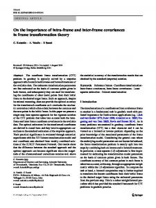
where i ranges from 0 to n−1 (i.e., indexing the ith diagonal). We then find the index p of the first peak from the left in this signal (Fig. 1(b)). The value p represents the average length, in frames, of the cardiac cycle. While at this point we have an estimate of the overall heart rate, we do not know, if given a particular frame i, the time offset from i at which the heart returns to the same position. If it were exactly p for all frames, then we would expect that di,i+p < di,i+p−1 and di,i+p < di,i+p+1 . However, we expect perturbations from this due both to changing heart rate and to how the IVUS frame capture rate imposes a discretization on the real-valued heart rate in every cycle. To find a more accurate offset from each frame, we trace a path v along the off-diagonal valley which represents the cardiac cycle length locally at each frame (Fig. 1(a)). This is accomplished through a dynamic programming step that begins at d1,p and traces down and to the right. That is, each step may proceed one entry downward, one entry to the right, or one entry down-right diagonally.2 Tracing terminates when the path 2 In
0 −0.05
(b)
n−i
1 X c(i) = − di+1,j , n − i j=1
(a)
c(i)
Given an n-frame pullback sequence, a symmetric proximity matrix D is constructed where each entry di,j represents the dissimilarity between frames i and j. In practice, almost any registration metric may be used to accomplish this; here we use normalized cross-correlation (NCC), though an ultrasound-specific metric would also be appropriate. While NCC returns values on the interval [−1, +1], we clamp these values to the interval [0, 1] and subtract the resulting value from one. This results in a matrix where (1) the main diagonal (i.e., identical frames) is everywhere zero and (2) all other entries are non-negative. While other registration metrics may be chosen, these two properties must be enforced. As the changes in image appearance due to the motion of the heart are far more rapid than the changes due to the pullback, these matrices exhibit a periodic appearance indicative of the cardiac cycle (Fig. 1(a)). We obtain a rough estimate of the heart rate over the entire recording with the function
practice, this tracing step operates only on a narrow band around the pth diagonal to prevent it from seeking the main diagonal. This band’s width may be set to a fraction of p so that it adjusts to the heart rate of the subject.
Fig. 1. (a) Dissimilarity matrix for the first 100 frames of a typical sequence, along with dynamic-programming path (dotted line), and (b) the c function for the same matrix. In (a), brighter points indicate greater inter-frame dissimilarity. v exits D near its lower-right corner, globally minimizing the sum of all matrix entries through which the path traverses. It remains to determine a set of frames, each captured at the same point in the cardiac cycle, which is associated with the point in phase when the heart is maximally motionless. We note that if the path we traced earlier passes through a point (i, j), this indicates that the heart obtains the same position in frame j as it did in frame i. In addition, if i and j are captured when the heart is moving slowly, the valley around (i, j) will be more pronounced. To accentuate this, we construct an X-shaped, inverted Gaussian kernel ( ³ 2 2´ +y − exp − x 2σ if|x| = |y| 2 Gσ (x, y) = (2) 0 otherwise, ˆ = D ⊗ Gσ . The matrix D ˆ where σ = dp/3e, and define D exhibits maxima in areas where a frame pair is associated by both high similarity and low motion.
# 1 2 3 4
n 1828 1945 1774 2283
δ 30.5 mm 32.4 mm 29.6 mm 38.0 mm
necg 135 116 109 140
nalg 135 115 110 140
µphase 54% 47% 47% 53%
σphase 8.1% 4.4% 12.0% 7.8%
Table 1. Comparison of pullback cases: n is the count of frames in the sequence, δ its physical length, necg and nalg the counts of frames gated by ECG and our algorithm, and µphase and σphase the mean and standard deviation of the fraction of the R-R cycle of the algorithm-selected frames. ˆ (derived from matrix in Fig. 1(a)), the Fig. 2. The matrix D dynamic-programming path (dotted line), the origin of the stepping process (4) along with associated steps (→), and the final frame pairs representing the gated sequence (4, ◦). To find a single phase-associated frame pair which we are also most confident is at the maximally-stable point in the ˆ along v to find a global cardiac cycle, we trace through D maximum, (s0 , t0 ). We use this starting point and v to proˆ collecting ceed step-wise upward and downward through D, the frames which will comprise our gated sequence (Fig. 2). The downward step sequence is as follows. 1. Let i ← 0. 2. The point on the diagonal below (si , ti ) is (ti , ti ). Locate the column j where v intersects row ti . If this does not exist, then we have reached the end of the sequence and may stop. Otherwise, let (si+1 , ti+1 ) = (ti , j). 3. Following a simple gradient ascent, adjust the position ˆ (this again of (si+1 , ti+1 ) to the local maximum of D helps account for heart rate/sampling variations). 4. Let i ← i + 1 and continue to Step 2. Stepping upward proceeds analogously. Assuming that after these steps the series of off-diagonal points we collected are ordered (u0 , v0 ), (u1 , v1 ), ..., (um , vm ) chronologically, then the frame numbers in our gated sequence are indicated by {u0 , v0 , u1 , v1 , ..., um , vm }. 3.2.1. Computational Complexity The primary source of complexity in the algorithm described so far is the construction of the dissimilarity matrix, D; this is an O(n2 ) operation in the number of frames. However, we note that the algorithm only operates on a narrow band around the main diagonal. The width of this band is dependent on the length of the cardiac cycle as well as the IVUS frame rate. Hence, if we let ρ be an estimate of the minimum heart rate (in beats/min) we expect to encounter in any subject, and let φ be the frame rate (in frames/s), then comparing a frame to
l m only its 2 60φ successors reduces the complexity of matrix ρ formation to O(n). (Note that the multiplication by 2 is to ˆ provide padding in the convolution to find D.) 4. RESULTS For the purposes of comparison with our non-ECG method, four IVUS pullbacks along with synchronized ECG signals were recorded in vivo in healthy swine. Properties of the frames picked by our method were then compared against those picked by ECG. These results are summarized in Table 1. In Fig. 3, the relationship between the algorithm- and ECG-picked frames is illustrated in more detail. Note that the discrepancy between the number of frames picked by the two methods and the histogram outliers are due to the phase offset between the methods, and are expected. The “spread” of the histograms is also expected, as the 970 Hz ECG signals must be resampled onto the 30 Hz frame sequences, leading to quantization effects. In general, though, lower σphase values indicate better reproduction of ECG behavior. As our ultimate goal is the reconstruction of pullback volumes, we visually compare these gating methods in Fig. 4. 5. CONCLUSION We have described an image-based frame gating method for IVUS pullback sequences. This method relies on the analysis of dissimilarity matrices derived from pairwise frame comparisons. The algorithm’s R-R fraction selection varies slightly by subject (47-54%), as we would expect from prior research. We note that such variability could not be obtained by blind ECG triggering based on a fixed R-R fraction. While we have chosen to pick the most visually-stable points in the sequence as our gating points, these tended to be at roughly the same fraction of the R-R cycle (∼ 50%). This being the case, truer ECG emulation could be accomplished by temporally shifting the algorithm-selected frames appropriately. However, as previous studies have hinted (Sec. 2), ECG may not be a reliable standard to aspire to.
0.5
Frame count
0.4 0.3 0.2 0.1 0
0
0.2
0.4
0.6
0.8
1
0.6
0.8
1
Phase 0.5
Frame count
0.4 0.3 0.2 0.1 0
0
0.2
0.4 Phase
0.5
Frame count
0.4 0.3 0.2 0.1 0
0
0.2
0.4
0.6
0.8
1
Phase
Fig. 4. Case 1: Time-axis slices from ungated (top), ECGgated (middle), and algorithm-gated (bottom) pullback volumes. As the latter two represent different fractions of the cardiac cycle, some features may differ in appearance.
0.5
Frame count
0.4
[2] B. Lu, S.-S. Mao, N. Zhuang, H. Bakhsheshi, H. Yamamoto, J. Takasu, S. C. K. Liu, and M. J. Budoff, “Coronary artery motion during the cardiac cycle and optimal ECG triggering for coronary artery imaging,” Invest Radiol, vol. 36, no. 5, pp. 250– 256, 2001.
0.3 0.2 0.1 0
0
0.2
0.4
0.6
0.8
1
Phase
Fig. 3. Number of frames selected per fraction of cardiac phase in each of (from top to bottom) Cases 1-4. The y-axes are normalized by the number of frames for comparability. As our method relies on the visible manifestation of heart motion in the sequence, portions of a pullback where no such motion is apparent are incapable of being analyzed. Fortunately, this is a rare occurrence as the vessel wall is almost always in view. In addition, we have not tested our method on pathological cases (e.g., those with irregular heartbeat) and hence have not modeled how these would affect performance. Future work will involve further validation and refinement of our method to account for such special cases. 6. REFERENCES [1] S. M. O’Malley, M. Naghavi, and I. A. Kakadiaris, “Imagebased frame gating for stationary-catheter IVUS sequences,” in Workshop on Computer Vision for Intravascular & Intracardiac Imaging, Copenhagen, Denmark, October 2006.
[3] A. U. Coskun, Y. Yeghiazarians, S. Kinlay, M. E. Clark, O. J. Ilegbusi, A. Wahle, M. Sonka, and J. J. Popma, “Reproducibility of coronary lumen, plaque, and vessel wall reconstruction and of endothelial shear stress measurements in-vivo in humans,” Catheter Cardio Inte, vol. 60, no. 1, pp. 67–78, 2003. [4] N. Bruining, C. von Birgelen, P. J. de Feyter, J. Ligthart, W. Li, P. W. Serruys, and J. R. T. C. Roelandt, “ECG-gated versus nongated three-dimensional intracoronary ultrasound analysis: Implications for volumetric measurements,” Cathet Cardiovasc Diagn, vol. 43, no. 3, pp. 254–260, March 1998. [5] H. Zhu, K. D. Oakeson, and M. H. Friedman, “Retrieval of cardiac phase from IVUS sequences,” in SPIE Medical Imaging: Ultrasonic Imaging and Signal Processing, February 2003, vol. 5035, pp. 135–146. [6] S. K. Nadkarni, D. Boughner, and A. Fenster, “Image-based cardiac gating for three-dimensional intravascular ultrasound imaging,” Ultrasound Med Biol, vol. 31, no. 1, pp. 53–63, January 2005. [7] S. A. de Winter, R. Hamers, M. Degertekin, K. Tanabe, P. A. Lemos, P. W. Serruys, J. R. T. C. Roelandt, and N. Bruining, “Retrospective image-based gating of intracoronary ultrasound images for improved quantitative analysis: The Intelligate method,” Catheter Cardio Inte, vol. 61, no. 1, pp. 84–94, January 2004.