committed cells (Metcalfe, 1987), and final differentiation of cells within a ..... by Drs Jane Dodd and Thomas Jessell (Columbia), antibodies against the glial ...
1059
Development 120, 1059-1070 (1994) Printed in Great Britain © The Company of Biologists Limited 1994
Immortalizing oncogenes subvert the establishment of granule cell identity in developing cerebellum Wei-Qiang Gao* and Mary E. Hatten The Rockefeller University, 1230 York Avenue, New York, NY 10021-6399, USA *Present Address: Department of Neuroscience, Genentech, Inc., South San Francisco, CA 94080, USA
SUMMARY After implantation into the external germinal layer of early postnatal cerebellum, primary external germinal layer progenitor cells gave rise exclusively to granule neurons. In contrast, all major classes of cerebellar cells were observed following implantation of embryonic day 13 cerebellar precursor cells into the external germinal layer. These results suggest that granule cells arise from precursors with a restricted potential. In contrast to results with the primary external germinal layer population, cell lines established from external germinal layer cells, by infection with a retrovirus containing the SV40 large T-antigen
oncogene, gave rise to several cerebellar cell types upon implantation. These included granule neurons, one subclass of stellate interneurons, Golgi cells, Bergmann glia and astrocytes. From these results, we conclude that early postnatal external germinal layer progenitors are normally fated to a granule cell identity and that expression of the SV40 large T-antigen oncogene subverts mechanisms that control granule neuron fate.
INTRODUCTION
and Hillman, 1969) of the cerebellum emerging over the past several decades. The basic plan of the cerebellar cortex includes three layers—two neuronal layers, the Purkinje cell layer (PCL) and the internal granule cell layer (IGL), and a superficial, plexiform layer, the molecular layer (ML). The PCL consists of a single row of Purkinje neurons situated at the upper margin of the IGL. The Purkinje neuron is easily distinguished by its large cell soma, elaborate, ascending dendrites, and single, descending axon. The IGL contains a vast number of granule neurons, calculated in the human cerebellar cortex to number 1011 (Kandel et al., 1991). The numerous, small granule neurons elaborate short, radiating dendrites in the IGL and project a unique ‘T-shaped’ axon up into the ML. The granule cell axons, termed parallel fibers, are densely stacked through the depth of the ML. Within each of three cerebellar layers, interneurons can be discerned by their location and pattern of neuritic arborization. In the ML, as described by Ramon y Cajal (1911), the small, stellate interneurons can be seen. There are two subtypes of stellate cells, horizontally oriented stellate cells in the most superficial aspect of the ML and spiny stellate cells within the core and deeper aspect of the ML. In the PCL, the mediumsized basket cells radiate long, slender dendrites up into the ML and project horizontal axons across the PCL, forming dense pericellular baskets around Purkinje cell bodies. In the IGL, two types of interneurons can be seen. One, the Lugaro cell, is situated just beneath the Purkinje cells, extending long, horizontal processes that contact the descending Purkinje cell axons. The other, the Golgi cell, projects thin, ascending dendrites into the ML, and a thick skirt of axons down into the
Current models of vertebrate neural fate specification derive from the hematopoietic system, where cell fate is established by the proliferation of pluripotent stem cells (Spangrude et al., 1988; Till and McCulloch, 1961), partial commitment of precursor cells to sublineages, amplified division of partially committed cells (Metcalfe, 1987), and final differentiation of cells within a given sublineage (Nicola and Johnson, 1982; Ogawa et al., 1983). Evidence for a hemopoiesis-like model for vertebrate neural cells has been obtained in studies on the neural crest cell population, including the sympathoadrenal sublineage (reviewed by Anderson, 1989). A general role for local signals in the establishment of CNS neural identity has been inferred from studies on neurological mutant mice (Heintz et al., 1993; Sidman, 1972), from CNS precursor cell transplantation studies (McConnell and Kaznowski, 1991), from the identification of diffusible factors that influence cell specification (Jessell and Melton, 1992) and from in vitro studies showing that close appositions among CNS precursor cells promote precursor cell division and restrict cell fate (Gao et al., 1991). The cerebellar cortex is perhaps the best studied region of the CNS. For nearly a century, all of the cerebellar cell types have been recognized and their pattern of synaptic connections known. Much of this wealth of information was described by Ramon y Cajal from Golgi studies (1911), with information on the development (Altman and Bayer, 1978; Miale and Sidman, 1961; Altman and Bayer, 1985), anatomy (Palay and ChanPalay, 1974), fiber tracts (Brodal, 1993) and circuitry (Llinas
Key words: granule neuron, cerebellum, oncogene, cell lines, transplantation
1060 W.-Q. Gao and M. E. Hatten IGL. In addition to these six types of cerebellar neurons, two classes of astroglial cells can be seen - the Bergmann glia, located just above the PCL with numerous, radial processes coursing to the pial surface, and astrocytes of the IGL. Although cerebellar glial cells (Hatten and Liem, 1981) and Purkinje cells (Ross et al., 1989) can be readily identified by cellular antigen markers, other cerebellar cells are classically identified by their size, laminar position and pattern of neuritic arborization. Among cerebellar neurons, the granule cell presents an opportune model for studying CNS neuronal specification. Unlike the other five cerebellar neurons, the granule cell arises in a displaced proliferation zone, the external germinal layer (EGL) (Ramon y Cajal, 1911). Experimental chick/quail chimeras provide evidence that, whereas the other cerebellar neurons originate from the caudal aspect of the mesencephalon, the EGL arises from the rostral portion of the metencephalon via a complex series of transverse migrations. The distinct origin of the EGL (Hallonet et al., 1990; Hallonet and Le Douarin, 1992; Martinez and Alvarado-Mallart, 1989) raises the question as to whether granule cell identity is specified by regulatory factors, localized to the superficial germinal zone. In vitro studies with purified EGL precursors support the conclusion that the close apposition of granule cell precursors within the EGL restricts the fate of these progenitor cells to a granule cell identity (Gao et al., 1991). In the present study, we have further examined whether the EGL provides local signals that restrict precursor cells to a granule cell identity, by re-implanting labeled EGL precursors into the EGL and examining their laminar positioning and neuritic arborization after short survival times (1-7 days). Another general approach to understanding the control of neural fate is to study progenitor cells immortalized by oncogene transfer. Although experiments in non-neuronal systems, including the hemopoietic system (Klinken et al., 1988), have cautioned against the use of immortalized cells in lineage analyses, the demonstration that immortalized CNS neuronal precursors would incorporate into developing brain, and differentiate into identifiable cell classes has suggested that immortalized CNS cells provide a convenient means of studying developmental processes in brain (for reviews see Cepko, 1988; Lendahl and McKay, 1990). However, as previous CNS cell lines have been generated by immortalizing a mixture of progenitor cells (e.g. Renfranz et al., 1992), it has not been possible to assess the effects of oncogene expression on the specification of the primary cell of a given CNS sublineage. In the current study, we transferred the tsA58 allele of the SV40 large T antigen oncogene into purified early postnatal EGL precursor cells and compared the development of primary EGL cells with infected cells both in vitro and in vivo. To compare the fate of the cells, we implanted primary EGL cells and immortalized EGL cells into cerebellar EGL on postnatal day 6. After implantation, whereas primary EGL cells gave rise exclusively to granule neurons, immortalized cells gave rise to granule neurons, stellate interneurons, neurons and astroglia. In the same transplantation assay, precursor cells taken from the cerebellar primordium on embryonic day 13 (E13) generated Purkinje neurons, interneurons, granule neurons and astroglia after implantation into the early postnatal EGL. These results indicate that whereas E13 cells show multiple fates,
early postnatal EGL cells have a restricted potential, giving rise only to granule neurons. The control of precursor cell potential is apparently subverted by the expression of the SV40 large Tantigen oncogene as immortalized cells generate multiple programs of cell differentiation. MATERIALS AND METHODS Preparation of early postnatal EGL precursor cells and astroglia and of embryonic day 13 cerebellar cells The present experiments were carried out with primary cells from C57Bl/6J mouse cerebella harvested on the 5-6 postnatal days (P5P6). EGL cells were purified as described previously (Gao et al., 1991; Hatten, 1985). Astroglial cells were purified from the same preparations used to harvest EGL cells, as described by Hatten, (1985). Cells from the E13 cerebellar analage were prepared as described by Hatten and Sidman, (1978). Briefly, cerebella were incubated sequentially in 0.08% and 0.25% trypsin in CMF-PBS containing 0.02% EDTA for 15 minutes at 37°C each., after which soybean trypsin inhibitor (0.05 mg/ml in CMF-PBS containing 0.05 mg/ml DNase) was added. The tissue was then triturated and dissociated into single cells. Implantation of EGL cells, astroglia, E13 cerebellar cells and immortalized EGL cells into P6 cerebellum Prior to implantation, purified EGL cells, astroglial cells, E13 cerebellar cells or GC-B6 cells (see below) were labeled with green fluorescent latex microbeads (Lumafluor, Inc., NJ) in vitro for 1 hour at a dilution of 1:300 in the culture medium. The cells were then washed with medium or CMF-PBS for several times, collected in a test tube and stained with PKH-26 at a concentration of 4 µM for 5 minutes (Zynaxis, Inc.; see Gao et al., (1992)). The labeled cells were then washed several times and suspended in DMEM + 9 mg/ml glucose in the presence of 20 mM Hepes buffer (pH 7.4). Approximately 25,000 cells were implanted into the EGL of P5-6 C57Bl/6J mice (Gao and Hatten, 1993). Prior to the cell implantation procedure, P6 mouse pups were rendered unconscious by chilling the animals at 4°C for 1-2 minutes and placed in a Stoelting stereotaxic device fitted with a neonatal rat adapter and a vertical holder for a Hamilton syringe (Wood Dale, IL). The skin overlying the midbrain and hindbrain was rinsed with alcohol, a small incision was made in the skin and the Hamilton syringe needle was lowered gently through the incision to a position just beneath the meninges (just above the EGL). The animals insensitivity to the surgical procedure was judged by their lack of movement. Approximately 1 µl of the cell suspension (2.5×10⁷ cell/ml) was injected slowly on each side of the cerebellum, after which the syringe was removed, the skull was rinsed with a solution of penicillin-streptomycin (0.25%), and the skin was replaced and sealed with Vetbond (Henry Schein Inc). The animal was then warmed to 35.5°C and returned to the litter. After survival of 1-7 days, animals were anaesthetized with ketamine prior to perfusion with 4% paraformaldehyde in 0.1 M phosphate buffer (pH 7.4). The cerebella were removed by dissection and post fixed in the same fixative prior to being washed in PBS and embedded in 3% agar gel. Serial sections (90-100 µm) were cut with a vibratome and labeled cells were visualized with epifluorescence microscopy using a Zeiss Axiophot microscope fitted with phase contrast, Nomarksi and epifluorescent illumination, Plan-neofluor 20× and 40× objectives and an Axiophot camera module. To examine the extent of PHK-26 dye leakage and re-uptake, we double labeled cells with PKH-26 and microbeads and followed the incorporation of labeled cells. In a cell sample of several thousand cells, we did not observe a cell that was not double labeled, suggesting that the dye was not transferred from implanted, double-labeled cells to endogenous, unlabeled cells. As a further control, we labeled primary EGL cells with PKH-26, killed them by freezing and thawing
Establishment of granule cell identity 1061 four times at −80°C, and implanted the dead, labeled cells into the P6 EGL. In those experiments, no PKH-26 labeled cells were seen distal to the site of injection, confirming previous findings (Gao et al., 1992; Gao and Hatten, 1993; Horan and Slezak, 1989) that the PKH-26 dye does not transfer to other cells in intact tissue. In some experiments, cells were visualized with an Axiovert 135 microscope with DIC and epifluorescent illumination, Plan-Neofluor 20× objective, and a computer-driven (z-axis, Ludl system) stage. Images were acquired with a high-sensitivity Biorad MRC-600 scanning, imaging head controlled with an 80386 host computer with scan control and imaging acquisition and analysis system, using two detectors for acquisition of simultaneous Nomarksi and fluorescence imaging. A 15 mW argon/krypton mixed gas multi-line mode laser with lines at 488, 568 and 647 was used for imaging of blue line excitation (FITC, Rhodamine 1,2,3). Images were stored on an optical disk, Ethernet-linked to a Silicon Graphics Iris computer system and printed on a Sony U-811 Video Printer System. Immortalization of EGL cells using retroviral constructs Purified EGL cells from P5-6 mouse cerebella were infected with conditioned medium from psi 2 cells packaging the retrovirus encoding tsA58/U19T antigen and neomycin, at a density of 107/ml in the presence of 8 µg/ml polybrene (Sigma) in a untreated culture dish. The virus-producing cells were kindly provided by R. McKay (NIH). After being infected twice (6 hours each time), the virus-containing medium was replaced with BME plus 10% horse serum, 5% fetal bovine serum, 9 mg/ml glucose, and cells were placed at 35°C for 2 days then transferred to a polylysine coated dish (500 µg/ml). Several days later, cultures were selected in the above medium containing G418 (200 µg/ml). The selection medium was replaced every 2 days, and G418-resistant colonies were observed in 2 weeks and picked with cloning rings. The clones were then expanded, frozen, subcloned, and cultured in DMEM plus 10% newborn calf serum. In the present experiments, although similar results were obtained from one of the other clones, only the data from subclone GC-B6 were described and used for transplantation. Immunocytochemistry Immunocytochemical labeling of primary EGL cells was as described previously (Gao et al., 1991). In the present experiments, GC-B6 cells were plated in 16-well lab-tek slides coated with poly-D-lysine (0.1 mg/ml) (35°C, overnight) in either DMEM plus 10% newborn calf serum or serum-free medium (Redu-Ser II from Upstate Biotech Inc.) at 39°C for 16-24 hours before being fixed with 4% paraformaldehyde for 30 minutes. The cells were then incubated with antibodies against N-CAM (Thiery et al., 1977), kindly provided by Dr Christo Goridis (Marseilles), L-1 (Rathjen and Schachner, 1984), provided by Dr Carl Lagenauer (Pittsburgh), TAG-1 (Dodd et al., 1988), provided by Drs Jane Dodd and Thomas Jessell (Columbia), antibodies against the glial filament protein (GFAP; Hatten and Liem, 1981), provided by Dr Ronald Liem (Columbia), or with monoclonal antibody R24a, which is against GD3, generously provided by Dr James Goldman (Columbia). Antibodies were applied at 4°C overnight at 1:2-1:1000 in the presence of 3% normal goat serum, after which the cells were washed three times with CMF-PBS, then incubated with rhodamine or FITC-conjugated second antibodies (1:100) (Tago, Inc.) for 30 minutes at room temperature. For cytoplasmic antigens, 0.1% Triton X-100 was added to the staining saline. The preparations were then mounted in Gel/Mount (Bio-Meda Corp.) and visualized with a Zeiss Axiophot microscope fitted with phase contrast, Nomarksi and epifluorescent illumination and Plan-neofluor 20×, or 40× objectives and an Axiophot camera module. Co-culture of the immortalized EGL cells with the primary EGL cells EGL cells were purified (Hatten, 1985) from P5-6 mouse cerebella and plated as a monolayer on a poly-D-lysine (500 µg/ml) coated 16-
well Lab-tek slide (1×10⁶ cells/well) in serum-free medium (Redu-Ser II from Upstate Biotech Inc.) for 1 day. The GC-B6 cells were removed from the culture dish with trypsin-EDTA (Gibco), labeled with PKH-26 (Gao et al., 1992), washed, and plated (3,000 cells/well) on the top of the monolayer culture of primary EGL cells at low density in serum-free medium. 24-48 hours later, the culture was fixed with 4% paraformaldehyde in phosphate buffer (pH 7.4) for 30 minutes, and mounted in gel/mount (Biomedical Co.). The implanted cells were visualized with a Zeiss Axiophot microscope fitted with phase contrast, Nomarksi and epifluorescent illumination and Planneofluor 40× objectives and an Axiophot camera module.
RESULTS Previous in vitro analyses of primary EGL cells in reaggregate cultures (Gao et al., 1991; Gao et al., 1992) demonstrated that local interactions among EGL precursor cells promote granule cell neurogenesis and differentiation. A striking aspect of these studies was the finding that proliferating cells in homotypic reaggregate cultures had a restricted cell fate, expressing cellular antigen markers and morphological features of granule cells, but not other classes of cerebellar neurons (Gao et al., 1991). To examine the fate of purified EGL cells in vivo, we double-labeled purified EGL cells with fluorescent microbeads and PKH-26, injected them just beneath the pia of P6 animals, allowed the animals to survive for short periods of time (1-7 days), and examined the morphology and location of labeled cells in the cerebellar cortex. Using fluorescent and confocal microscopy it was found that labeled cells remained at the injection site during the first 1-24 hours in vivo, with some cells undergoing mitosis in the superficial aspect of the EGL, as evidenced by labeled, mitotic figures (not shown). 24-48 hours after implantation, primary EGL cells, characterized by their small (4-6 µm) size and globular shape, descended into the deeper aspect of the EGL, where they extended long fibers, 100-200 µm in length, parallel to the surface of the anlage (Fig. 1A). This sequence of developmental events confirmed the hypothesis of Ramon y Cajal, who suggested that granule cell axon extension precedes the inward migration of the cell soma (Ramon y Cajal, 1911). A second stage of development commenced 48 hours after implantation, the polarization of the cell soma to extend a descending process perpendicular to the plane of parallel fiber extension (Fig. 1B). Following extension of this descending or ‘leading process’ (Edmondson and Hatten, 1987; Rakic, 1971; Ramon y Cajal, 1911), many of the implanted cells started to migrate along Bergmann glial fibers, requiring approximately 10-12 hours to reach the internal granule cell layer (IGL). During migration, the trailing process of labeled cells displayed an ascending ‘T-shape’ (Fig. 1C), characteristic of the granule cell axon. At low magnification, in a coronal plane of section, several dozen labeled cells could be seen migrating through the molecular layer (ML) and settling in the IGL (Fig. 1D). All of these cells expressed the morphology described in classical studies of Golgi-impregnated tissue (Ramon y Cajal, 1911). After settling within the IGL, labeled cells extended four to six, short radiating dendrites, with branched claw-like endings, characteristic of mature granule cells. Among the more than 80 animals injected with several thousand labeled EGL cells each,
1062 W.-Q. Gao and M. E. Hatten
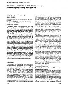
Fig. 1. Primary EGL cells differentiated exclusively into granule cells after re-implantation into the EGL of P6 mouse cerebellum. (A) 2 days after implantation, labeled cells extended long parallel fibers. (B) 3-4 days after implantation, labeled cells extended a descending migratory process and began to transit the molecular layer (ML), displayed morphological features of migrating cells (C). 6 days after implantation, labeled cells are positioned beneath the Purkinje cell layer, in the internal granule cell layer (IGL). The ascending axon of the labeled neuron formed a ‘T-shape’ characteristic of mature granule cells. Within the IGL, the cell extended 4-6 short, dendritic processes. (D) At lower magnification, all labeled cells in the field showed features of developing granule neurons. Fluorescence microscopy. Bar, 15 µm.
and analyzed by serial sectioning of the brain, more than 99% of the cells that underwent differentiation expressed a granule neuron identity, positioning their soma in the IGL, forming 46 short dendrites, and extending an ascending ‘T-shaped’ parallel fiber into the molecular layer. Less than 0.5% of the differentiated cells we counted assumed the glial phenotype, consistent with the level of contamination of the cell preparation with glial cells. No other classes of cerebellar neurons
were observed after injection of labeled EGL cells (Table 1), suggesting that EGL precursor cells were normally fated to a granule cell identity. By confocal microscopy, approximately 10-20% of the cells we injected into the host tissue differentiated as described. The same general distributions of cell types, were obtained in each of the 80 animals we injected with labeled cells. As a control, we implanted labeled, primary astroglial cells purified from
Establishment of granule cell identity 1063 Table 1. Differentiation of implanted cells in developing cerebellum Classes of labeled cells observed Cells implanted Primary EGL cells Primary astroglial cells GC-B6 cells* E13 cerebellar cells
Granule Purkinje Bergmann neuron neuron Interneurons glia Astroglia 99 0
0 0
0 0