Perfusion 2004; 19: 25 ¡ 32
In vitro assessment of the unloading and perfusion capacities of the PUCA II and the IABP Stijn Vandenberghe1 , Patrick Segers1 , Hans Josemans2 , Jan-Paul Van Loon3 , Gerhard Rakhorst4 and Pascal Verdonck1 1
Institute Biomedical Technology, Ghent University, Belgium; Arrow International, Weesp, The Netherlands; 3 Intra-Vasc.NL b.v., Groningen, The Netherlands; 4 Department of Biomedical Engineering, University of Groningen, The Netherlands 2
The PUCA II pump is a minimally invasive intra-arterial left ventricular assist device that can be used as an alternative for the intra-aortic balloon pump (IABP). In this study, we assessed the cardiac unloading and organ perfusion capacities of both PUCA II and IABP in an in vitro set up, consisting of a heart simulator and a silicone arterial tree, mimicking anatomical geometry and flow distribution. The IABP was positioned in the descending aorta, while the PUCA II was tested both in ‘trans-aortic’ and ‘abdominal’ positions. All devices were driven by the
same Arrow AutoCat IABP driver at different pump rates. Apart from flow, arterial pressure and pulse pressure, we also calculated haemodynamic indices for myocardial oxygen supply and demand. The ‘abdominal’ PUCA II assist and the IABP both provide mild unloading of the heart, and a limited improvement of arterial pressure and flow. The ‘trans-aortic’ PUCA II assist greatly enhances flow and pressure, but does not unload the heart properly in the tested configuration. Perfusion (2004) 19, 25 ¡ 32.
Introduction
by most commercially available IABP drivers and drivers for pneumatic cardiac assist devices. Both the IABP and the PUCA II are expected to support the patient’s heart by increasing the blood flow, unloading the myocardium and improving end-organ and coronary perfusion. We used an in vitro set up to assess these characteristics from a commercially available IABP and from the PUCA II. The latter was placed both in the normal trans-aortic position (designated ‘aortic’), where it runs through the aortic valve in the left ventricle, and in an ‘abdominal’ position, where its main benefit is expected to be an increase in abdominal organ perfusion.
Patients suffering from progressive cardiac failure are often supported with an intra-aortic balloon pump (IABP) when the disease comes to an endstage. The IABP is a counterpulsation device that was first used by Kantrowitz1 and has already been in use for three decades to stabilize heart failure patients or for the treatment of acute cardiac failure. The pump is used for short- or medium-term application and its main advantage is the fast, minimally invasive insertion that can be done in a cathlab, or even by trained ER staff. The pulsatile catheter (PUCA) pump is a device that combines the counterpulsation principles of the IABP with an actual blood pump. The newest version of this device (PUCA II) consists of a thin-walled catheter with one single tilting valve incorporated into the catheter wall. The catheter can be introduced transarterially and shifted retrograde into the aorta or the left ventricle. The catheter is connected to a custom-made membrane pump that can be actuated Address for correspondence: Stijn Vandenberghe, Hydraulics Laboratory, Ghent University, Institute Biomedical Technology, Sint-Pietersnieuwstraat 41, 9000 Gent, Belgium. E-mail:
[email protected]
# Arnold 2004
Materials In vitro set up The in vitro model is composed of a pulse duplicator system and a silicone rubber arterial tree, simulating the systemic circulation2 (Figure 1). The pulse duplicator consists of two silicone sacs, representing the left atrium and ventricle, actuated by a computer-controlled pneumatic driver to obtain physiological intraventricular pressures. A bileaflet and a bovine pericardial valve are used as mitral and 10.1191/0267659104pf710oa
In vitro comparison of PUCA II and IABP S Vandenberghe et al.
26
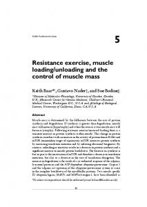
Figure 1 Top: picture of the PUCA II catheter and membrane pump and a detailed drawing of the tilting valve. Bottom: overview of the model set-up. 1. collection container, 2. buffer reservoir, 3. centrifugal pump, 4. lung reservoir with weir, 5. left atrium, 6. left ventricle, 7. PUCA II catheter and membrane pump in trans-aortic position, 8. variable resistor combined with an overow reservoir.
aortic valves, respectively. The elastic arterial tree consists of a tapered aorta with eight branches representing the main arteries to the head, the arms, the abdominal organs and the legs, on both left and right sides. Arterial dimensions are derived from the literature so as to mimic a 1.8-m tall male, 85 kg.3 The arteries in the abdominal region were bundled and simply represented by equivalent renal arteries, whose cross-sectional areas were adapted accordingly. Each branch ends in a resistor with a compressible foam and a venous overflow system, which allows volumetric measurement of the mean flow through every branch. The test fluid, a 60 ¡ 40% water-glycerine solution (kinematic viscosity: 3.7 mm2 /s, density 1093 kg/m3 ) was channelled from the overflow system to a collector, where it was pumped up again into a preload reservoir that mimics the lungs. The flow distribution over the entire model was set according to literature values,4 where the major portion of the flow ( Í 50%) goes to the abdominal region in patients at rest.
PUCA II and IABP The PUCA-II catheter (Intra-vasc.NL b.v., Groningen, The Netherlands) consists of a 28-cm long, 21 Fr reinforced polyurethane shaft that incorporates a stainless steel inflow cage at the tip and an outflow valve located 10 cm distally.5,6 The catheter is connected to a pneumatically driven, single port, valveless membrane pump with a stroke volume of 40 mL, which is placed paracorporeally. The catheter’s tilting valve is designed specifically for the trans-aortic position, as to direct blood flow from the left ventricle (LV) to the membrane pump during the aspiration phase, and to direct the blood into the aorta during pump ejection. Trans-aortic position (PUCAao). The PUCA II catheter was introduced in the right subclavian artery and positioned through the aortic valve using the standard technique with a guiding pressure catheter.7 To seal the set up, the subclavian artery was tied up around the PUCA II, thus occluding one of the eight branches.
In vitro comparison of PUCA II and IABP S Vandenberghe et al.
27
Abdominal position (PUCAabd). To simulate the use of the PUCA II as an organ perfusion pump, the same catheter was introduced through the left femoral artery and shifted retrograde until the valve was positioned at the bifurcation site of the renal arteries. In this position, the femoral artery was occluded. Intra-aortic balloon setup (IABP). An 8 Fr Narrowflex intra-aortic balloon catheter (Arrow Int., Reading, PA, USA) was inserted via the left femoral artery for the IABP experiment. The balloon (40-mL stroke volume, 26-cm length and 15-mm inflated diameter) was positioned in the descending aorta and the femoral artery was occluded for sealing purposes. Measurements Both the intra-aortic balloon and the PUCA II were driven by an AutoCat IABP driver (Arrow Int., Reading, PA, USA). The required ECG feedback for the driver was provided by the computer control of the pulse duplicator. All experiments were performed with the AutoCat driver set to ‘operator’ mode, while an experienced IABP operator adjusted the inflation and deflation timing to obtain optimal counterpulsation with the devices in a 1:1 mode. All three devices (PUCAao, PUCAabd, IABP) were tested with the cardiac simulator running at two levels of ventricular contractility (low and high) at three heart rates (60, 90 and 120 bpm). The pressure level of the pneumatic controller of the pulse duplicator was adjusted to obtain the different contractility levels, i.e., systolic aortic pressure of 70 mmHg (low contractility) and 120 mmHg (high contractility) during control conditions (no support). As such, six different haemodynamic conditions were obtained for each device, resulting in a total of 18 runs (three devices, two contractilities, three heart rates). Because our simulator is not subject to biovariability, no measurements were repeated. Instead we performed control measurements (CTRL) with the device shut off at the beginning and end of each run and compared these to assess the stability of our in vitro set up. Consequently, each run consisted of the following three steps: CTRL, pumping in 1:1 mode, CTRL. For each step, pressure data were sampled at 200 Hz and captured for at least 5 s when a steady state condition was present. Pressure was measured in the LV (P LV ) with a pigtail catheter and a DTX» pressure transducer (BD, Franklin Lakes, NJ, USA) and in the ascending aorta (P ao ) with a Millar highfidelity transducer (Millar Instruments Inc., Houston, TX, USA). Flow distribution over the model
Figure 2 Flow distribution over the 8 arterial branches when applying the different devices. The CTRL set is taken from the IABP experiment. All these data were captured at a heart rate of 120 BPM and the high contractility setting. ao¾ aortic, abd¾ abdominal, R ¾ right, L¾ left.
(Figure 2) was measured with the overflow system. Total mean flow was calculated by summation of mean regional flows. To assess cardiac unloading and perfusion capacities of the assist devices, cardiac oxygen demand and supply were estimated from the LV and aortic pressure curves.8,9 The tension time index (TTI) is a marker for oxygen demand, and is calculated as the area under the LV pressure curve during systolic ejection multiplied by the heart rate (HR) to express it per minute. The diastolic time index (DTI) is a measure of perfusion or oxygen supply and is defined as the area under the aortic pressure curve during diastole, also multiplied by HR. TTI¾
P LV dt × HR
(mmHg × s=min)
Pao dt × HR
(mmHg × s=min)
systole
DTI ¾ diastole
Results The variability in the flow distribution appeared to be small. The control values at the beginning and end of each run were compared for LV and aortic pressure. The maximum differences found were 3% and 5% of the average, respectively. This demonstrates the stability of the set up and allowed use of the average of the control measurements for each run for further processing.
In vitro comparison of PUCA II and IABP S Vandenberghe et al.
28
The flow distribution over the whole arterial tree and the influence of the different devices is given in Figure 2 for the high contractility condition and a HR of 120 bpm, with the CTRL data taken from the IABP experiment. IABP increases total flow, but the largest effect is obtained with the PUCA II in the trans-aortic position (PUCAao). The effect of IABP or PUCA II support on ventricular and aortic pressure is demonstrated in Figure 3, where cardiac cycle samples are shown for the three different heart rates during the low contractility condition. Data indicated as CTRL (panel A) are again taken from the IABP experiment (with the pump off). Aortic pressure in the IABP experiment shows a high diastolic peak, being the result of a steep pressure rise and fall at the beginning and the end of diastole, respectively. This sudden pressure decrease at the end of diastole leads to a very low aortic (afterload) pressure at the onset of ejection. Aortic pressure behaves similarly in the PUCAabd experiment, except that the obtained pressure peak is lower and the rise and fall of diastolic pressure is less steep. In contrast, PUCAao data display only a slight bump in diastolic aortic pressure (Figure 3, panel C). This is the result of a much slower pressure rise and fall, leading to an elevated end diastolic pressure. The shape of the LV pressure waveform is not altered by the IABP, but it is shifted to a lower level compared with CTRL,
similar to what happens with the PUCA II in the abdominal position. In the PUCAao experiment, however, LV pressure has a flattened waveform, keeping the pressure constant throughout systole. The pressure increase in diastole results in an elevated MAP. In Figure 4 (panel B), this is presented as the change in MAP compared with the CTRL value (which was obtained in the same test series), thus allowing assessment of the effect of switching the pump on. There is a very high increase in MAP by use of the PUCA II in the trans-aortic position (range 20 mmHg) and a moderate increase by using it abdominally or by the IABP (about 10 mmHg). The pressure rise seems to depend on the level of contractility, where all devices have more difficulties in further increasing the pressure in the presence of a highly contractile LV (averaging all control measurements, MAP is 4997 mmHg for the low contractility versus 7498 mmHg for the high contractility experiments, p B 0.05). Panel A of Figure 4 leads to similar conclusions for the total flow: PUCAao provides the highest increase in total flow, which is again most obvious in the low contractility experiments where flow levels are lower than in the higher contractility setting (averaged CTRL flow is 1.390.15 and 2.09 0.17 L/min in the low and high contractility conditions, respectively; p B 0.05). Note that the IABP
Figure 3 Raw aortic (black) and left ventricular (grey) pressure data from the different experiments. The data shown was captured at the low contractility setting, at different heart rates (60, 90, and 120 BPM). The CTRL data are taken from the IABP experiment. In the CTRL panel is illustrated how TTI (º O2 demand) and DTI (º O2 supply) are calculated.
In vitro comparison of PUCA II and IABP S Vandenberghe et al.
29
Figure 4 Effect of the devices on total ow, mean arterial pressure (MAP), diastolic time index (DTI), and tension time index (TTI). The results are expressed for all the tested heart rates and contractilities. The top panels display for each experiment the difference between the respective CTRL value and the value obtained during assist, thereby illustrating the changes that occur by switching on a device. For the bottom panels, these differences are expressed relative to the CTRL value.
and PUCAabd also seem to demonstrate a contractility dependency for the flow. PUCA II in the trans-aortic position yields higher DTI ( º oxygen supply) values than PUCAabd or the IABP, as shown in Figure 4 (panel C). Data are presented relative to their respective CTRL values and show that using the PUCA II in a low contractile ventricle at high heart rates can almost double the oxygen supply (98% increase). The TTI is an indication of how well the ventricle is unloaded; a decreased TTI results from reduced LV pressure and/or shorter exposure of the myocardium to high pressure, resulting in decreased wall stress and oxygen consumption. Our results (Figure 4, panel D) indicate that IABP and the abdominally placed PUCA II reduce the oxygen demand of the LV, while on the contrary, PUCA II in the trans-aortic position slightly increases the demand, thus loading the ventricle. Especially at 120 bpm, there is a noteworthy increase in TTI of 25.5% under low contractility conditions. The influence of contractility (more modest changes in high contractility conditions) on DTI and TTI can be appreciated from Figure 4. To characterize the effect of the devices on aortic pressure, we also calculated the changes induced by
the different devices in end diastolic pressure (Figure 5, top panel) and pulse pressure (bottom panel). The use of the PUCA II in the trans-aortic position results in a large increase in end-diastolic aortic pressure, while the IABP is capable of dragging this pressure down. The opposite is seen in the pulse pressure, which is reduced by the PUCAao and increased by the IABP and (to a lesser extent) PUCAabd. The pulse pressure is, in some cases, even doubled by IABP application (highest increase¾ 217%), which is in accordance with the raw data from Figure 3. Again, the induced changes are less pronounced in the high than in the low contractility conditions (Figure 5).
Discussion Overall, our results show that the PUCA II pump, when used in the abdominal position, yields similar haemodynamic effects as obtained with a standard IABP. Both assist methods increase mean systemic arterial pressure and flow to the same extent, and their impact on oxygen supply (approximated by DTI) and oxygen demand (TTI) is also comparable. This is not surprising, since, per se, they use the same principle of aortic counterpulsation to aug-
In vitro comparison of PUCA II and IABP S Vandenberghe et al.
30
Figure 5 End diastolic pressure changes and pulse pressure changes obtained in the different experiments, for all the tested heart rates and contractilities.
ment coronary perfusion and flow: a certain volume is injected into the aorta during diastole and retrieved again during systole. The major difference is that the IABP displaces gas (helium) while the PUCA II pumps blood, which results in larger inertial forces. This can be noticed in the raw data of Figure 3, where the pressure rise and fall are less steep in the PUCAabd experiment compared with the IABP. Consequently, the IABP is capable of increasing the pressure more rapidly during diastole, and decreasing it quickly again at the end of diastole, before the contraction starts. The PUCAabd shows little effect on end diastolic pressure. When PUCA II is used in the trans-aortic position, it performs better as an assist device in terms of displaced blood volume, but it augments the end diastolic pressure, thereby increasing the load on the heart as quantified by TTI. It has been shown that increased end diastolic pressure causes a later opening of the aortic valve, at a higher intraventricular pressure, resulting in increased stroke work of
the ventricle.10 This incapability of dragging down the end diastolic pressure has the advantage that the increase in DTI is much higher, which should have favourable effects on coronary perfusion. Although the diastolic pressure peak is not very high in the PUCAao experiment, the pressure stays high throughout the whole of diastole as well as during systole. For PUCAabd and IABP, the diastolic peak pressure is high, but pressure varies much more over the diastolic period, and is gradually built up in systole, starting from a low end diastolic pressure. Consequently, the MAP is highest in the PUCAao experiment, and since total vascular resistance stays constant in the in vitro set up, the gain in total flow is highest for the PUCA II in the trans-aortic position. In terms of haemodynamic performance, there is clearly a difference between IABP and PUCAabd on one hand, and PUCAao on the other. These differences originate from the fact that with PUCA II in the trans-aortic position, blood is aspirated from the left ventricle, while IABP and PUCAabd work directly in the aorta, allowing a fast pressure drop there. The advantage of PUCAao is that it actually moves blood from the left ventricle to the aorta instead of just displacing it in and out of the aorta. Therefore, the PUCAao actually pumps along with the natural circulation, thus more drastically increasing the flow and the mean arterial pressure. It can also be observed from the pressure waveforms in Figure 3 that LV pressure is nearly constant throughout systole in the PUCAao experiment. In this setting, PUCA II does not decrease the LV pressure more than IABP or PUCAabd, but it smears out the waveform more equally over systole. Consequently, applying PUCA II directly in the LV does not unload the ventricle as one might anticipate, which was already indicated by the increase in TTI. We hypothesize that the suction pressure in the membrane pump is not high enough for optimal assist, since the blood instigates substantial inertia that has to be overcome. An important issue with the trans-aortic application of the PUCA II is the trans-valvular position of the catheter, which may augment valvular regurgitation of valves that are often stenotic in the clinical setting. Although the placement itself is relatively simple,7 it also results in an obstruction of the ejection of the LV, which may also explain the lack of unloading. In addition, in previous experiments,11 we had calculated that the tilting valve of the PUCA II catheter directs approximately 80% of the membrane pump output into the aorta (80% efficiency), meaning that a small amount of blood is returned to the left ventricle during cardiac diastole.
In vitro comparison of PUCA II and IABP S Vandenberghe et al.
31
This observation, together with valvular regurgitation, contributes to the fact that the diastolic aortic pressure does not peak as high in the PUCAao experiment as in the other experiments. While the correct functioning of PUCA II in the trans-aortic position requires the tilting valve, this is not a necessity when it is used abdominally. The only argument in favour besides manufacturing standardization is that the flow could be directed more organ-specific by placing the tilting valve at a targeted arterial bifurcation. This effect, however, was not observed in the measured flow distributions (Figure 2), where we were not able to detect any specific abdominal gain.
Study limitations This study reports the haemodynamic impact of three different cardiac assist methods, all counterpulsation based. Since it is an in vitro study, important biological and physiological feedback mechanisms are neglected, thus, only the pure mechanical effects are visible. This means that the changes in MAP are probably overestimations of the real clinical situation, where an increase in MAP, if unphysiological, will be countered by a decrease in vascular resistance and, consequently, a higher increase in flow. We quantified the unloading capacities using TTI and DTI. Although these indices do not give an absolute indication of the oxygen consumption or supply, we believe that they at least allow an indication as to which of the tested devices, and in which mode, will have the highest impact on oxygen demand and supply. Also, our pulse duplicator shows an unphysiological heart rate dependency, where increases in heart rate result in decreased ventricular pressure and stroke volume. The flow levels in our study are rather low, mainly because of a rather high systemic vascular resistance. However, comparable low flows ( B 2 L/min) have also been reported clinically.12 Moreover, we
have found previously that it is mainly the pressure level and pressure gradient over the catheter that determines the performance of the PUCA II, rather than the absolute flow levels in the model.11 The observed differences in IABP and PUCA II performance between the low and high contractility conditions, reported in this study, are therefore the consequence of the different pressure levels at which the model operates and that the devices undergo.
Conclusion We compared the haemodynamic effect of an IABP with a PUCA II pulsatile catheter, which were used both in a trans-aortic and an abdominal position. We found that the latter application has no directly measurable haemodynamic advantage over the traditional IABP, when both devices use the same driver. When the PUCA II is used in the trans-aortic position, however, it yields an important augmentation of flow and arterial pressure, and, thus, possibly coronary perfusion. The disadvantage of this application is that it does not unload the heart in terms of the TTI. Further studies, using a specifically designed and more powerful pneumatic driver, are needed to resolve this shortcoming.
Acknowledgements The authors wish to express their gratitude to Alain Storms for his assistance with the AutoCat driver and to Ilse Van Tricht for patiently managing the overow system. They are also grateful to Marcel Antheunis and Martin Van Daele for their technical assistance. This research was funded by a specialization grant of the Institute for the Promotion of Science and Technology in Flanders (IWT-993171, S Vandenberghe) and by a postdoctoral fellowship from the Fund for Scientic Research ¡ Flanders (FWO Vlaanderen, P. Segers).
References 1 Kantrowitz A, Tjonneland S, Freed PS, Phillips SJ, Butner AN, Sherman JL Jr. Initial clinical experience with intraaortic balloon pumping in cardiogenic shock. JAMA 1968; 203: 113 ¡ 18. 2 Segers P, Dubois F, De Wachter D, Verdonck P. Role and relevancy of a cardiovascular simulator. CVE 1998; 3: 48 ¡ 56. 3 Westerhof N, Bosman F, De Vries CJ, Noordergraaf A. Analog studies of the human systemic arterial tree. J Biomechanics 1969; 2: 121 ¡ 43.
4 Wade OL, Bishop JM. Cardiac output and regional blood ow. London: Blackwell, 1962. 5 Mihaylov D. Development of the PUCA pump ¡ a trans-arterial ventricular assist device. Groningen: Rijksuniversiteit Groningen, 2000. 6 Rakhorst G, van Loon JP, Elstrodt J, van der Plaats A, Verkerke GJ. Measuring pump performance. Med Device Technol 2001; 12: 18 ¡ 20. 7 Mihaylov D, Kik C, Elstrodt J, Verkerke GJ, Blanksma PK, Rakhorst G. Development of a new introduction
In vitro comparison of PUCA II and IABP S Vandenberghe et al.
32 technique for the pulsatile catheter pump. Artif Organs 1997; 21: 425 ¡ 27. 8 Hoeft A, Sonntag H, Stephan H, Kettler D. Validation of myocardial oxygen demand indices in patients awake and during anesthesia. Anesthesiology 1991; 75: 49 ¡ 56. 9 Reitan JA, Martucci RW, Levine NA. A computer evaluation of the ratio of the diastolic pressure-time index to the time-tension index from three arterial sites in dogs. J Clin Monit 1986; 2: 95 ¡ 99.
10 Nichols WW, Pepine CJ. Ventricular/vascular interaction in health and heart failure. Compr Ther 1992; 18: 12 ¡ 19. 11 Vandenberghe S, Van Loon JP, Segers P, Rakhorst G, Verdonck P. In vitro evaluation of PUCA II intraarterial LVAD. Int J Artif Organs 2003; 26(8): 743 ¡ 52. 12 Linde C, Gadler F, Edner M, Nordlander R, Rosenqvist M, Ryden L. Results of atrioventricular synchronous pacing with optimized delay in patients with severe congestive heart failure. Am J Cardiol 1995; 75: 919 ¡ 23.