Visual Neuroscience (2000), 17, 331–343. Printed in the USA. Copyright © 2000 Cambridge University Press 0952-5238000 $12.50
Inhibitory basket cell synaptic input to layer IV simple cells in cat striate visual cortex (area 17): A quantitative analysis of connectivity
JULIAN M.L. BUDD School of Cognitive and Computing Sciences, University of Sussex, Brighton, BN1 9QH, UK (Received May 12, 1999; Accepted December 7, 1999)
Abstract In the absence of a direct and specific marker for basket cells, the aim of this paper was to use available data to estimate the density of basket cell synaptic input to smooth and spiny neurons within layer IV of cat striate visual cortex (area 17). A linear quantitative analysis of layer IV basket cell connectivity data suggests that on average basket cells (1) comprise 25–35% of all GABAergic neurons in layer IV (3552– 4736 cells mm23 ), (2) account for 30– 41% of all putative inhibitory dendritic synapses of layer IV spiny stellate cells (145–195 synapses cell21 ) and a similar proportion of layer IV basket cells (25–37%, 71–107 synapses cell21 ), and (3) provide each layer IV spiny cell with 13– 45 axons and each layer IV basket cell with 6–29 axons. These estimates suggest that basket cells may be less common and provide a smaller proportion of the dendritic synaptic input to layer IV spiny and smooth neurons than previously thought. In addition, the analysis indicates that a layer IV spiny stellate cell may receive on average as many synapses and axons from layer IV basket cells as from lateral geniculate relay cells. Based on this potential numerical similarity, a geniculate-basket synaptic pairing in a spine-shaft microcircuit is hypothesized. This microcircuit could implement a type of local (dendritic) push–pull interaction underlying subfield antagonism. Keywords: GABA, Lateral geniculate nucleus (LGN), Receptive field, Visual cortex
selectivity, intracortical inhibition mediated by the neurotransmitter gamma-aminobutyric acid (GABA) is essential for maintaining other simple RF properties such as subfield antagonism (Sillito, 1975; Palmer & Davis, 1981; Ferster, 1988; Douglas et al., 1991; Hirsch et al., 1998) and the dynamic modulation of cortical excitability (see Douglas & Martin, 1991). GABA is released by a heterogeneous group of intrinsic nonpyramidal cells with smooth or sparsely spinous dendritic morphology (see Fairén et al., 1984). The exact number of different types of GABAergic interneuron in cat visual cortex is not known but is expected to be low (for a discussion, see Peters & Jones, 1984). Three of the most common types of smooth cell described in the literature (Somogyi, 1989) (see Fig. 1) are (1) axoaxonic or chandelier cells, which synapse only with the axon initial segment of spiny cells (Somogyi et al., 1982; Freund et al., 1983; Somogyi et al., 1985); (2) basket cells, which synapse with the somata and proximal dendrites of smooth and spiny neurons (Martin et al., 1983; Somogyi et al., 1983; Kisvárday et al., 1985, 1987; Somogyi & Soltész, 1986; Gabbott et al., 1988; Tamás et al., 1997a); and (3) bitufted or neurogliaform cells, including double bouquet morphology, that synapse mainly with the middle and distal dendrites of spiny cells and with smooth cells (see Somogyi, 1989). All types are reactive to GABA antibodies (Somogyi, 1989). The most extensively studied of these three types is the basket cell (see Kisvárday, 1992), which has been widely proposed to
Introduction What is the microcircuitry underlying the receptive-field (RF) properties of layer IV “simple cells” in cat striate cortex? In layer IV, dorsal lateral geniculate nucleus (dLGN) afferent axons with unoriented RFs synapse with cortical simple cells which possess strongly oriented and directional RFs (Hubel & Wiesel, 1962). For nearly four decades, increasingly sophisticated anatomical and physiological approaches have been applied to understanding the pattern of synaptic connections responsible for this transformation in visual response selectivity. Yet the full nature of the connectivity involved remains a mystery and the topic controversial. The simplest explanation is that simple cell orientation and motion direction selectivity arises from the precise linear alignment of geniculate RF centers (Hubel & Wiesel, 1962; Reid & Alonso, 1995; Ferster et al., 1996). But others argue that orientation and motion direction selectivity emerges from a more broadly tuned geniculate signal shaped by intracortical inhibition and amplified by intracortical recurrent excitation (Douglas & Martin, 1991; Pei et al., 1994; Somers et al., 1995; Douglas et al., 1995). However, regardless of the mechanism used to generate orientation and motion direction
Address correspondence and reprint requests to: Dr. J. Budd, School of Cognitive and Computing Sciences, Sussex University, Brighton, East Sussex BN1 9QH, UK. E-mail:
[email protected]
331
332
Fig. 1. Simplified schematic diagram summarizing the positions of putative inhibitory synapses (filled circles) formed with spiny stellate cells derived from different types of layer IV GABAergic neuron: axoaxonic (chandelier) cells synapse exclusively with axon initial segment (thick line), basket cells synapse with the somata and proximal dendrites, and bitufted or neurogliaform (including double bouquet) cells synapses with middle and distal dendrites of spiny cells (see Somogyi, 1989).
have an important role in maintaining simple cell RF properties (Kisvárday et al., 1985; Hata et al., 1988; Martin, 1988; Bonds, 1989; Douglas et al., 1991; see Kisvárday, 1992). But what is the degree of basket cell synaptic input to layer IV simple cells? At present the frequency of basket cells or their presynaptic boutons cannot be directly quantified: only a few intrinsic cortical cells per preparation can be fully labelled by intracellular dye injection (see Peters et al., 1991), and there is no direct and exclusive molecular marker for basket cells (see Naegele & Katz, 1990). Therefore, the aim of this paper is to estimate using available empirical data the density of basket cell synaptic input to layer IV smooth and spiny neurons in cat striate cortex (area 17). Most GABAergic neurons in cat visual cortex are believed to be basket cells. Neurons with basket cell morphology are frequently labelled during intracellular dye-injection studies of cortical neurons both in vivo and in vitro (Martin & Whitteridge, 1984; Naegele & Katz, 1990; Ahmed et al., 1997; Azouz et al., 1997). According to the only estimate, 51% of all GABA cells in the superficial layers of cat area 17 are basket cells (Kisvárday, 1992). This estimate, originally reported as 17% but acknowledged to have been incorrectly calculated (Kisvárday, personal communication), was calculated under the assumption that all GABAergic axosomatic synapses originate from basket cells. However, it is unclear whether this assumption holds in layer IV where the neuropil composition is slightly different from the superficial layers of area 17 (Beaulieu & Colonnier, 1983, 1985). Recent estimates also suggest that the basket cell is the main source of putative inhibitory synaptic input in layer IV (Ahmed et al., 1994, 1996, 1997). These estimates are based on the best statistical fit (nonlinear least-squares) between the distributions of bouton cross-sectional area (c.s.a.) sampled from identified cell types (e.g. basket cells) and those of unknown origin sampled from
J.M.L. Budd sections of labelled postsynaptic layer IV neurons (e.g. spiny stellate). Due to the small size of each sample, the raw sample distributions of bouton c.s.a. were modelled by a normal or log–normal function. According to this method, basket cells account for 84% (normal distributions of bouton c.s.a., Ahmed et al., 1994) and 57% of putative inhibitory synapses made with the dendrites of layer IVA spiny stellate cells (log–normal distributions, Ahmed et al., 1996) and 79% of putative inhibitory synapses on the dendrites of layer IV basket neurons (log–normal distributions, Ahmed et al., 1997). Besides its complexity, however, the method produces estimates that are sensitive to the choice (compare normal or log–normal estimates) and goodness of fit of the function used to model the raw bouton c.s.a. distribution of identified boutons (e.g. 23 basket cell boutons). In addition, the lack of boutons from other GABAergic cell types in these studies is acknowledged to overestimate basket cell synaptic density (Ahmed et al., 1994, 1996, 1997). The robustness of these estimates was not examined (i.e. sensitivity to error). Taken together, these estimates suggest that the basket cell is the dominant component of the GABAergic inhibitory local circuitry of layer IV. But both approaches may exaggerate basket cell number and synaptic input to layer IV neurons, respectively. In the present study, a linear analysis of layer IV basket cell connectivity data was used to quantify the degree of synaptic input from basket cells to layer IV cells in cat striate cortex. The analysis suggests a lower proportion of basket cells and density of innervation in layer IV than previously estimated. In addition, the estimates suggest a potential quantitative similarity between inhibitory basket and excitatory geniculocortical connectivity to layer IV spiny stellate (simple) cells. The functional implications of this possible similarity are explored in terms of pathway-specific inhibition and subfield antagonism. A hypothesis is put forward that geniculocortical afferent and basket cell synapses may be spatially paired in a spine-shaft microcircuit. Some results have been previously reported in abstract (Budd, 1995). Summary of numerical data Layer IV spiny stellate neurons have a “simple” RF organization (Anderson et al., 1994a). These cells receive multiple synapses from GABAergic basket cells onto their soma and proximal dendrites (Kisvárday et al., 1985) and multiple dendritic synapses from X- or Y-type dLGN afferent axons (Freund et al., 1985a,b). Basket cells also receive multiple synapses from these dLGN afferent axons onto their somata and dendrites (Martin et al., 1983; Kisvárday et al., 1985; Freund et al., 1985a,b) such that visually evoked stimulation is likely to produce a rapid, reliable, and powerful disynaptic inhibition of spiny stellate neurons (see Fig. 2). A previous linear analysis quantified geniculocortical connectivity in cat (Peters & Payne, 1993), and here a similar approach is taken to quantify layer IV basket cell connectivity. The available empirical data relating to the general neuronal and synaptic composition of cat striate cortex layer IV are summarized and calculated below (see Table 1). These data are then used, with as few assumptions as possible, to estimate the mean density of layer IV basket cells and the mean density of their synaptic input to layer IV smooth and spiny neurons (see Table 2). Finally, the robustness of these estimates is evaluated, including a comparison of layer IV basket cell connectivity with that of large basket cells in other layers and regions of cat visual cortex (see Table 3).
Quantitative connectivity of layer IV basket cells
Fig. 2. Simplified schematic diagram of feedforward excitatory lateral geniculate and inhibitory layer IV basket cell connections with spiny stellate cells in layer IV of cat area 17. A geniculate afferent axon makes multiple mainly axospinous synapses with the proximal dendrites of spiny stellate cells and multiple synapses with somata and dendrites of basket cells. Basket cells in turn form multiple axodendritic and axosomatic synapses with spiny stellate cells and other basket cells (not shown). The axonal connections of spiny stellate cells are not shown.
333 with GABA-negative targets, presumably spiny cells, in layer IV (Bueno-Lopez et al., 1989). No figure was given for the proportion of axosomatic synapses with GABAergic cells. In addition, Beaulieu and Somogyi (1990) reported that 11% of 27 GABA-positive boutons formed axosomatic synapses with spiny cells and none with GABAergic neurons in layer IV. The parameter values from Beaulieu and Colonnier (1985) and Bueno-Lopez et al. (1989) are used in the following analysis. These values are similar and drawn from a larger sample than Beaulieu and Somogyi (1990), even though the other studies do not either distinguish between the cell bodies of smooth or spiny neurons, or report on axosomatic contacts made with GABAergic cells, respectively. Estimates of the total synaptic input to different types of layer IV cell are available. This information can be used to quantify the proportion of synaptic input from layer IV basket cells. Excitatory layer IVA spiny stellate cell dendrites receive more than five thousand synapses (4770 RA & 477 FS types; Anderson et al., 1994b). Geniculate afferent axons contribute about 5% of RA type synapses per spiny neuron (Peters & Payne, 1993). The remainder of putative excitatory synaptic input derives mainly from nearby layer IV spiny neurons and the recurrent collateral projections of layer VI pyramidal axons (Ahmed et al., 1994). Layer IV basket cells receive a similar total number of synaptic inputs and proportion of synaptic types (4383 RA & 288 FS types; Ahmed et al., 1997).
Layer IV neuropil
Basket cell connectivity in layer IV
The adult cat primary visual cortex (area 17) has a mean surface area of 399 mm 2 (Anderson et al., 1988). Layer IV of area 17 has an average thickness of 0.346–0.405 mm (Table 1, line 2), and can be subdivided. Layer IVA consists of medium or large, fusiform0 ovoid cell bodies (63,149 cells mm23 ), whereas layer IVB consists of more densely packed medium-sized, conical0ovoid cell bodies (67,078 cells mm23 ) (O’Leary, 1941; Lund et al., 1979; Beaulieu & Colonnier, 1983). Averaging across sublayers suggests a mean neuronal density of 65,114 cells mm23 in the monocular region of area 17. Twenty-one percent of area 17 neurons are GABApositive (Gabbott & Somogyi, 1986). This proportion suggests a mean density of 13,674 GABAergic neurons mm23 and, by subtraction from the total mean density of neurons, 51,440 nonGABAergic (spiny) neurons mm23 in layer IV. The synaptic density in layer IVB (304 3 10 6 synapses mm23 ) is slightly greater than in layer IVA (293 3 10 6 synapses mm23 ) (Beaulieu & Colonnier, 1985). Averaging across sublayers suggests a mean synaptic density of 298.5 3 10 6 synapses mm23 in the monocular region of area 17. Two main types of synapse are found in layer IV: flatsymmetric (FS or Gray’s Type 2) and round-asymmetric (RA or Gray’s Type 1). Almost all (99%) FS type synapses in cat visual cortex are GABA-positive (Beaulieu & Somogyi, 1990), implying they have an inhibitory function while GABA-negative RA type synapses have an excitatory function. Fifteen percent of all synapses in layer IV are GABA-positive (Bueno-Lopez et al., 1989). This proportion suggests a mean density of 44.8 3 10 6 GABAergic synapses mm23 in layer IV. Putative inhibitory synapses in layer IV are formed with a variety of postsynaptic elements. Of around 3600 FS synapses sampled from all layers and regions of area 17, Beaulieu and Colonnier (1985) identified that 7.4% of those located in layer IVA and 9.2% of those in layer IVB were axosomatic, although the type of postsynaptic cell body (spiny or smooth) was not recorded. Similarly, 6.5% of 146 GABA-positive boutons formed synapses
In the subsequent calculations, data are taken from two functionally and structurally characterized small basket or “clutch” cells (“CC1” and “CC2”) located in layer IV of the monocular region of cat area 17 (Kisvárday et al., 1985). Three other basket cells from the same study are excluded from the main part of the analysis because (1) the connectivity was not fully examined (cell “CC3”, Kisvárday et al., 1985), (2) the estimated pattern of connectivity may be unrepresentative due to nonrandom sampling (cell “CC4”, Somogyi & Soltész, 1986), or (3) the vast majority of axonal arbor was located in layer III (cell “BC1”, see Fig. 1 in Somogyi et al., 1983; Martin et al., 1983). The pattern of clutch cell connectivity is similar to basket cells in other layers of cat visual cortex (see Table 3). The axon arbor of cell CC1 produces 2733 boutons within layer IV (95% of the total number), and each bouton makes on average 1.25 synapses (CC1 and CC2 synapse-to-bouton ratios). The product of these two values suggests that a single basket cell axon forms 3416 synapses within layer IV. When soma size is taken into account, this figure is compatible with those obtained for basket cells in other layers (Somogyi et al., 1983; Kisvárday et al., 1987; Tamás et al., 1997a). Electron microscopy (CC1, n 5 184; CC2, n 5 99) reveals that (Kisvárday et al., 1985) 18% (cell CC2) and 24% (cell CC1) of clutch cell synapses are formed with the cell bodies of spiny cells (GABA-negative), and 0.5% (CC1) to 1.0% (CC2) with the cell bodies of smooth cells. The remainder of the synapses are formed with a variety of other postsynaptic structures: 5–8% with unidentified shafts, 8–9% with smooth dendritic shafts, 8.5–10% with smooth cell processes, 61.5– 61.9% with spiny dendritic processes, and 82.0–86.5% with spiny cell processes. Although layer III pyramidal cell basal dendrites extend into layer IV and can receive multiple contacts from clutch cell axons, it is believed that the main target for clutch cell axons are layer IV spiny cell dendrites (Kisvárday et al., 1985).
334
J.M.L. Budd
Table 1. Summary of numerical data on layer IV neuropil and basket cell Line
Parameter
Value
Source
A. Layer IV neuropil Volume 1 Mean surface area of one hemisphere of area 17 2 Mean thickness of layer IV 3 Mean volume of layer IV Neuronal composition 4 Mean neuronal density of layer IV
5 Proportion of GABA neurons 6 Mean density of layer IV GABA neurons 7 Mean density of layer IV nonGABA neurons Synaptic composition 8 Mean synaptic density of layer IV
9 10 11 12 13 14
Proportion of GABA synapses Mean density of layer IV GABA synapses Proportion of GABA synapses with spiny somata Density of GABA synapses with spiny somata Proportion of axosomatic FS synapses Density of axosomatic FS synapses
Synaptic input to dendrites of spiny and smooth cells 15 Mean number of FS synapses per spiny stellate 16 Mean number of geniculate synapses per spiny cell 17 Mean number of FS synapses per basket cell
399 mm 2 a. 0.346 mm b. 0.405 mm 138–162 mm 3
Anderson et al. (1988) Beaulieu & Colonnier (1983) Peters & Yilmaz (1993) line A1 3 line A2
63,149 67,078 Mean: 21% 13,674 51,440
Beaulieu & Colonnier (1983)
(IVA) (IVB) 65,114 cells mm23 cells mm23 cells mm23
Gabbott & Somogyi (1986) line A4 3 line A5 line A4 2 line A6
304 3 10 6 (IVA) 293 3 10 6 (IVB) Mean: 298.5 3 10 6 synapses mm23 15% 44.8 3 10 6 synapses mm23 6.5% 2.91 3 10 6 synapses mm23 7.4–9.2% 3.32– 4.12 3 10 6 synapses mm23
Bueno-Lopez et al. (1989) line A8 3 line A9 Bueno-Lopez et al. (1989) line A10 3 line A11 Beaulieu & Colonnier (1985) line A10 3 line A13
477 160–200 288
Anderson et al. (1994b) Peters & Payne (1993) Ahmed et al. (1997)
Beaulieu & Colonnier (1985)
B. Basket cell data Number and pattern of basket cell connections 1 Number of boutons per basket cell in layer IV 2 Mean number of synapses per basket cell bouton 3 Number of synapses per basket cell in layer IV 4 Proportion of basket cell synapses with a. spiny somata b. smooth somata c. spiny shafts d. smooth shafts e. unknown shafts f. spines g. axon initial segments h. all somata i. spiny dendrites j. spiny cells k. smooth cells Divergence of basket cell axon 5 Number of basket cell synapses per connection with a spiny neuron
6 Mean number of spiny cells contacted by a basket cell axon 7 Number of basket cell synapses per connection a. postsynaptic basket cell b. postsynaptic double bouquet or dendritic-targeting cell 8 Mean number of smooth cells contacted by a basket cell axon a. postsynaptic basket cells only b. postsynaptic double bouquet or dendritic-targeting cells only
2733 1.25 3416.25 CC1 24% 0.5% 31.3% 8% 5% 30.6% 0.6% 24.5% 61.9% 86.5% 8.5%
CC2 18% 1% 35.2% 9% 8% 26.3% 2.5% 19% 61.5% 82% 10%
6–8 6–15 15 Range: 6–15 synapses 189– 493 cells
Kisvárday et al. (1985) Kisvárday et al. (1985) line B1 3 line B2 Kisvárday et al. (1985)
(a 1 b) (c 1 f ) (a 1 c 1 f 1 g) (b 1 d) Somogyi et al. (1983) Kisvárday et al. (1987) Tamás et al. (1997a) (line B4j 3 line B3) 4 line B5 Tamás et al. (1998)
12 synapses 2 synapses (line B4k 3 line B3) 4 line B7 24–29 cells 145–171 cells
Table 2. Summary of calculations and estimates based on different assumptions a Line Parameter
Value Assumption 1
Density and number of basket cells 1 Neuronal density (basket cells mm23 )
335
3961– 6350 29– 46% 1:8–1:13 547,000–1,026,000
Table 1, line A12 4 (Table 1: line B3 3 line B4a) Table 1, line A14 4 (Table 1: line B3 3 line B4h) (line 1 4 Table 1, line A6) 3 100 Table 1, line A7 4 line 1 line 1 3 Table 1, line A3
27–36%
13.5–21.7 3 10 6 30– 48%
line 1 3 Table 1, line B3 line 1 3 Table 1, line B5 (line 5 4 Table 1, line A10) 3 100
193–272 synapses 13– 45 axons 145–195 synapses 30– 41% 0.91–0.97 synapses
216–365 synapses 14– 61 axons 162–261 synapses 34–55% 1.30–2.09 synapses
(line 5 3 Table 1, line B4j) 4 Table 1, line A7 line 7 4 Table 1, line B5 (line 5 3 Table 1, line B4i) 4 Table 1, line A7 (line 9 4 Table 1, line A15) 3 100 line 10 4 Table 1, line A16
75–118 synapses
84–159 synapses
(line 5 3 Table 1, line B4k) 4 Table 1, line A6 line 12 4 Table 1, line B7
6–9 axons 39–59 axons 71–107 synapses 25–37%
6–13 axons 42–80 axons 79–143 synapses 28–50%
26–35% 1:11–1:15 490,000–765,000
Basket cell contribution to layer IV neuropil 5 Synaptic density (synapses mm23 )
12.1–16.2 3 10 6
Synaptic input to layer IV spiny neurons 7 Mean number of BC synapses per cell 8 Mean number of BC axons per cell 9 Mean number of BC dendritic synapses per cell 10 Proportion of FS dendritic synapses 11 Number of BC synapses per geniculate synapse with dendrites Synaptic input to layer IV smooth neurons 12 Mean number of BC synapses per cell 13 Mean number of BC axons per cell a. postsynaptic basket cells only b. postsynaptic double bouquet or dendritic targeting cells only 14 Mean number of BC dendritic synapses per cell 15 Proportion of FS dendritic synapses a
Note: BC 5 basket cell.
Assumption 2
3552– 4736
2 Proportion of GABA cells 3 Ratio of basket to layer IV spiny cells 4 Total number of basket cells in area 17
6 Proportion of GABA synapses
Source
(line 5 3 Table 1, line B4d) 4 Table 1, line A6 (line 14 4 Table 1, line A17) 3 100
336
J.M.L. Budd
Table 3. Effect of variation of basket cell pattern of connectivity on estimates of spiny cell synaptic input Proportion of basket cell synapses a Basket cell source Somogyi et al. (1983) layer III0IV “BC1” c layer III “BC2” layer III “BC3” Kisvárday et al. (1987) layer V0VI Tamás et al. (1997a) d “BC1” “BC3” layer II–III mean (of six) Mean e Range
Basket cell synapses onto spiny cell dendrites
Spiny somata ~Psps !
Spiny dendrites ~Pspd !
Ratio ~Pspd 0Psps !
n (synapses cell21 )
%FS b
40.0% 29.4% 37.3%
50.0% 66.7% 49.0%
1.25 2.27 1.31
71 128 74
14.8% 26.9% 15.6%
20.1%*
79.4%*
3.95
224
46.9%
35.5%* 61.5%* 49.9%* 35% 20.1– 61.5%
64.7%* 38.4%* 50.1%* 59% 38.4–79.4%
1.83 0.62 1.00 1.96 0.62–3.95
104 35 57 111 35–224
21.8% 7.4% 11.9% 23% 7.4– 46.9%
a Asterisk indicates that only a combined figure for both smooth and spiny target types was reported. In these cases, it has been assumed that the ratio of Pspd 0Psps will be similar for spiny target types only. b %FS gives n as a percentage of the flat-symmetric (FS) type synapses onto spiny stellate dendrites (Ahmed et al., 1994). c Actual proportions not given in Somogyi et al. (1983) but in earlier report by Martin et al. (1983). d Cells BC2 and BC3 represent most extreme patterns of connectivity in this sample of six basket cells (see Table 1 in Tamás et al., 1997a). c Mean values are based on cells from Somogyi et al. (1983) and Kisvárday et al. (1987), and mean of cells from Tamás et al. (1997a).
It is not known how many synapses a clutch cell axon forms on average with a single layer IV neuron. However, in other layers basket cells form 6–15 synapses with a single pyramidal cell (Somogyi et al., 1983; Kisvárday et al., 1987; Tamás et al., 1997a). If a layer IV basket cell makes this number of synapses per connection, then each clutch cell would contact 187– 493 spiny neurons. This range of axon divergence is in accord with light microscopic estimates [434 (CC1) and 311 (CC2) somata, Kisvárday et al., 1985]. In addition, a basket cell axon forms on average of two synapses per double bouquet cell, and 12 synapses per basket cell (Tamás et al., 1998). If all smooth cells were basket type, then a clutch cell would contact 24–29 smooth cells or, if all smooth cells were double bouquet type, 145–171 smooth cells. It should be noted that a basket cell also form autapses with its own soma and dendrites (Tamás et al., 1997b). Calculations and estimates In the following calculations, estimates are generated using two different assumptions regarding the relationship between the basket cell and layer IV neuropil. “Assumption 1” considers the case where basket cells account for all GABAergic synapses made with the cell bodies of spiny neurons (see Table 2, Assumption 1). This assumption does not include axosomatic connections made by basket cells with GABAergic neurons. “Assumption 2” (see Kisvárday, 1992) considers the case where basket cells account for all flat-symmetric axosomatic connections in the neuropil (see Table 2, Assumption 2). However, this assumption may lead to overestimates of basket cell density as around 1% of FS type synapses are GABA-negative (Beaulieu & Somogyi, 1990). Both assumptions lead to maximum mean-valued estimates because an unknown number of putative inhibitory axosomatic synapses are formed by other GABAergic neuronal types (Tamás et al., 1997a) and extrinsic afferent fibers (DeLima & Singer, 1986; Freund & Meskenaite, 1992).
Density, proportion, and total number of basket cells in layer IV To estimate the density of layer IV basket cells, the mean density of putative inhibitory axosomatic synapses in the neuropil is divided by the mean number of axosomatic synapses formed by a basket cell axon within layer IV. Under Assumption 1, the estimated density is 3552– 4736 basket cells mm23 (Table 2, line 1) or 26–35% of GABAergic cells in layer IV (Table 2, line 2). From the density of layer IV spiny cells (51,440 cells mm23 ), this estimate suggests that there are approximately 11–15 layer IV spiny neurons for each basket cell (Table 2, line 3). Under Assumption 2, the estimated density is 3961– 6350 basket cells mm23 or 29– 46% of GABAergic cells in layer IV. This estimate implies a ratio of 8–13 layer IV spiny cells per basket cell. Data suggest at least one basket cell for each geniculocortical afferent axon. To estimate the total number of layer IV basket cells in area 17 of one hemisphere, the density of basket cells is multiplied by the volume of layer IV. Under Assumption 1, the estimated total number is 490,000–765,000 basket cells, and under Assumption 2, 547,000–1,026,000 basket cells (Table 2, line 4). The estimated total number of layer IV basket cells in area 17 is greater than, in some cases double or more, the total number of Xand Y-type geniculocortical afferents projecting to layer IV (360,000 axons, Peters & Payne, 1993). Basket cell synaptic contribution From the estimated density of layer IV basket cells, it is possible to extrapolate the mean density of basket cell synapses in the neuropil, and to derive the mean basket cell synaptic input to layer IV spiny and smooth neurons. Neuropil To estimate the total number of layer IV synapses made by basket cell axons, the mean number of synapses per basket cell
Quantitative connectivity of layer IV basket cells axon is multiplied by the density of layer IV basket cells. Under Assumption 1, this calculation gives an estimate of 12.1–16.2 3 10 6 synapses mm23 (Table 2, line 5), which is equivalent to 27– 36% of all GABAergic synapses in layer IV (Table 2, line 6). Under Assumption 2, basket cell axons make an estimated total of 13.5–21.7 3 10 6 synapses mm23 or 30– 48% of all layer IV GABAergic synapses. These values suggest that basket cells form between a third and a half of all the inhibitory synapses in layer IV. The mean density of synapses made by basket cell axons estimated here is between the same and double the estimated mean density of geniculocortical synapses in layer IV (10–12 310 6 synapses mm23 ; Peters & Payne, 1993). Synaptic input to spiny neurons Basket cells synapse with the axon initial segment, somata, dendritic shafts, and spines of spiny neurons (Somogyi et al., 1983; Kisvárday et al., 1985; Tamás et al., 1997a). To maximize the degree of innervation, it is assumed that layer IV spiny neurons are the only spiny cell target of layer IV basket cell axons. Under Assumption 1, each layer IV spiny cell receives an average of 193–272 synapses from basket cells (Table 2, line 7), with 145– 195 synapses formed with dendrites (Table 2, line 9). Under Assumption 2, the values are increased to 216–365 synapses and 162–261 synapses, respectively. Compared to the mean number of geniculocortical synapses with layer IV spiny neuron dendrites (Peters & Payne, 1993), there are 0.91–0.97 (Table 2, line 11; Assumption 1) and 1.30–2.09 basket cell synapses per geniculocortical synapse (Assumption 2). If the synaptic input to layer IVA spiny stellate dendrites (Anderson et al., 1994b) is representative of layer IV spiny neurons as a whole, then basket cells contribute 30– 41% (Table 2, line 10; Assumption 1) and 34–55% (Assumption 2) of the inhibitory dendritic synaptic input to spiny neurons. How many basket cell axons converge onto a single layer IV spiny neuron? To estimate basket cell axon convergence, the mean number of basket cell synapses per spiny neuron is divided by the mean number of synapses per basket cell connection with a spiny neuron. Under Assumption 1, 13– 45 basket cell axons converge onto a single spiny neuron (Table 2, line 8). Under Assumption 2, 14– 61 basket cell axons converge onto a single spiny neuron. This range of estimates, like the number of synapses, overlaps with the range of the number of geniculocortical afferent axons estimated to converge onto simple and complex RF cells in cat area 17 (10–30, Tanaka, 1983; Reid & Alonso, 1995). Synaptic input to smooth neurons Basket cells also synapse with the cell bodies and dendrites of smooth and sparsely spinous GABAergic neurons (Somogyi et al., 1983; Kisvárday et al., 1985; Tamás et al., 1997a). It is assumed here that layer IV smooth neurons are the only smooth cell targets of basket cell axons. Under Assumption 1, each layer IV smooth cell receives an average of 75–118 synapses from basket cells (Table 2, line 12), with 71–107 synapses formed with dendrites (Table 2, line 14). Under Assumption 2, the values are 84–159 synapses and 79–143 synapses, respectively. If the synaptic input to layer IV basket cell dendrites (Ahmed et al., 1997) is representative of layer IV smooth neurons as a whole, then basket cell axons contribute 25–37% (Table 2, line 15; Assumption 1) and 28–50% (Assumption 2) of the inhibitory dendritic synaptic input to smooth and sparsely spinous neurons. This degree of dendritic synaptic input is very similar to the proportion of inhibitory synaptic input for spiny neurons from basket cell axons (e.g. Assumption 1 in Table 2, 25–37% cf. 30– 41%).
337 A basket cell is likely to receive synaptic input from other basket cells. An estimate of the number of basket cell axons converging onto a single postsynaptic basket cell depends on whether basket cell axons prefer to contact only particular types of smooth cell. If basket cell axons contact only other basket cells, then (as axon convergence will equal divergence) 24–29 basket cell axons converge onto a single basket cell. If randomly connected, using the same strategy as applied earlier for spiny neuron synaptic input, then 6–9 basket cell axons (Table 2, line 13; Assumption 1) or 7–13 basket cell axons converge onto a single layer IV basket cell (Assumption 2). This range of values (6–29 axons) overlaps with the range of basket cell (13– 45, Assumption 1) and geniculocortical axons converging onto a single spiny neuron (10–30) in layer IV. Is Assumption 2 valid? For Assumption 2 to be valid, the density of basket cells obtained under Assumption 1 should account for all FS type axosomatic synapses made with smooth cells. Subtracting the mean number of basket cell synapses made with dendrites (Table 2, line 14, Assumption 1) from the total basket cell synaptic input (Table 2, line 12, Assumption 1) suggests that basket cell axons contribute an average of 4–12 axosomatic synapses per smooth cell. However, as each cell body receives 50 or more FS type synapses (Ahmed et al., 1997; IVA 51.5 and IVB 62.5, Beaulieu & Colonnier, 1983, 1985), basket cells account for only 9–24% of the total inhibitory axosomatic input. Even if the cell bodies of smooth cells generally receive only half this number of FS type synapses (Hamos et al., 1983), basket cell axons would account for less than half of the FS type axosomatic synapses made with smooth cells. These calculations suggest that Assumption 2 (Kisvárday, 1992) substantially overestimates basket cell density and synaptic input to smooth and spiny neurons. Consequently, more weight should be given to the estimates obtained under Assumption 1. Effects of parameter variation The estimates presented above are based on two basket cells. The same number of basket cells were also sampled for labelled boutons in the nonlinear estimates of synaptic input by Ahmed et al. (1994, 1996, 1997). However, the low sample size may not reflect the true variability of key parameters such as the mean number of synapses made per layer IV basket cell and the pattern of connectivity. The effect of varying these two parameters is now examined in order to establish the robustness of the estimates given above. Fig. 3 shows that the estimated mean density of basket cells is inversely proportional to the mean number of axosomatic synapses made by a basket cell axon. For example, 1000 axosomatic synapses per basket cell axon suggests around 20% (Assumption 1) and 25–30% (Assumption 2) of GABAergic neurons in layer IV are basket cells, whereas 500 axosomatic synapses per basket cell axon suggests 40% (Assumption 1) and 50– 60% (Assumption 2). Both light microscopy of clutch cell axon CC1 (39.6% or 1083 boutons apposed to cell bodies, Kisvárday et al., 1985) and the pattern of connectivity of other basket cells in cat visual cortex (19– 69%, see Kisvárday, 1992; Tamás et al., 1997a) suggest a higher proportion of axosomatic synapses per basket cell axon than used here. A more frequent targeting of cell bodies by basket cell axons would indicate a lower density of layer IV basket cells than given in Table 2. Table 3 examines the effect of variation in the pattern of basket cell connectivity on estimates of basket cell synaptic input to layer IV spiny neurons. The degree of variation in the pattern of con-
338
J.M.L. Budd
Fig. 3. Effects of varying the mean number of axosomatic synapses made by a basket cell on the estimated density of basket cells in layer IV. The arrows define the empirical range of 300–1500 axosomatic synapses per cell based on electron-microscopic analysis of HRP-filled clutch cells. This range of variation is equivalent to (a) 9– 44% axosomatic synapses with the same number of synapses per basket cell as in Table 1, or (b) 1225–7895 total number of synapses per basket cell with the same proportion of axosomatic synapses per basket cell as quoted in Table 1.
nectivity is based on ten other basket cells from cat visual cortex (Martin et al., 1983; Somogyi et al., 1983; Kisvárday et al., 1987; Tamás et al., 1997a). In Table 3, the calculations are independent of the total number of synapses made by a basket cell axon (see Appendix A), and the estimates compare only with those obtained under Assumption 1 in Table 2. Table 3 shows that these basket cell axons form on average around twice (mean 1.96, range 0.62–3.95) as many synapses with the dendrites than the cell bodies of spiny neurons. This ratio equates to a mean of more than a hundred synapses with the dendrites of a layer IV spiny neuron (mean 111, range 35–224 synapses cell21 ) or 23% of FS type synapses made with spiny stellate dendrites (range 7.4– 46.9%). If applied to layer IV, this pattern of connectivity would suggest that the dendrites of a layer IV spiny cell receive less synaptic input from basket cells than estimated in Table 2 (Assumption 1). Table 3 also highlights the difference between the estimates obtained here and those of Ahmed et al. (1996). For example, to attain 57% or 272 basket cell synapses with the dendrites of a layer IV spiny cell (Ahmed et al., 1996), the ratio of basket cell synapses made with the dendrites to cell bodies of spiny cells would need to be 4.8. This ratio value implies that only 16% of basket cell synapses form with cell bodies, below that usually acceptable for classification as a basket cell (see Kisvárday, 1992). Taken together, the results from varying these two parameters support the estimates based on clutch cell data. Thus, both sets of results suggest that the basket cell is a less dominant component of the layer IV microcircuitry in cat striate cortex than suggested by previous studies.
Discussion The analysis suggests that most GABAergic neurons in layer IV are not basket cells and most putative inhibitory synapses in layer IV do not originate from basket cells. This conclusion is contrary to previous estimates. Moreover, Assumption 2 (Kisvárday, 1992) is unlikely to hold. Consequently, the rest of the discussion will
concentrate on estimates obtained under Assumption 1. The main results are summarized in Fig. 4.
Evaluation of analysis Before proceeding, it is necessary to evaluate the analysis presented above. All the calculations are linear, and variation in one or more parameter value will cause proportional changes in the corresponding numerical estimates. Therefore, the reliability of the estimates depends upon the accuracy of empirical parameter values available in the literature and the validity of the assumptions made. It is important to note that the estimates of neuronal and synaptic density of cat area 17 (Beaulieu & Colonnier, 1983, 1985) and measurements such as cortical thickness and surface area preceded the widespread acceptance of unbiased estimation methods such as the disector principle (see Gundersen, 1986). Thus, these values are subject to verification. A number of reasons suggest that most parameter values used are reliable. For example, the strong agreement between the proportions of flat-symmetric and GABAergic axosomatic synapses in layer IV (Beaulieu & Colonnier, 1985; Bueno-Lopez et al., 1989) suggests that the mean density of axosomatic synapses used is probably accurate. In addition, the values used for the total mean density of neurons in layer IV and mean thickness of layer IV (Beaulieu & Colonnier, 1983) are similar to those obtained in comparable studies (e.g. Peters & Yilmaz, 1993). However, some other parameter values are less secure. For example, the main data are derived from only two clutch cells. However, these two cells were fully reconstructed and examined in considerable detail (Kisvárday et al., 1985), which suggests that the number of synapses and their pattern of connectivity is likely to be accurate. When somatic size is taken into account, the number of synapses per clutch cell axon is comparable to the number made by basket cells in other layers (3773 boutons for large layer V basket cell, Kisvárday et al., 1987; 4442 boutons for large layer III basket cells, Tamás et al., 1997a). Moreover, the effects of
Quantitative connectivity of layer IV basket cells
339
Fig. 4. Summary of main numerical estimates of layer IV basket cell connectivity (see text for details). The estimates in italics are from previous studies of geniculocortical connectivity: *: Peters & Payne (1993), and **: Tanaka (1983), Reid & Alonso (1995).
parameter variation were systematically explored for the mean number of axosomatic synapses formed by a basket cell axon on basket cell density estimates (see Fig. 3) and the pattern of connectivity of basket cells on spiny neuron innervation estimates (see Table 3). The results of both parameter variations emphasize the robustness of the estimates using clutch cell data and suggest, if anything, lower rather than higher estimates of basket cell density and basket cell innervation of spiny cells. How are the results affected if most basket cells in layer IV do not have compact arbors, like clutch cells, but possess the longrange horizontal collaterals of large basket cells in other layers (Kisvárday et al., 1985; Somogyi & Soltész, 1986; Gabbott et al., 1988)? The calculations here are dependent on the mean number of axosomatic synapses made by a basket cell axon but independent of the spatial distribution of these synapses within layer IV. The proportion of each type of basket cell in layer IV is unknown. So it has been assumed that there is no significant difference in the number of synapses per axon between clutch and large basket cell types. Could inhibitory axosomatic synapses derive from sources other than layer IV basket cells? Large basket cells located in the deep and superficial layers have radial axon collaterals which cross layer IV but produce few, if any, boutons in layer IV (Somogyi et al., 1983; Kisvárday et al., 1987; Tamás et al., 1997a). This suggests that basket cell axosomatic connections in layer IV will generally derive from basket cells in layer IV and not other layers. However, other types of smooth cell axon also synapse with somata. For example, 7–9% of some types of double bouquet cell synapses are formed with somata (Somogyi & Cowey, 1981; Freund et al., 1986; c.f. Tamás et al., 1997a), and other types of smooth cell, which mainly target dendrites, form between 2.5 and 4.5% of their synapses with cell bodies (DeFelipe & Fairén, 1988; Tamás et al., 1997a). In nearly all cases, the somata possessed spiny cell morphology. In addition, there are extrinsic sources such as basal forebrain fibers (DeLima & Singer, 1986; Freund & Meskenaite, 1992), although these are generally believed to be numerically small. Inclusion of these additional sources would, by lowering the number of axosomatic synapses accounted for by basket cells, reduce the current estimates of density of basket cells and basket cell innervation of spiny cells.
How accurate are the estimates of basket cell innervation of layer IV spiny cells? The calculations ignored, as spiny postsynaptic targets, the apical dendrites of deep layer pyramidal cells that ascend through or arborize in layer IV and the basal dendrites of layer III pyramidal cells that descend into layer IV (Lund et al., 1979; Martin & Whitteridge, 1984; Martin, 1988). Ignoring these additional spiny cell dendritic targets maximizes the estimated mean number of basket cell synapses per layer IV spiny neuron. However, electron microscopy suggests that the vast majority of dendrites contacted by clutch cell axons belong to layer IV spiny neurons (Kisvárday et al., 1985). This problem does not apply to the calculations of basket cell synaptic input of layer IV smooth cells because the dendrites of deep and superficial layer smooth and sparsely spinous cells rarely extend into layer IV (see Peters & Regidor, 1981). Comparison with previous numerical estimates The current estimates of the mean proportion of basket cells and the mean proportion of basket cell synaptic input to spiny neurons in layer IV are significantly lower than previous estimates (see Fig. 4). The analysis presented here suggests that 25–35% of layer IV GABAergic neurons are basket cells, while the proportion in superficial layers is 51% (corrected estimate of Kisvárday, 1992). This difference could be accounted for by methodological reasons [e.g. the assumption used by Kisvárday (1992) was found not to hold here] or laminar differences in neuropil composition. However, an explanation based on laminar differences may be unlikely because Tamás et al. (1997a) data suggest that a similar estimate of 26% of GABAergic neurons in upper layers are basket cells (Tamás, personal communication). This estimate implies that basket cell density may be fairly uniform across layers. In comparison, parvalbumin, a molecular marker for basket and chandelier cell types (Hendry et al., 1989; DeFelipe et al., 1989), is present in 78% of GABAergic cells in layer IV and 32% in superficial layers of cat visual cortex (Demeulemeester et al., 1991). These data support the contention that basket cells in the superficial layers represent less than a third of all GABAergic neurons, contrary to Kisvárday’s (1992) corrected estimate. However, these data also suggest more than double the proportion of layer IV basket cells
340 estimated here (Assumption 1), although this difference may be explained by the presence of other parvalbumin-positive GABAergic cell types in layer IV. The current analysis estimates that basket cells provide a mean of 145–195 synapses or 30– 41% of inhibitory synaptic input to spiny cell dendrites, and 71–107 synapses or 25–37% of inhibitory synaptic input to smooth cell dendrites. These estimates are lower than the respective estimates of 57% to layer IVA spiny stellate dendrites (Ahmed et al., 1996) and 79% to layer IV basket cell dendrites (Ahmed et al., 1997). Four potentially significant factors may account for the discrepancies in estimates. First, 5–8% of the synapses of clutch cell axons were made with unidentified dendritic shafts (Kisvárday et al., 1985) and were not included in the analysis. But even if all these unidentified shafts belonged to spiny neurons, the values would only increase the mean number of synapses by a small amount to 36– 47%, which is still lower than the 57% estimate of Ahmed et al. (1996). Second, sublaminar averaging in the calculations may disguise the fact that layer IVA spiny stellates receive more synapses from basket cells than do spiny cells in layer IVB. At present, it is difficult to validate this explanation meaningfully. However, the density of basket type cells may be roughly uniform across layers (this report; Tamás, personal communication). In addition, to provide each layer IVA spiny stellate cell with 272 dendritic synapses (57% of total, Ahmed et al., 1996) and maintain the overall density of basket cell synapses in the layer IV neuropil, the dendrites of each layer IVB spiny cell would receive on average only 65 synapses from basket cells. Unless such a strong difference in basket cell synaptic input exists between sublayers, the analysis suggests the estimates given in Ahmed et al. (1996) are too high. Third, microelectrode bias toward larger neurons may inflate the frequency of basket cells encountered in both in vivo and in vitro intracellular studies (Martin & Whitteridge, 1984; Naegele & Katz, 1990). Fourth, cell type misclassification may occur when based on light microscopy alone (for a discussion, see Tamás et al., 1997a). Implications for pathway-specific inhibition in layer IV The membrane-specific synaptic targeting by different GABAergic cell types may reflect their functional role in regulating distinct extrinsic and intrinsic excitatory pathways, “pathway-specific inhibition hypothesis” (see Somogyi, 1989). A key role of layer IV basket cells, for example, may be to specifically inhibit geniculate synaptic inputs to spiny neurons. This role is suggested from the similarities in the size and form of both small basket cell and Y-type dLGN axon arbors (Gilbert & Wiesel, 1983 cf. Kisvárday et al., 1985; see also macaque parvocellular innervation of layer IVCb, Kisvárday et al., 1986). In addition, both types of axon produce relatively large terminal boutons that preferentially synapse with the proximal dendrites of spiny neurons (Freund et al., 1985b; Dehay et al., 1991; Ahmed et al., 1994, 1997). In contrast, afferent axons from other extrinsic sources have relatively small terminals, tend to contact the more distal dendrites of spiny neurons, and are thought to be minor sources of terminations (LeVay, 1986; Henry et al., 1991; see Ahmed et al., 1994). However, the hypothesis is vague about the details of the comparative connectivity of the two types of pathway, such as whether or not each excitatory geniculocortical dendritic synapse is paired with an inhibitory basket cell dendritic synapse. Also, no attempt has been made to explain exactly how pathway-specific inhibition might contribute to the RF properties of simple cells.
J.M.L. Budd Based on the quantitative analysis of layer IV connectivity presented here, it is possible to make more explicit predictions about the possible nature and function of pathway-specific inhibitory connections. First, a similar mean number of synapses are made with the dendrites of a single layer IV spiny neuron by both basket cells and geniculocortical afferent axons. In addition, a similar mean number of basket cell and geniculocortical afferent axons converge onto a single layer IV spiny neuron. Although many interpretations of these estimates are possible (including chance), the simplest is that each geniculocortical synapse may be spatially paired with a basket cell synapse on a postsynaptic spiny cell (synapse pairing hypothesis). This arrangement might exist in order to maximize the degree of inhibitory control of dLGN-based excitation. Second, there are as many or more layer IV basket cells than the total number of geniculate afferent axons. Given the similarity in the size and form of the two types of axon arbor, this result might be taken to imply that the connections of a single afferent axon are matched by those of a single basket cell axon. However, axon pairing is unlikely because a single Y-type geniculate afferent, for instance, makes more synapses and probably more connections per axon than a single layer IV basket cell axon (cf. Peters & Payne, 1993). Thus, the synaptic connections of a single layer IV basket cell axon could be paired with the synaptic inputs of a number of different dLGN afferent axons. However, this prediction is based on a comparison of averaged values (of only two basket cells) and implicitly assumes a constant relationship between basket cell and geniculate axon connections to individual layer IV spiny cells. Given that only a few layer IV basket cells were available for analysis, it is unclear whether the numerical relationship between basket cell and geniculate axon connections remains constant with changes in retinal eccentricity or among animals. By contrast, the synapses of intrinsic excitatory axons are unlikely to be paired with those of basket cell axons. The average synaptic contribution from layer IV spiny stellate and layer VI pyramidal cells to a layer IV spiny cell greatly outnumbers that from basket cells (Ahmed et al., 1994). Moreover, only layer IV spiny stellate axons contact the same membrane domain of spiny cells as basket cell axons (proximal dendrites); layer VI pyramidal cell axons tend to target the more distal dendrites of spiny cells (Ahmed et al., 1994). Thus, apparently only geniculocortical afferent and not intrinsic excitatory axons match the qualitative and quantitative characteristics of basket cell axon connectivity with layer IV spiny cells. For a basket cell to effectively inhibit an individual geniculocortical synaptic input, their synapses must be located close to each other on the postsynaptic cell. The main postsynaptic target for geniculocortical axons are dendritic spines (LeVay & Gilbert, 1976; Freund et al., 1985a). Geniculocortical synapses, however, are infrequently paired with flat-symmetric synapses on the same spine neck or head (Dehay et al., 1991). Flat-symmetric synapses are much more common on dendritic shafts (Beaulieu & Colonnier, 1985). Basket cell inhibition is mediated by postsynaptic GABA-A receptors (Tamás et al., 1997a; Tarzcy-Hornoch et al., 1998), and computer simulations suggest that GABA-A shunting inhibition is generally more effective when the inhibitory synapse is located at the base of the spine on the parent shaft (on the path to the cell body) than on the spine head or neck (Qian & Sejnowski, 1990). Anatomical evidence for such an arrangement can be found in detailed maps of the synaptic inputs to layer IVA spiny stellate dendrites (cf. Figs. 5–8 in Anderson et al., 1994b). This type of functional microcircuit could implement, for example, a precise and localized (dendritic) push–pull interaction
Quantitative connectivity of layer IV basket cells
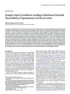
Fig. 5. A basket-geniculate synapse-pairing microcircuit may implement subfield antagonism in simple cells. Geniculate-basket axospinousaxodendritic synapse pairs respond to stimuli at the same RF center position but of opposite sign polarity. In the example here, an “oncenter” (1) geniculate axon synapses with the soma and proximal dendrites of the basket cell and an “off-center” (2) geniculate axon synapses with a dendritic spine of the spiny stellate cell. The basket cell synapses with the parent dendritic shaft of this spine to locally inhibit the “offcenter” geniculate signal.
underlying subfield antagonism in simple cells (see Palmer & Davis, 1981; Ferster, 1988). Subfield antagonism results from a strong, transient increase in GABA-A based shunting inhibition (BorgGraham et al., 1998; Hirsch et al., 1998). Fig. 5 shows a hypothetical circuit where an “on-center” geniculate axon synapses with the cell body and dendrites of a basket cell, and an “off-center” geniculate axon for the same RF position synapses with a dendritic spine of a spiny stellate neuron. The basket cell axon then synapses with the dendritic shaft near the spine base of the spiny stellate cell receiving the geniculate input. This pattern of connectivity is different from Heggelund’s (1981) simple cell model because the antagonism results from RF centers of the same position (not spatially offset) but of opposite rather than the same polarity.
Acknowledgments I thank Eberhard Buhl, Stephen Eglen, Zoltán Kisvárday, and especially Bertram Payne for their comments on earlier versions of this paper, and Gabor Tamás for his advice. I also thank the two anonymous referees for their helpful comments. This work was partially supported by UK Engineering and Physical Sciences Research Council (EPSRC).
References Ahmed, B., Anderson, J.C., Douglas, R.J., Martin, K.A.C. & Nelson, J.C. (1994). Polyneuronal innervation of spiny stellate neurons in cat visual cortex. Journal of Comparative Neurology 341, 39– 49.
341 Ahmed, B., Anderson, J.C., Douglas, R.J., Martin, K.A.C. & Nelson, J.C. (1996). The inhibitory innervation of spiny stellate cells of layer 4A of the cat visual cortex. Journal of Physiology (London) 495, 61P. Ahmed, B., Anderson, J.C., Martin, K.A.C. & Nelson, J.C. (1997). Map of the synapses onto layer 4 basket cells of the primary visual cortex of the cat. Journal of Comparative Neurology 380, 230–242. Anderson, J.C., Douglas, R.J., Martin, K.A.C. & Nelson, J.C. (1994a). Synaptic output of physiologically identified spiny stellate neurons in cat visual cortex. Journal of Comparative Neurology 341, 16–24. Anderson, J.C., Douglas, R.J., Martin, K.A.C. & Nelson, J.C. (1994b). Map of the synapses formed with the dendrites of spiny stellate neurons of cat visual cortex Journal of Comparative Neurology 341, 25–38. Anderson, P.A., Olavarria, J. & Van Sluyters, R.C. (1988). The overall pattern of ocular dominance bands in cat visual cortex. Journal of Neuroscience 8, 2183–2200. Azouz, R., Gray, C.M., Nowak, L.G. & McCormick, D.A. (1997). Physiological properties of inhibitory interneurons in cat striate cortex. Cerebral Cortex 7, 534–545. Beaulieu, C. & Colonnier, M. (1983). The number of neurons in the different laminae of the binocular and monocular regions of area 17 in the cat. Journal of Comparative Neurology 217, 337–344. Beaulieu, C. & Colonnier, M. (1985). A laminar analysis of the number of round-asymmetric and flat-symmetric synapses on spines, dendritic trunks, and cell bodies in area 17 of the cat. Journal of Comparative Neurology 231, 180–189. Beaulieu, C. & Somogyi, P. (1990). Targets and quantitative distribution of GABAergic synapses in the visual cortex of the cat. European Journal of Neuroscience 2, 296–303. Berman, N.J., Douglas, R.J., Martin, K.A.C. & Whitteridge, D. (1991). Mechanisms of inhibition in cat visual cortex. Journal of Physiology (London), 440, 697–722. Bonds, A.B. (1989). Role of inhibition in the specification of orientation selectivity of cells in the cat striate cortex. Visual Neuroscience 2, 41–55. Borg-Graham, L.J., Monier, C. & Frégnac, Y. (1998). Visual input evokes transient and strong shunting inhibition in visual cortical neurons. Nature 393, 369–373. Budd, J.M.L. (1995). Density of clutch cells in cat striate cortex: An estimate. Brain Research Association Abstracts 12, 66. Bueno-Lopez, J.L., Beaulieu, C. & Somogyi, P. (1989). Proportions of GABA-immunopositive and GABA-negative synaptic elements in layer 4 of cat’s striate cortex. European Journal of Neuroscience (Suppl.) 2, 106. DeFelipe, J. & Fairén, A. (1988). Synaptic connections of an interneuron with axonal arcades in the cat visual cortex. Journal of Neurocytology 17, 313–323. DeFelipe, J., Hendry, S.H.C. & Jones, E.G. (1989). Visualization of chandelier cell axons by parvalbumin immunoreactivity in monkey cerebral cortex. Proceedings of the National Academy of Sciences of the U.S.A. 86, 2093–2097. Dehay, C., Douglas, R.J., Martin, K.A.C. & Nelson, J.C. (1991). Excitation by geniculocortical synapses is not “vetoed” at the level of dendritic spines in cat visual cortex. Journal of Physiology (London) 440, 723–734. DeLima, A.D. & Singer, W. (1986). Cholinergic innervation of the cat striate cortex: A choline actyltransferase immunocytochemical analysis. Journal of Comparative Neurology 250, 324–338. Demeulemeester, H., Arckens, L., Vandesande, F., Orban, G.A., Heizmann, C.W. & Pochet, R. (1991). Calcium binding proteins and neuropeptides as molecular markers of GABAergic interneurons in the cat visual cortex. Experimental Brain Research 84, 538–544. Douglas, R.J. & Martin, K.A.C. (1991). A functional microcircuit for cat visual cortex. Journal of Physiology (London) 440, 735–769. Douglas, R.J., Martin, K.A.C. & Whitteridge, D. (1991). An intracellular analysis of the visual responses of neurones in cat visual cortex. Journal of Physiology (London) 440, 659– 696. Douglas, R.J., Koch, C., Mahowald, M., Martin, K.A.C. & Suarez, H.H. (1995). Recurrent excitation in neocortical circuits. Science 269, 981–985. Fairén, A., DeFelipe, J. & Regidor, J. (1984). Nonpyramidal neurons: General account. In Cerebral Cortex, Vol 1, Cellular Components of the Cerebral Cortex, ed. Peters, A. & Jones, E.G., pp. 201–253. New York, New York: Plenum Press. Ferster, D. (1988). Spatially opponent excitation and inhibition in simple cells of the cat visual cortex. Journal of Neuroscience 8, 1172–1180.
342 Ferster, D., Chung, S. & Wheat, H. (1996). Orientation selectivity of thalamic input to simple cells of cat visual cortex. Nature 380, 249–252. Freund, T.F., Martin, K.A.C., Smith, A.D. & Somogyi, P. (1983). Glutamate decarboxylase-immunoreactive terminals of golgi-impregnated axoaxonic cells and of presumed basket cells in synaptic contacts with pyramidal neurons of the cat’s visual cortex. Journal of Comparative Neurology 221, 263–278. Freund, T.F., Martin, K.A.C. & Whitteridge, D. (1985a). Innervation of cat visual areas 17 and 18 by physiologically identified X- and Y-type thalamic afferents. I. Arborization patterns and quantitative distribution of postsynaptic elements. Journal of Comparative Neurology 242, 263–274. Freund, T.F., Martin, K.A.C., Somogyi, P. & Whitteridge, D. (1985b). Innervation of cat visual areas 17 and 18 by physiologically identified X- and Y-type thalamic afferents. II. Identification of postsynaptic targets by GABA immunocytochemistry and Golgi impregnation. Journal of Comparative Neurology 242, 263–274. Freund, T.F., Maglóczky, Zs., Soltész, I. & Somogyi, P. (1986). Synaptic connections, axonal and dendritic patterns of neurons immunoreactive for cholecystokinin in the cat visual cortex of the cat. Neuroscience 19, 1133–1159. Freund, T.F. & Meskenaite, V. (1992). g-aminobutyric acid-containing basal forebrain neurons innervate inhibitory interneurons in the neocortex. Proceedings of the National Academy of Sciences of the U.S.A. 89, 738–742. Gabbott, P.L.A. & Somogyi, P. (1986). Quantitative distribution of GABAimmunoreactive neurons in the visual cortex (area 17) of the cat. Experimental Brain Research 61, 323–331. Gabbott, P.L.A., Martin, K.A.C. & Whitteridge, D. (1988). Evidence for the connections between a clutch cell and a corticotectal neuron in area 17 of the cat visual cortex. Proceedings of the Royal Society B (London) 233, 385–391. Gilbert, C.D. & Wiesel, T.N. (1983). Clustered intrinsic connections in cat visual cortex. Journal of Neuroscience 3, 1116–1133. Gundersen, H.J.G. (1986). Stereology of arbitrary particles. Journal of Microscopy 143, 3– 45. Hamos, J.E., David, T.L. & Sterling, P. (1983). Four types of neuron in layer IVab of cat cortical area 17 accumulate 3 H-GABA. Journal of Comparative Neurology 217, 449– 457. Hata, Y., Tsumoto, T., Sato, H., Hagihara, K. & Tamura, H. (1988). Inhibition contributes to orientation selectivity in visual cortex of cat. Nature 335, 815–817. Heggelund, P. (1981). Receptive-field organization of simple cells in cat striate cortex. Experimental Brain Research 42, 89–98. Hendry, S.H.C., Jones, E.G., Emson, P.C., Lawson, D.E.M., Heizmann, C.W. & Streit, P. (1989). Two classes of cortical GABA neurons defined by differential calcium binding protein immunoreactivities. Experimental Brain Research 76, 467– 472. Henry, G.H., Salin, P.A. & Bullier, J. (1991). Projections from area 18 and 19 to cat striate cortex: Divergence and laminar specificity. European Journal of Neuroscience 3, 186–200. Hirsch, J.A., Alonso, J-M., Reid, R.C. & Martinez, L.M. (1998). Synaptic integration in striate cortical simple cells. Journal of Neuroscience 18, 9517–9528. Hubel, D.H. & Wiesel, T.N. (1962). Receptive fields, binocular interaction and functional architecture in the cat’s visual cortex. Journal of Physiology (London) 160, 106–154. Kisvárday, Z.F. (1992). GABAergic networks of basket cells in the visual cortex. Progress in Brain Research 90, 385– 405. Kisvárday, Z.F., Martin, K.A.C., Whitteridge, D. & Somogyi, P. (1985). Synaptic connections of intracellularly filed clutch neurons, a type of small basket neuron in the visual cortex of the cat. Journal of Comparative Neurology 241, 111–137. Kisvárday, Z.F., Cowey, A. & Somogyi, P. (1986). Dendritic and axonal patterns of a type of GABA-immunoreactive neuron and its synaptic relationship to spiny stellate cells in layer IVC of the monkey striate cortex. Neuroscience 19, 741–761. Kisvárday, Z.F., Martin, K.A.C., Friedlander, M.J. & Somogyi, P. (1987). Evidence for interlaminar inhibitory circuits in the striate cortex of the cat. Journal of Comparative Neurology 260, 1–19. LeVay, S. (1986). Synaptic organisation of claustral and geniculate afferents to the visual cortex in the cat. Journal of Neuroscience 6, 3564–3575. LeVay, S. & Gilbert, C.D. (1976). Laminar patterns of geniculocortical projection in the cat. Brain Research 113, 1–19. Lund, J.S., Henry, G.H., Macqueen, C.L. & Harvey, A.R. (1979). An-
J.M.L. Budd atomical organization of the primary visual cortex (area 17) of the cat. A comparison with area 17 of the macaque monkey. Journal of Comparative Neurology 184, 599– 618. Martin, K.A.C. (1988). Wellcome Prize lecture: From single cells to simple circuits in the cerebral cortex. Quarterly Journal of Experimental Physiology 73, 637–702. Martin, K.A.C., Somogyi, P. & Whitteridge, D. (1983). Physiological and morphological properties of identified basket cells in the cat’s visual cortex. Experimental Brain Research 50, 193–200. Martin, K.A.C. & Whitteridge, D. (1984). Form, function and intracortical projections of spiny neurones in the striate visual cortex of the cat. Journal of Physiology (London) 353, 463–504. Naegele, J.R. & Katz, L.C. (1990). Cell surface molecules containing N-acetylgalactosamine are associated with basket cells and neurogliaform cells in cat visual cortex. Journal of Neuroscience 10, 540–557. O’Leary, J.L. (1941). Structure of the area striata of the cat. Journal of Comparative Neurology 75, 117–145. Palmer, L.A. & Davis, T.L. (1981). Receptive-field structure in cat striate cortex. Journal of Neurophysiology 46, 260–276. Pei, X., Vidyasagar, T.R., Volgushev, M. & Creutzfeldt, O.D. (1994). Receptive-field analysis and orientation selectivity of polysynaptic potentials of simple cells in cat striate cortex. Journal of Neuroscience 14, 7130–7140. Peters, A. & Regidor, J. (1981). A reassessment of the forms of nonpyramidal neurons in area 17 of cat visual cortex. Journal of Comparative Neurology 203, 685–716. Peters, A. & Jones, E.G. (1984). Classification of cortical neurons. In Cerebral Cortex, Vol 1, Cellular Components of the Cerebral Cortex, ed. Peters, A. & Jones, E.G., pp. 107–121. New York, New York: Plenum Press. Peters, A., Palay, S.L. & Webster, H.DeF. (1991). The Fine Structure of the Nervous System: Neurons and their Supporting Cells, 3rd edition. New York, New York: Oxford University Press. Peters, A. & Payne, B.R. (1993). Numerical relationships between geniculocortical afferents and pyramidal cell modules in cat primary visual cortex. Cerebral Cortex 3, 69–78. Peters, A. & Yilmaz, E. (1993). Neuronal organization in area 17 of cat visual cortex. Cerebral Cortex 3, 49– 68. Qian, N. & Sejnowski, T.J. (1990). When is an inhibitory synapses effective? Proceedings of the National Academy of Sciences of the U.S.A. 87, 8145–8149. Reid, R.C. & Alonso, J.-M. (1995). Specificity of monosynaptic connections from thalamus to visual cortex. Nature 378, 281–284. Reid, R.C. & Alonso, J.-M. (1996). The processing and encoding of information in the visual cortex. Current Opinion in Neurobiology 6, 475– 480. Sillito, A.M. (1975). The contribution of inhibitory mechanisms to the receptive-field properties of neurons in the striate cortex of the cat. Journal of Physiology (London) 250, 305–329. Somers, D.C., Nelson, S.B. & Sur, M. (1995). An emergent model of orientation selectivity in cat visual cortical simple cells. Journal of Neuroscience 15, 5448–5465. Somogyi, P. (1989). Synaptic organization of GABAergic neurons and GABA-A receptors in the lateral geniculate nucleus and visual cortex. In Neural Mechanisms of Visual Perception, ed. Lam, D.K.-T. & Gilbert, C.D., pp. 35– 62. Houston, Texas: Portfolio. Somogyi, P. & Cowey, A. (1981). Combined golgi and electron microscopic study on the synapses formed by double bouquet cells in the visual cortex of the cat and monkey. Journal of Comparative Neurology 195, 547–566. Somogyi, P., Freund, T.F. & Cowey, A. (1982). The axo-axonic interneuron in the cerebral cortex of the rat, cat and monkey. Neuroscience 7, 2577–2607. Somogyi, P., Kisvárday, Z.F., Martin, K.A.C. & Whitteridge, D. (1983). Synaptic connections of morphologically identified and physiologically characterized large basket cells in the striate cortex of cat. Neuroscience 10, 261–294. Somogyi, P., Freund, T.F., Hodgson, A.J., Somogyi, J., Beroukas, D. & Chubb, I.W. (1985) Identified axo-axonic cells are immunoreactive for GABA in the hippocampus and visual cortex of the cat. Brain Research 332, 143–149. Somogyi, P. & Soltész, I. (1986). Immunogold demonstration of GABA in synaptic terminals of intracellularly recorded, horseradish peroxidasefilled basket cells and clutch cells in the cat’s visual cortex. Neuroscience 19, 1051–1065.
Quantitative connectivity of layer IV basket cells Tamás, G., Buhl, E.H. & Somogyi, P. (1997a). Fast IPSPs elicited via multiple synaptic release sites by different types of GABAergic neurone in the cat visual cortex. Journal of Physiology (London) 500, 715–738. Tamás, G., Buhl, E.H. & Somogyi, P. (1997b). Massive autaptic selfinnervation of GABAergic neurons in cat visual cortex. Journal of Neuroscience 17, 6352– 6364. Tamás, G., Somogyi, P. & Buhl, E.H. (1998). Differentially interconnected networks of GABAergic interneuron in visual cortex of the cat. Journal of Neuroscience 18, 4255– 4270. Tanaka, K. (1983). Cross-correlation analysis of geniculostriate neuronal relationships in cats. Journal of Neurophysiology 49, 1301–1318. Tarczy-Hornoch, K., Martin, K.A.C., Jack, J.J.B. & Stratford, K.J. (1998). Synaptic interactions between smooth and spiny neurones in layer 4 of cat visual cortex in vitro. Journal of Physiology (London) 508, 351–363.
Appendix A: Derivation of calculations used in Table 3 In the Table 2 (Assumption 1) calculation, the mean number of basket cell synapses made with the dendrites of a spiny neuron is estimated using the total number of synapses per basket cell axon parameter. For most of the ten additional basket cells, the value of this parameter is not available. However, the estimate can be made independently of the total number of
343 synapses per basket cell axon. Let the density of basket cells, B, be given by the equation (see Table 2, line 1, Assumption 1): B 5 Taxo 0~m 3 Psps !,
(A1)
where Taxo is the density of GABAergic axosomatic synapses made with the cell bodies of spiny cells (Table 1, line A12), m is the total number of synapses made by a basket cell axon, and Psps is the proportion of basket cell synapses made with the cell bodies of spiny cells (Table 1, line B4a). The number of basket cell synapses made with the dendrites of a spiny neuron n is found from the equation (see Table 2, line 9, Assumption 1): n 5 ~B 3 m 3 Pspd !0S,
(A2)
where Pspd is the proportion of basket cell synapses made with spiny cell dendritic processes (spines and spiny shafts) (Table 1, line B4i) and S is the density of spiny neurons in layer IV (Table 1, line A7). Substituting eqn. (1) into (2) simplifies (as m cancels) to n 5 ~Taxo 0S! 3 ~Pspd 0Psps !, which is used in Table 3.
(A3)