Sensors 2009, 9, 3386-3404; doi:10.3390/s90503386 OPEN ACCESS
sensors ISSN 1424-8220 www.mdpi.com/journal/sensors Article
Intelligent Foreign Particle Inspection Machine for Injection Liquid Examination Based on Modified Pulse-Coupled Neural Networks Ji Ge *, YaoNan Wang, BoWen Zhou and Hui Zhang College of Electrical and Information Engineering, Hunan University, China; E-Mail:
[email protected] (Y-N.W.) * Author to whom correspondence should be addressed; E-Mail:
[email protected] Received: 25 March 2009; in revised form: 22 April 2009 / Accepted: 23 April 2009 / Published: 7 May 2009
Abstract: A biologically inspired spiking neural network model, called pulse-coupled neural networks (PCNN), has been applied in an automatic inspection machine to detect visible foreign particles intermingled in glucose or sodium chloride injection liquids. Proper mechanisms and improved spin/stop techniques are proposed to avoid the appearance of air bubbles, which increases the algorithms’ complexity. Modified PCNN is adopted to segment the difference images, judging the existence of foreign particles according to the continuity and smoothness properties of their moving traces. Preliminarily experimental results indicate that the inspection machine can detect the visible foreign particles effectively and the detection speed, accuracy and correct detection rate also satisfying the needs of medicine preparation. Keywords: Intelligent inspection machine; foreign particle detection; modified PCNN; injection quality inspection; image processing; illumination styles
1. Introduction Injection liquid clarity is one of the vital indexes in clinical treatment using fluids. The presence of visible foreign particles, which can not be metabolized by human beings in injection liquids is prohibited [1]. However, different kinds of foreign substances such as rubber chips, chemical fibers, glass fragments, calcium carbonate and other crystalline particles [2] appear due to problems with
Sensors 2009, 9
3387
injection bottle quality, packing procedures, collisions, filtration or filling. They can cause thrombus, phlebitis, tumor, anaphylactic reaction or even death when these kinds of particles are injected into the vein. Thus, foreign particles in injection liquids are now a matter of social concern which is often reported in the mass media. Thus, it was reported recently that more than 75% medicine recalls were related to the presence of foreign substances. Pharmaceutical corporations have been taking active measures against this problem to avoid damage to their public image and economic losses caused by recalls. Nevertheless, according to the statistics [3] taken by China Pharmaceutical Industry Association (CPIA), 99.6% pharmaceutical corporations in China use a light inspection method carried out by workers [1]: inspectors put the injection bottle under a special lamp housing, rotate and turn it over gently, suspending any visible foreign substance present in the transfusion and then deciding whether it is acceptable or not based on their inspection experience. This method is simple, but the inspection efficiency and repeatability are poor, with omission rates increasing synchronously due to workers’ tiredness. A visible foreign particles inspection system was previously developed and has been utilized in an actual product line [4,5]. In this system, an injection container such as an ampoule is rotated at high speed and stopped abruptly. The ampoule forms a vortex due to inertia. The moving foreign substances can be distinguished from stationary scratches on the surface of the ampoule by applying image processing techniques. Detection in rectangular plastic bottles of medicinal solutions based on realtime image processing are proposed in [6]. Some experiments have been performed on discrimination of particle in size and shape [7] and visualization of the spatial behavior of particles [8]. Developmental research for detecting impurity particles in a water supply has been described in [9]. Currently we are developing an Automatic Inspection Machine (AIM) equipped with CCD cameras. Since an ampoule or a vial is small and simple in shape, having a smooth surface, it is easy to gather any foreign substances present in the center by forming a vortex in the solution and thus distinguish them from scratches and other defects of the surface of the container. Nevertheless, in our machine, the detection target is mass transfusion and it is commonly contained in a glass or plastic bottle. Sometimes the cross sections of the bottle are circular, oval, rectangular or hexagonal and they can have a complicated surface such as embossed symbols and graduations on the surface, which makes it difficult to create a vortex and distinguish the foreign particles from uneven surface features. Hence, image segmentation and recognition of foreign particles is the key module of AIM. A large number of different image segmentation techniques are currently available. Basic algorithms like thresholding, edge detection and region growing are described in [10]. Although simple to implement, these methods usually have serious shortcomings when employed in practical situations. The most obvious case is thresholding, which can only be used to segment an image if the segments consist of non-overlapping pixel intensity ranges, which is rarely the case. Edge detection and region growing can tolerate some intensity overlap between the different segments, but can fail if the edges between the segments are not sharp enough. Several newer and more advanced techniques have been proposed to address these problems. One interesting class of methods are the graph based methods [11] which has yielded several impressive results [12,13]. PCNN is a biologically inspired type of neural network, which based on the experimental observations of synchronous pulse bursts in the cat and the monkey visual cortex [14]. Compared with previous classical Artificial Neural Networks model, PCNN can be applied to image processing
Sensors 2009, 9
3388
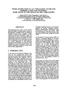
without training process. However, it needs to properly set a number of numerical control parameters for which it is usually not known a priori how to select the best values for a given application. The optimum values typically depend on attributes of the images to be segmented, e.g. pixel intensity ranges, contrasts and noise levels. As a consequence, values successful for one set of images may not be useful for other images with different intensity ranges, contrasts and noise levels. A number of different solutions have been suggested to address this problem, including the use of adaptive connection weights between neighboring neurons [15], automatic adaptation by genetic algorithms [16], reinforcement learning [17] and neural networks [18]. This paper proposes an adaptive segmentation method based on a modified PCNN, which multithresholds determined by the water region area in a histogram. Those control parameters can be achieved adaptively. A number of injection images are segmented. According to continuity and smoothness properties of extracted objects’ moving traces, the inspection machine judges whether this injection is acceptable. Furthermore, improved spin/stop technique of controlling motors and illumination styles are applied through a large quantity of experiments to reduce the influence of air bubbles. The experimental results show that the inspection machine is superior to proficient workers, and can detect visible foreign particles effectively with satisfactory speed and accuracy. In the following paragraphs, Section 2 introduces the architecture of the PCNN model. Section 3 gives intelligent inspection machine’s overview. Section 4 brings forward key algorithms of foreign substances detection. Section 5 is devoted to the experiments and analysis of their results. Section 6 gives the conclusion. 2. Architecture of the PCNN Model PCNN is a biologically inspired type of neural network, which is based on the experimental observations of synchronous pulse bursts in the cat’s visual cortex by Eckhorn et al. and was adapted for image processing by Johnson [19]. Figure 1. Structure of a PCNN neuron.
2.1. PCNN Neuron Model and Parameters Determination The PCNN is significantly different from other artificial neural network models in both its structure and operation. There is no training phase involved. Each neuron in the processing layer is directly tied to an input, in this case an image pixel or a set of neighboring image pixels. These are the feeding
Sensors 2009, 9
3389
inputs, and they are also linked to nearby neurons, the linking inputs. The feeding inputs are iteratively processed together with the linking inputs producing a pulse train. The PCNN neuron consists of three parts [19,20]: the dendritic tree, the linking modulation, and the pulse generator, as shown in Figure 1. The corresponding neuron’s functions are: Fij [ n] e F n Fij [ n 1] S ij VF M ijkl Ykl [ n 1]
(1)
Lij [n] e L n Lij [n 1] VL Wijkl Ykl [n 1]
(2)
U ij [n] Fij [n] (1 Lij [n])
(3)
kl
kl
1 if Yij [n] 0
U ij [n] ij [n 1] otherwise
ij [n] e ij [n 1] V Yij [n] n
(4) (5)
where, Fij - feeding input; Lij - linking input; Uij - internal state; Sij - external stimulus; θij - dynamic threshold; M , W - synaptic weights matrix to neuron; VF, VL and Vθ - normalizing constant; αF, αL and αθ - negative decay constants of leaky integrator; β - linking strength between neurons; n - iteration times; Yij - output. A PCNN is a two-dimensional non-training neural network in which each neuron in the network corresponds to one pixel in an input image. The neuron receives its input (e.g. intensity) as an external stimulus. However, each neuron also connects with its neighboring neurons, receiving local stimuli from them. These stimuli are combined in an internal activation system, and are accumulated until they exceed a dynamic threshold. This will result in a pulse output, which is called natural fire. Through an iterative process, the algorithm produces a temporal series of binary images as outputs. Due to the linking of neighboring neurons, segments of the image consisting of pixels of similar intensity values tend to pulse together, which is called captured fire. The output of a PCNN can therefore be used for image segmentation by taking the pixels corresponding to synchronously pulsing neurons as segments. Nevertheless, the performance of image segmentation based on PCNN depends on the suitable PCNN parameters. It is necessary to adjust the various threshold parameters of its mathematical model manually and then can achieve the optimum processing. As Karvonen [21] mentioned, a very large set of data should be required to optimize PCNN parameters, which is unfeasible in most applications. Hence, in order to determine PCNN parameters adaptively, this paper suggests an adaptive segmentation method based on a modified PCNN, which multi-thresholds determined by water region area in histogram. The implementation of the algorithm is computationally simple and can be used in foreign particles real-time detection in injections. The detailed process is given as following. 2.2. Multi-Threshold Acquisition Using “Water Region Area” Method A novel threshold auto-detection algorithm in image histograms is proposed. In accordance with the intuitional features of the histogram, the peaks of the histogram are considered as watersheds, each
Sensors 2009, 9
3390
valley including two neighboring peaks and a valley bottom points. We call the maximal water capacity in each valley the “water region area”. Step 1: Draw image histogram and normalize them. Step 2: Seek all peaks and valley bottom points in the histogram. Step 3: Calculate the water region area from the left valley bottom point. Define Θ that lies within [0.01, 0.05]. The smaller Θ is, the more threshold points we will get. When the water region area is larger than Θ, the corresponding valley bottom point will be kept in threshold array Tm. Meanwhile, the corresponding left side peak point will be kept in peak points array Pm. Otherwise the valley will be taken as invalid. At this situation, comparing the two peaks’ values located in the valley’s two sides: (1) If the left peak point is larger than the right one, it will be treated as the new left peak point. While the next right peak point will be the new right peak one, the smaller between the current and the next valley bottom point will be regarded as the new valley bottom point. (2) Otherwise, the right peak point, the right valley bottom point and the next right peak point will be regarded as new left peak point, new valley bottom point and new right peak point respectively and then the new water region area will be calculated again. Step4: Iteratively execute Step3 until all valley bottom points have been processed and then we can get the threshold array Tm (m = 1, …, M and T1