transcription factor for expression .... 0s1d4. 14Sâ'Y. 100%. N.tch#{149}. -. 227. Ntu.tCh#{149}$. 0. Gop. 0. C..r'.t1s. Sb.t&t.ttoflS .... high transcript levels in RNA.
Vol.
2, 401-407,
August
Cell
1991
Growth
& Differentiation
401
Interferon Regulatory Factor 1 Is a Myeloid Differentiation Primary Response Gene Induced by Interleukin 6 and Leukemia Inhibitory Factor: Role in Growth Inhibition’
Abbas Abdollahi, Kenneth A. Lord, Barbara Liebermann,2 and Dan A. Liebermann2 Department School
of Biochemistry
of Medicine,
and Biophysics,
Philadelphia,
Pennsylvania
University
Hoffmanof Pennsylvania
19104
Abstract To better understand the immediate early genetic response of myeboid cells to terminal differentiation and growth inhibitory stimuli, we have recently isolated complementary DNA clones of myeboid differentiation primary response (MyD) genes, activated in the absence of protein synthesis in Ml myeboid precursor cells following induction for terminal differentiation and growth arrest by conditioned media of mouse lungs, a potent physiological source of hemopoietic differentiation inducers. In this study, it is shown that one particular MyD complementary DNA clone, expressed highly in normal precursor enriched bone marrow cells, encodes for interferon regulatory factor 1 (IRF-1), a positive transcription factor for expression of the j9-interferon (IFN-) gene. Using a clone of Ml cells inducible for terminal differentiation by both interleukin 6 (IL-6) and leukemia inhibitory factor (LIF), two multifunctional cytokines recently identified as physiological inducers of hemopoietic cell differentiation, it has been shown that IRF- 1 expression is rapidly induced by IL-6 and LIF in the absence of protein synthesis and is followed by a later increase in the bevels of IFN-$ mRNA, observed to be largely dependent on protein synthesis. Also, it is shown that the growth inhibition associated with IL-6 or LIF induced terminal differentiation could be partially abrogated via the use of IRF-l antisense oligomers or IFN-fl antiserum. Taken together, these findings imply a regulatory cascade, where induction of terminal myeloid differentiation by IL-6 or LIF triggers the immediate early activation of IRF-l, leading to the later induction of IFN-fi, in turn playing an autocrine robe in growth inhibition. Introduction Terminal differentiation of eukaryotic cells involves two interrelated cellular processes, the regulated progression of cells through successive stages of cell differentiation and growth inhibition, which ultimately results in growth arrest. A profound example of this process, which continues throughout life, is the complex process of blood
cell formation, whereby a hierarchy of hemopoietic progenitor cells in the BM3 proliferate and differentiate along multiple, distinct cell lineages, including the proliferation and differentiation of myeboid precursor cells into mature granubocytes and macrophages (for overview, see Ref.
1). The establishment of in vitro culture systems for the clonal development of bone marrow cells (2, 3) and the availability of the Ml myeboid leukemia cell line, which can be induced for differentiation (Mi D+) by physiobogical myebopoietic differentiation inducers (4), provide an excellent
biological
ogy of normal
system
to study
cell growth
that afflict it, upon oncogenesis 9). Recently, it has been shown multifunctional cytokines (10-12), etic differentiation ferentiation and
the
molecular
and differentiation,
biol-
and lesions
and its progression (5that IL-6 and LIF, two also act as hemopoi-
inducers which induce terminal growth arrest of Mi cells (13-15).
dif-
To better understand the immediate sponse of myeboid cells to terminal
early genetic differentiation
reand
growth
recently
this
inhibitory
stimuli,
we
have
used
experimental system to isolate and characterize cDNA clones of MyD genes (16-18), activated in the absence of protein synthesis in Mi cells following induction for terminal differentiation and growth arrest by conditioned media of mouse lungs, a potent physiological source of hemopoietic expression
differentiation also was shown
precursor
enriched
in M1D+
cells
inducers in primary
(9, 19). cultures
BM cells, resembling
induced
their
for differentiation
In the present study, it is revealed MyD cDNA clone, highly expressed
MyD gene of myeboid
expression
(16-18).
that one particular in normal precursor
enriched bone marrow cells, encodes for IRF-i , a positive transcription factor for expression of the IFN-13 gene (20-
22).
Using
a clone
differentiation
of Mi
by either
study
physiologically
cells,
it is shown,
for the
absence
of protein
in the
terminal cytokmnes, induction
cells
lL-6
induced
differentiation
first
time,
plays an autocrine
the later
that
synthesis
myeboid differentiation and that a regulatory of terminal myeboid
IL-6 or LIF triggers 1, leading to the
inducible
for
terminal
or LIF as a model
immediate induction
role in growth
IRE-i
upon
system
to
of myeboid is activated
induction
of
by either one of these cascade exists, whereby differentiation by either early activation of IFN-fl, which
of IRFin turn
inhibition.
The abbreviations used are: BM, bone marrow; Ml D+, Mi differentiation competent; IL-6, interleukin 6; LIF, leukemia inhibitory factor; IFN, interferon; IRF-i, interferon regulatory factor i; cDNA, complementary 3
Received 4/2/91. 1 This work was
supported
by the
National
Institutes
and the American Cancer Society (B. H-L.). 2 To whom requests for reprints should be addressed.
of Health
(D.
A. L.)
DNA; PCR, standard
MyD, myeloid differentiation polymerase chain reaction; saline
citrate.
primary response; SDS, sodium dodecyl
kb,
kilobase(s); sulfate; SSC,
402
IRF-i
in Growth
Inhibition
of Differentiating
Myeloid
Cells
1
co.plSt
a. 1.
NYO3SSI
t,,t#{149}rt .ror
NIJSIR?11 1,,tt.1 0s1d4. Gop
5cr. 14S”’Y
-
23 100%
Optts1sd
10
Sor.
N.tch#{149}. 0
I
fictor
.q,1.try
C..r’.t1s
30
Sb.t&t.ttoflS
30
.0$A.
237 227
-
ttqniftcI”C#{149} Ntu.tCh#{149}$
21.01 0 0
-
00
30
40
70
tilitit lilt IIIttlIttt Itt lttt cGAAcccAAcccAAccGAAcecGGceGAGTrGcGccGAGcTcAcccGAGcTcGccAcAGGkccccAGcATcr I
40
30 #{149}0
IltttIIIItI
O 0
70 100
SO 110
O 120
100 1)0
140
111111 ItItit
110
120
1)0
130
140
140
170
130 100
100 100
170 300
210
IIIIIII ti lilt itt tililti II 100
100 220
200
210
220
2)0
240
0
iltill t I I Itt
GArTOArTCCAAc 230
b.
1.
240
0YD321 *2*10111
jr,t.rt.rOO
$0#{149}
initial
Scor.
a..ido. Gap.
1d.nttY
-
I
210 100%
10
r.q%1*t0ry
20
1S)0
co.pl.t
$IqntftcaflC#{149} iS1uatch#{149}
40
1S30
30
1040
0
1 aMA, 210 210
Sob.tttuttOts
30
1040
so
factor
Scors
Opti.1iSd Match.. Cot..-catlVS
70
1$S0
110
0 0
40
1170
100
10.72 -
1S0
120
1)0
140
TccTckGc.cc.rrGGcAGTcc.rcAGckGGcccAGGGAAAAGGcGGGrrG.rGAGcGccTrGGcG1Gkcrcrr
tllIItt
?ccTcAcccc.rrGGckG.Ttx.TcAGckGGccc,,GGGM.kAGGcGGG.rTG.rGkGcGccrrGGcGTGAcrcrr 1000
1q10 130
1020 140
1020 170
1040 iSO
1S0 100
10 200
210
ittIIItIItIIItt 1070
1000
100
2000
o1
.
2010
2020
20)0
2
LyBrKiMi
kb
.
-2.1
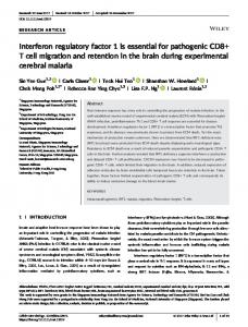
of MyD32 as a cDNA clone of mouse IRF-1 (a and in various murine tissues (c). The results of homology searches against the GenBank with My032 nucleic acid end sequences are shown. In both alignments, the top sequence corresponds to My032 cDNA. a, identity ofthe MyD32 5K end to the 5’ end ofIRF-1. b, identity of the inverse complement of the My032 KS end with 3’ end sequences of IRF-1. c, elevated expression of !RF-1 in highly myelopoietic myeloid precursor enriched bone marrow (BM) cells, as compared to other murine tissues. Ly, lymphocytes; Br, brain; Ki, kidney; Mu, muscle. Direct sequence determination of both ends of the MyD32 cDNA clone insert and homology searches were performed as indicated in “Materials and Methods.” Highly myelopoietic BM cells, consisting primarily of cells of the myeloid lineage, and enriched with myeloid precursors, as well as other Fig.
1.
Identification
b) and expression
murine
prepared
tissues
with
were
total
obtained
from
RNA (5 gig/lane)
CD-i
mice
and exposed
(16).
to X-ray
RNA
blot
was
film for 48 h.
Results Identification of MyD32 as a cDNA Clone Encoding for IRF-1. As shown in Fig. 1, a and b, the end sequences of one particular MyD cDNA clone, MyD32, were found to be identical to end sequences of IRF-1, a positive transcription factor for expression of the IFN-/3 gene (20-22). Analysis of MyD32/IRF-1 expression in several murine tissues (Fig. ic) has revealed relatively high transcript levels in RNA obtained from highly myebopoietic and myeloblast enriched BM cells immediately after effusion from femurs of sodium caseinate injected mice (9). In contrast, no detectable expression of MyD32/IRF-1 was
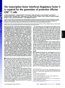
observed in several other murine tissues, including lymphocytes, brain, kidney, and muscle. Analysis of the Expression of IRF-1 and IFNupon Induction of Ml Terminal Differentiation by IL-6 or LIF. To investigate the robe of IRF-1 in growth inhibition associated with physiologically induced terminal differentiation of hemopoietic cells, advantage has been taken of a clone of Mi myeloid precursor cells (Mi D+ clone 6) inducible for terminal differentiation by both lL-6 and LIF. As shown in Fig. 2a, both LIF and bL-6 similarly inhibited the growth of Ml cells. Both of these myeloid differentiation inducers induced, to similar extents, a spectrum of early to late differentiation markers, including C3 receptors, cell attachment, lysozyme synthesis, and mature macrophage-like cells. We wished to ascertain whether expression of IRF-i is activated upon induction of Mi terminal differentiation by bL-6 and/or LIF, and if so, whether its activation is a primary response to stimulation of the cells with these two differentiation inducing cytokmnes. As shown in Fig. 2b, !RF-1 expression was induced within 1 h following stimulation of M1D+ cells with IL-6 or LIF, exhibiting biphasic kinetics of expression, with maximal induction at 1 h and a decline (3 h) followed by increased levels of steady-state mRNA in terminally differentiated cells. Cycboheximide, a potent protein synthesis inhibitor, did not inhibit, and even supermnduced, the early increase in the steady-state bevel of IRF-i mRNA, indicating that it is an immediate early response to stimulation of the cells with lL-6 or LIF. Since IRF-i has been implicated in the regulation of IFN-j3 expression (20-22), it was also of interest to examine whether !FN-f3 expression is induced, and whether its expression is primary or secondary to the terminal differentiation program induced by lL-6 or LIF. In contrast to the expression of IRF-1, an increase in the level of bFN- mRNA was detected only following 6-h stimulation with bL-6 or LIF. It can also be seen that, unlike the increase in the level of IRF-i mRNA, this later increase in the level of bFN- mRNA was inhibited to a large extent by cycboheximide, suggesting that it is dependent on protein synthesis. It should be pointed out that no increase in the steady-state levels of IRF-1 and IFN-j3 mRNAs was observed in a clone of WEHI-3B D cells following stimulation by lL-6 or LIF, which neither induced differentiation nor inhibited the growth of these cells. To investigate the regulation ofIRF-1 and IFN-13 induction following stimulation of Mi cells with IL-6 or LIF, run-on transcription assays were performed with nuclei isolated from Ml D+ cells before and 1 or 6 h after stimulation. Transcription of both !RF-1 and IFN-3 was below detection levels prior to stimulation with IL-6 or LIF; IRE-i transcription increased to detectable levels 1 h following stimulation, and IFN-13 transcription was detected only after 6 h (Fig. 2c). Taken together, these observations, along with previous work where IRF-i was shown to regulate IFN-f3 expression (20-22), are consistent with the notion that immediate early activation of IRE-i by bL-6 or LIF plays a role in the later induction of !FN-i.
Robe of IRF-l and IFN-$ in Growth Inhibition Associated with Terminal Myeboid Differentiation Indued by bL-6 or LIF. To ascertain the role of the putative IRF-l/ IFN-/3
regulatory
cascade
in growth
inhibition
associated
Cell Growth
& Differentiation
403
a. i nhi bition
Growth
Differentiation
associated
12 LI)
10
0
properties
Control receptors
q3 Cell
8
attachment
L.L&
LEE
(%)
1.2
57
54
(%)
0.8
67
69
U)
8)
C.)
6
0
4
Lysozye (ug.equiv/5x10 cell
8)
.0
2
E
z
0.14
type
(%)
Blast Intermediate
0 0
1
2
Days
>99
0
C. __________ LIF
I L6
_____________________ Unind. 1L6 lh
0 lh
3h
6h
Id
3d
6h
0
lh
3h
6h id
Fig. 2. Growth M1D+ myeloid differentiation
units/mI Differentiation
conditioned
associated
Actin
I)
H3
.
If
-
associated for terminal determined
media).
properties
Viable
represent
properties differentiation with cells
cell the
number
values
(a), by seeded
was determined
of assays
with
inhibition SD of up
and Methods.” in oMaterials
dose
and
curves,
Analysis
To increase
product, and ADNA to indicated probes. was performed run-on assays
response
to ±15%. Methods,”
sensitivity and
one-fifth
in “Materials levels
of detection
as internal marker X-ray film exposure
using nuclear were conducted
as indicated
of steady-state of
the
of IRF-i
by trypan
performed
of steady-state products
were
and (and
4 days
Methods.” actin)
IFN-$
levels (b) and transcription (c), in of the growth inhibition and or LIF (300 units/mI purified, 10
and analysis of IRF-i/IFNsteady-state mRNA IL-6 or LIF. CX, cycloheximide. Characteristics at i0 cells/mI with or without IL-6 (50 ng/mI)
blue
after
dye
addition
exclusion, of
the
counting inducers,
All values
mRNA
was
transcripts,
represent
performed
means with
PCR was used with
electrophoresed.
Control
samples
of at least
5 gig/lane not
reverse
indicated cells
with
time
points.
C3 receptors
differentiation was determined blast cells, cells at intermediate of the linear differentiation and three
oftotal
1-zg aliquots
at the in which
could first be detected after 12 h, cell attachment after 1 day, and lysozyme and mature cells after 2 days. Morphological on May-Grunwald-Giemsa stained cytospin smears by counting at least 300 cells and scoring the proportion of immature monocyte stages of differentiation, and mature macrophages. Cytokine concentrations used represent the optimum growth
6h
#{236}#{212}S#{149}#{149}S#{149} S #{149}. 2 . 2
inhibition and differentiation cells (clone 6) induced associated properties were
in Cos cell
:
lh
.
________
=T1
lFN
IRF1
3d 6h k
LIF
6h
S
#{176}...#{149}
________________
Actin
56 33
in culture
b.
IRF1
11
8
57 35