■ trauma update Section Editors: David J. Hak, MD, MBA & Philip F. Stahel, MD
Intramedullary Nailing of Proximal Third Tibial Fractures: Techniques to Improve Reduction David J. Hak, MD, MBA
Abstract: Obtaining and maintaining an acceptable reduction of proximal third tibial fractures can be problematic. Deforming forces acting on the proximal fragment and the spaciousness of the intramedullary canal at this level contribute to this challenge during intramedullary nailing. Several surgical techniques have been developed to address this problem, including the use of a more lateral and proximal starting point, adjunctive plate fixation, blocking screws, semiextended nailing, and most recently the use of a retropatellar portal approach. Familiarity with these techniques is critical to achieve satisfactory results when nailing proximal third tibial fractures.
I
ntramedullary nailing of simple diaphyseal tibial shaft fractures usually results in near anatomic reduction, as the intramedullary nail fills the intramedullary canal. In contrast, accurate reduction of tibial fractures that are near the proximal metaphyseal junction are notoriously problematic when treated by intramedullary nailing (Figure 1). In the absence of special techniques to achieve and
maintain accurate reduction, extra-articular proximal third tibial fractures treated with an intramedullary nail will commonly be malreduced in valgus, apex anterior, and have posterior displacement of the distal segment. Two studies published in 1995 highlighted the difficulty of achieving an adequate reduction when nailing proximal tibial fractures. Lang et al1 reported on 32 extra-articular
Dr Hak is from Denver Health/University of Colorado, Denver, Colorado. Dr Hak has no relevant financial relationships to disclose. Correspondence should be addressed to: David J. Hak, MD, MBA, Denver Health/University of Colorado, 777 Bannock St, MC 0188, Denver, CO 80204 (
[email protected]). doi: 10.3928/01477447-20110526-19
532
ORTHO0711Hak.indd 532
1
2
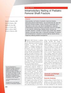
Figure 1: Malreduction following intramedullary nailing of a proximal third tibial fracture. Figure 2: The distal location of the Herzog bend of this older unreamed Synthes tibial nail caused posterior and distal displacement of the shaft segment during nail insertion.
fractures of the proximal third of the tibia treated with an intramedullary nail. At followup, 84% of their patients had angulation ⬎5⬚ in the frontal or sagittal plane, and 59% had ⭓1 cm displacement at the fracture site. They noted that valgus, apex anterior, and residual displacement at the fracture site were common. Freedman and
Johnson2 reported malalignment (defined as a ⭓5⬚ angulatory deformity in any plane) in 7 of 12 (58%) proximal third tibial fractures treated with an intramedullary nail. At that time, the design of 1 of the most commonly used intramedullary nail had a distal Herzog bend (Figure 2). Henley et al3 outlined how a
ORTHOPEDICS | ORTHOSuperSite.com
6/24/2011 9:53:16 AM
■ trauma update
3
4A
4B
5A
5B
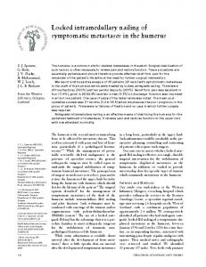
Figure 3: Apex anterior angulation is commonly seen in proximal tibial fractures treated with an intramedullary nail. This deformity occurs when the knee is flexed to obtain the entry site. Attachment of the patellar tendon to the proximal fracture segment results in apex anterior malalignment as the knee is flexed. Figure 4: On the AP view, the ideal entry site for a proximal tibial fracture nailing should be aligned with the lateral tibial eminence (A). On the lateral view, the ideal entry site should be more proximal (B). Figure 5: Lateral radiograph of a segmental tibial fracture (A). A 7-hole compression plate has been placed anterior to the path of the intramedullary nailing for reduction of the proximal portion of this segmental tibial fracture (B).
nail with a Herzog bend distal to the fracture site becomes wedged, displacing the distal segment posteriorly and distally as it is inserted. Since that time, contemporary intramedullary nails have been designed that have a more proximal Herzog bend with greater proximal interlocking options. Despite these improvements, difficulties remain in achieving a satisfactory reduction when nailing proximal tibial fractures. Two main factors complicate the reduction of extraarticular proximal tibial fractures: (1) the deforming forces acting on the proximal tibial segment; and (2) the spaciousness of the intramedullary canal at this level. Flexion of the knee is required to create a traditional intramedullary nail entry site in the proximal tibia. Because of the attachment of the patellar tendon to the proximal fracture
JULY 2011 | Volume 34 • Number 7
ORTHO0711Hak.indd 533
segment, apex anterior displacement occurs (Figure 3). In response to these problems, surgeons have developed several techniques to achieve an improved reduction when nailing proximal third tibial fractures, including: (1) starting point location; (2) adjunctive plating; (3) blocking screws; (4) semiextended nailing technique; and (5) retropatellar portal technique.
STARTING POINT LOCATION Buehler et al4 reported on the use of a more lateral and proximal entrance site to achieve reduction of proximal tibial shaft fractures. They also used a medially placed universal distractor and placed the interlocking screws with the knee in full extension using a special proximal interlocking jig. A more lateral and proximal entry site is helpful to avoid malreduction in proximal tibial fractures (Figure 4).
Proximally, the medial side of the tibia has been described as a chute that deflects the nail laterally.4 The central axis of the intramedullary canal is most commonly aligned with the lateral tibial eminence. Using a more proximal entry site will achieve a longer segment of nail within the proximal segment and usually place the nail’s Herzog bend completely within the proximal segment, rather than at or distal to the fracture site.
ADJUNCTIVE PLATING Several surgeons have proposed temporary or permanent placement of a small fragment plate to maintain reduction of the proximal fracture and allow the knee flexion required to insert an intramedullary nail.5-7 Clinically, both onethird tubular and small-fragment compression plates have been used. Locking plates provide another useful option.
A 4- to 6-hole plate is commonly used. The plate can be used temporarily and removed after the nail is successfully inserted and interlocked, or left in place to assist with maintaining the reduction. With the use of unicortical screws, the plate can be positioned along almost any surface. Good screw purchase can usually be obtained with a plate placed anteriorly in the area of thick cortical bone. Alternatively, the plate can be placed along the medial surface. In this case, bicortical screws may be placed from medial to lateral as long as they are anterior to the proximal path of the nail (Figure 5). Nork et al,7 in a series of 37 fractures of the proximal quarter of the tibia, discussed the use of supplemental unicortical plates for 13 of the fractures. In 3 cases, the plates were used temporarily as a reduction aid and removed, while in the other 10 cases they were left in
533
6/24/2011 9:53:17 AM
■ trauma update
6A
6B
8
7
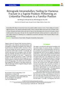
Figure 6: Valgus deformity is seen during initial insertion of an intramedullary nail of the proximal portion of this segmental fracture (A). The nail was removed and a blocking screw (arrow) placed just lateral to the central axis of the tibia to correct this deformity (B). Figure 7: To correct apex anterior angulation, a blocking screw should be placed just posterior to the intended ideal nail pathway, as shown in this lateral diagram. As the nail passes anterior to the blocking screw, reduction is achieved. Figure 8: To correct valgus deformity, a blocking screw is placed just lateral to the central axis of the tibia, as shown in this AP diagram. As the nail is passed medial to the blocking screw, reduction is achieved.
place. They placed plates both anteriorly and posteromedially with screws directed to avoid interference with the reamers and the intramedullary nail. They reported that the plates were effective in maintaining the reduction and did not adversely affect the healing of the fracture.
BLOCKING SCREWS Krettek et al8 described the use of Poller or blocking screws to improve reduction in metaphyseal fractures treated with intramedullary nailing. The blocking screws essentially reduce the size of the available nail pathway. Properly positioned screws can prevent malreduction as a nail is placed into a large metaphyseal space. Ricci et al9 reported on the use of blocking screws in 12 patients with proximal third tibial fractures treated with intramedullary nailing. They
534
ORTHO0711Hak.indd 534
found that the blocking screws were effective in obtaining and maintaining alignment of the fractures. Proper placement of blocking screws can be difficult. If they are placed too close to the intended ideal nail pathway, the nail may not be able to be passed, while if they are placed too far from the intended ideal nail pathway, they will not adequately aid reduction of the fracture. Blocking screws can be placed preemptively to prevent known deformity. Alternatively, if a malreduction occurs during placement of an intramedullary nail, the nail can be extracted, the blocking screw(s) placed, and the nail reinserted (Figure 6). Intraoperative fluoroscopy is routinely used to assess the optimal position for placement of a blocking screw. To prevent apex anterior deformity, a blocking screw is placed from medial to lateral
just posterior to the intended ideal posterior location of the intramedullary nail (Figure 7). To prevent valgus angulation, a blocking screw should be placed just lateral to the central axis of the tibia (Figure 8). As the nail is passed medial to the locking screw, the deformity is corrected. In contrast, to prevent varus angulation, which is less commonly seen in proximal tibial fractures, a blocking screw should be placed just medial to the central axis of the tibia.
SEMIEXTENDED NAILING TECHNIQUE Tornetta and Collins10 proposed using an extended incision, releasing the medial patellar retinaculum to allow subluxation the patella laterally to permit entry site creation and intramedullary nail insertion with the knee in only 15⬚ of flexion. By moving the patella out of the way, the en-
try site can be obtained with the knee in near full extension, with the awl or opening drill flush up against the trochlear groove of the femur (Figure 9).
RETROPATELLAR PORTAL TECHNIQUE Most recently, a retropatellar portal technique has been developed for tibial nail insertion (Figure 10). It provides the knee extension benefit of the semiextended nailing technique without the need for an extensile incision. In this approach, a suprapatellar incision is used and the quadriceps tendon fibers split longitudinally. A cannula is used to protect the patellar surface during passage of the entry drill, reamers, and tibial nail. While there are no reported long-term clinical outcomes of this technique, cadaveric investigations have shown it to be a safe technique.11,12
ORTHOPEDICS | ORTHOSuperSite.com
6/24/2011 9:53:18 AM
■ trauma update
TA, et al. Intramedullary nailing of proximal quarter tibial fractures. J Orthop Trauma. 2006; 20(8):523-528. 8. Krettek C, Stephan C, Schandelmaier P, Richter M, Pape HC, Miclau T. The use of Poller screws as blocking screws in stabilising tibial fractures treated with small diameter intramedullary nails. J Bone Joint Surg Br. 1999; 81(6):963-968.
9
10
Figure 9: An extensile medial knee arthrotomy is performed to allow lateral subluxation of the patella. By moving the patella out of the way, the knee can remain extended while an awl or drill is used to create the entry site. Figure 10: Lateral fluoroscopic image showing the retropatellar portal technique for tibial nailing.
CONCLUSION Obtaining and maintaining an acceptable reduction of proximal third tibial fractures can be problematic. Deforming forces acting on the proximal fragment and the spaciousness of the intramedullary canal at this level contribute to this challenge during intramedullary nailing. Several surgical techniques have been developed to address this problem, including the use of a more lateral and proximal starting point, adjunctive plate fixation, blocking screws, semiextended nailing, and most recently the use of a retropatellar portal approach. Familiarity with these techniques is critical to achiev-
JULY 2011 | Volume 34 • Number 7
ORTHO0711Hak.indd 535
ing satisfactory results when nailing proximal third tibial fractures.13
REFERENCES 1. Lang GJ, Cohen BE, Bosse MJ, Kellam JF. Proximal third tibial shaft fractures. Should they be nailed? Clin Orthop Relat Res. 1995; (315):64-74. 2. Freedman EL, Johnson EE. Radiographic analysis of tibial fracture malalignment following intramedullary nailing. Clin Orthop Relat Res. 1995; (315):25-33. 3. Henley MB, Meier M, Tencer AF. Influences of some design parameters on the biomechanics of the unreamed tibial intramedullary nail. J Orthop Trauma. 1993; 7(4):311-319. 4. Buehler KC, Green J, Woll TS, Duwelius PJ. A technique for intramedullary nailing of proximal
third tibia fractures. J Orthop Trauma. 1997; 11(3):218-223. 5. Kim KC, Lee JK, Hwang DS, Yang JY, Kim YM. Provisional unicortical plating with reamed intramedullary nailing in segmental tibial fractures involving the high proximal metaphysis. Orthopedics. 2007; 30(3):189-192. 6. Matthews DE, McGuire R, Freeland AE. Anterior unicortical buttress plating in conjunction with an undreamed interlocking intramedullary nail for treatment of very proximal tibial diaphyseal fractures. Orthopedics. 1997; 20(7):647-648. 7. Nork SE, Barei DP, Schildhauer
9. Ricci WM, O’Boyle M, Borrelli J, Bellabarba C, Sanders R. Fractures of the proximal third of the tibial shaft treated with intramedullary nails and blocking screws. J Orthop Trauma. 2001; 15(4):264-270. 10. Tornetta P III, Collins E. Semiextended position of intramedullary nailing of the proximal tibia. Clin Orthop Relat Res. 1996; (328):185-189. 11. Eastman J, Tseng S, Lo E, Li CS, Yoo B, Lee M. Retropatellar technique for intramedullary nailing of proximal tibia fractures: a cadaveric assessment. J Orthop Trauma. 2010; 24(11):672-676. 12. Gelbke MK, Coombs D, Powell S, DiPasquale TG. Suprapatellar versus infra-patellar intramedullary nail insertion of the tibia: a cadaveric model for comparison of patellofemoral contact pressures and forces. J Orthop Trauma. 2010; 24(11):665-671. 13. Flierl MA, Stahel PF, Morgan SJ. Surgical fixation of extraarticular tibia fractures: tips and tricks. Minerva Orthop Traumatol. 2009; 60(6):527-540.
Coming next issue...
sports medicine update
535
6/24/2011 9:53:19 AM