SPECIAL FEATURE
Introduction and characterization of a functionally linked metal ion binding site at the exposed heme edge of myoglobin Christie L. Hunter*, Robert Maurus*, Marcia R. Mauk, Hung Lee†, Emma L. Raven‡, Harry Tong§, Nham Nguyen, Michael Smith¶, Gary D. Brayer, and A. Grant Mauk储 Department of Biochemistry and Molecular Biology and Protein Engineering Network of Centres of Excellence, University of British Columbia, Vancouver, BC, Canada V6T 1Z3 Edited by Kenneth N. Raymond, University of California, Berkeley, CA, and approved January 22, 2003 (received for review November 4, 2002)
T
he use of site-directed mutagenesis to introduce new metal ion binding sites into proteins of known structure has been of interest since the work of Arnold and coworkers (1, 2) in which surface histidyl residues were introduced into cytochrome c (1) and somatotropin (2) to facilitate purification of these proteins by metal-affinity chelate chromatography. Whereas the sites for introduction of histidyl residues in these cases were identified by inspection of their structures, Hellinga and colleagues (3–8) developed computational methods for identifying sites in structurally defined proteins that are particularly amenable to introduction of metal ion binding sites (3–5) that they have applied successfully to several proteins (6–8). With the structural characterization of manganese peroxidase and definition of the coordination environment of the Mn2⫹ substrate near the edge of the heme prosthetic group (9), considerable interest has arisen in the introduction of similar divalent metal ion binding sites in other heme proteins. In particular, this type of site has been introduced on the surface of both cytochrome c peroxidase (10, 11) and lignin peroxidase (12). On the other hand, Poulos and coworkers (13) introduced a binding site for K⫹ ions into cytochrome c peroxidase to evaluate the role of metal ion binding sites in determining the location of the radical center of the compound I intermediate during catalytic turnover. As part of our work to manipulate the peroxidase activity of horse heart myoglobin (Mb) (14, 15), we have prepared a Mb variant in which two surface residues near the heme 6-propionate group have been replaced in an effort to introduce a metal ion binding site similar to that observed in the structure of manganese peroxidase. The effects of these substitutions and the resulting metal ion binding on selected functional properties of the variant have been evaluated, the www.pnas.org兾cgi兾doi兾10.1073兾pnas.0636702100
nature of Mn2⫹ binding to this site has been studied, and the structure of the Mn2⫹–Mb complex has been determined by x-ray crystallography. Materials and Methods Protein Preparation. The expression (16, 17), purification (17),
and mutagenesis (18) of horse heart Mb were performed as described.
Spectroscopy and Electrochemistry. The pKa for the acid–alkaline
transition of metmyoglobin (metMb) was determined by spectrophotometric pH titration with metMb samples (in 0.1 M NaCl) by varying pH from 6.0 to 11.5 with 0.1 M NaOH. The changes in the A583 were fitted to a single proton titration function (SCIENTIST, Version 2; MicroMath Scientific Software, Salt Lake City). Electronic absorption spectra were obtained with a Cary Model 219 spectrophotometer (Varian). Azide binding affinity was measured spectrophotometrically (19). Potentiometric titrations were performed with an optically transparent thin-layer electrode (20). Mb (100–150 M) was prepared in sodium phosphate buffer (ionic strength I ⫽ 0.1 M, pH 7.0, 25°C). Recrystallized Ru(NH3)6Cl3 (Alfa) (21) and 2-hydroxy-1,4-naphthoquinone (⬇30 M each) were used as mediators. Trace amounts of Rhus vernicifera laccase and catalase (Sigma) were added to the protein solution to ensure anaerobiosis and removal of H2O2. The midpoint reduction potential, Em, was calculated by fitting the ⌬ASoret as a function of solution potential to the Nernst equation. Circular dichroism spectra were recorded with a Jasco model J-720 spectropolarimeter as described (22). Protein samples (10 M metMb; 10 mM sodium phosphate buffer, pH 7.0) were placed in a cylindrical, water-jacketed quartz cell (0.1-cm path length), and thermal stability was evaluated by monitoring the change in ellipticity at 222 nm on increasing the sample temperature from 40°C to 85°C (50°C兾hr). The midpoint melting temperature (Tm) was determined from the first derivative of the resulting ellipticity vs. temperature plot. This paper was submitted directly (Track II) to the PNAS office. Abbreviations: Mb, myoglobin; metMb, metmyoglobin; I, ionic strength; ABTS, 2,2⬘-azinobis(3-ethylbenzthiazoline-6-sulfonic acid). Data deposition: The atomic coordinates have been deposited in the Protein Data Bank, www.rcsb.org (PDB ID codes 1NZ2–1NZ5). *C.L.H. and R.M. contributed equally to this work. †Permanent address: Department of Environmental Biology, University of Guelph, Guelph,
ON, Canada N1G 2W1. ‡Current
address: Department of Chemistry, University of Leicester, Leicester LE1 7RH, United Kingdom.
§Current
address: Argonne National Laboratory, Argonne, IL 60439.
¶Deceased 储To
October 4, 2000.
whom correspondence should be addressed. E-mail:
[email protected].
PNAS 兩 April 1, 2003 兩 vol. 100 兩 no. 7 兩 3647–3652
CHEMISTRY
A binding site for metal ions has been created on the surface of horse heart myoglobin (Mb) near the heme 6-propionate group by replacing K45 and K63 with glutamyl residues. One-dimensional 1H NMR spectroscopy indicates that Mn2ⴙ binds in the vicinity of the heme 6-propionate as anticipated, and potentiometric titrations establish that the affinity of the new site for Mn2ⴙ is 1.28(4) ⴛ 104 Mⴚ1 (pH 6.96, ionic strength I ⴝ 17.2 M, 25°C). In addition, these substitutions lower the reduction potential of the protein and increase the pKa for the water molecule coordinated to the heme iron of metmyoglobin. The peroxidase [2,2ⴕ-azinobis(3-ethylbenzthiazoline-6-sulfonic acid), ABTS, as substrate] and the Mn2ⴙ-peroxidase activity of the variant are both increased ⬇3-fold. In contrast to wild-type Mb, both the affinity for azide and the midpoint potential of the variant are significantly influenced by the addition of Mn2ⴙ. The structure of the variant has been determined by x-ray crystallography to define the coordination environment of bound Mn2ⴙ and Cd2ⴙ. Although slight differences are observed between the geometry of the binding of the two metal ions, both are hexacoordinate, and neither involves coordination by E63.
Table 1. Data collection and structure refinement statistics
K45E Structure determination parameters Space group P21 Unit cell dimensions a, Å 64.2 b, Å 28.8 c, Å 35.9 , ° 107.1 No. of unique reflections 10,978 Data completeness, % 77 Merging R factor, % 5.7 Maximum resolution, Å 1.7 Number of solvent molecules 68 Average thermal factors, Å2 Protein atoms 17.8 Solvent atoms 31.9 Final R factor, % 16.5 Final structure stereochemistry rms deviations from ideal values Distances, Å Bond (1–2) 0.021 Angle (1–3) 0.039 Planar (1–4) 0.056 Planar restraints, Å 0.016 Chiral volumes, Å3 0.047 Torsion angles, ° Planar (0° or 180°) 1.9 Staggered (⫾60°, 180°) 20.0
K45E兾 K63E
Mn2⫹ ⫹ Cd2⫹ ⫹ K45E兾 K45E兾 K63E K63E
P212121
P212121 P212121
29.0 35.8 125.8 — 14,180 79 9.7 1.6 43
29.3 35.8 125.2 — 11,123 87 7.3 1.8 49
28.9 35.6 125.1 — 11,392 77 8.3 1.7 55
18.0 28.0 18.1
18.3 30.8 19.9
19.3 31.3 17.5
0.021 0.041 0.057 0.014 0.035
0.021 0.038 0.054 0.013 0.046
0.020 0.041 0.055 0.015 0.042
1.9 19.2
1.7 19.2
2.0 20.0
Structure Determinations. Crystals of the K45E and K45E兾
K63E variants of horse heart metMb were grown by using the hanging-drop method (25°C). For the K45E variant, each 10-l hanging droplet contained 15 mg兾ml protein, 60% saturated ammonium sulfate, 20 mM Tris䡠HCl, and 1 mM EDTA (pH 7.3) and was suspended over a well containing 1 ml of 67% saturated ammonium sulfate, 20 mM Tris䡠HCl, and 1 mM EDTA. Hanging drops for the K45E兾K63E variant were the same except for adjustment to pH 8.3. The wells used in this case were also similar except for containing 65% saturated ammonium sulfate, 20 mM Tris䡠HCl, and 1 mM EDTA. For both variants, crystals grew to a maximal size of 0.4 ⫻ 0.4 ⫻ 0.2 mm in ⬇1 month. K45E variant crystals were isomorphous with those grown for wild-type metMb (22, 23). In contrast, the morphology and space group of crystals formed by the K45E兾K63E variant protein were different, as indicated by the space groups and unit cell parameters provided in Table 1. Soaking experiments to prepare Mn2⫹ and Cd2⫹ complexes with these variants were carried out by adding saturating quantities of MnSO4 and CdSO4 to droplets containing variant crystals. Such soaks were conducted over 2 and 7 days (25°C) for the Cd2⫹ and Mn2⫹ complexes, respectively. Metal ion binding was observed only for the K45E兾 K63E variant. Diffraction data sets were collected with a Rigaku R-Axis IIC imaging plate area detector system using Cu K␣ radiation generated by a Rigaku RU300 rotating anode. Intensity data were processed by using procedures described by Higashi (24) and Sato et al. (25) as implemented on the R-Axis instrument. Data collection and processing statistics are summarized in Table 1. 3648 兩 www.pnas.org兾cgi兾doi兾10.1073兾pnas.0636702100
Refinement of the K45E variant structure was initiated by using a truncated version of the wild-type Mb structure (22) as a starting model in which residue 45 was represented as an alanine. A subsequent Fo ⫺ Fc difference electron density map suggested the best fit for the glutamate side chain at residue 45, and this group was added to the refinement model. During the course of further refinement, omit, Fo ⫺ Fc, and 2Fo ⫺ Fc difference electron density maps covering the entire polypeptide chain were examined periodically and manually adjusted as necessary. Only the N-terminal residue G1 and the C-terminal residues Q152 and G153 were significantly disordered in electron density maps, and these were positioned in the most geometrically reasonable configurations. Observed water molecules were added to the refinement model if these participated in reasonable hydrogen bond interactions with protein atoms and refined with thermal factors ⬍60 Å2. Because the K45E兾K63E variant crystallized in an alternative space group (Table 1), it was necessary to use a molecular replacement approach in its structure solution (26). The search model used was that of recombinant wild-type Mb (22) with the side chains of residues 45 and 63 modeled as alanine. Subsequent structural refinement and placement of substituted side chains followed procedures described earlier for the K45E variant. Refinements of the Cd2⫹ and Mn2⫹ K45E兾K63E complexes were initiated by using the isomorphously crystallized unbound K45E兾K63E variant structure. For the Cd2⫹ complex structure, a Fo ⫺ Fc difference electron density map clearly indicated the presence of two binding sites, whereas the related map for the Mn2⫹ complex structure indicated the presence of only a single binding site. After the preliminary placement of bound metal ions, both metal ion-variant complex structures were refined as previously described. Data processing and refinement statistics for all variants and their metal complexes are shown in Table 1. Potentiometric Titrations. Binding of Mn2⫹ ions to wild-type
Mb and the variant was studied by monitoring the release of protons upon metal ion binding. Instrumentation (27), preparation of metal ion solutions (28), and data acquisition and analysis procedures (28, 29) were reported previously.
Mn2ⴙ-Mb 1H NMR Titrations. 1H NMR spectra were recorded with
a Bruker MSL-200 spectrometer operating in quadrature mode at 200 MHz (20°C). Transients (20,000) were collected with a superWEFT pulse sequence (30) for solvent suppression. Chemical shifts were referenced to DSS (2,2-dimethyl-2-silapentane-5sulfonate) through the residual water resonance. metMb samples were exchanged into deuterated sodium phosphate buffer [50 mM, pH 7.0 (uncorrected pH meter reading)]. Protein solutions (1 mM) were then titrated with 0–1.2 eq of MnSO4, and the NMR spectrum was recorded after each addition. The free induction decay was multiplied by an exponential function that introduced 20-Hz line broadening before Fourier transformation. Activity Measurements. Peroxidase activities of the Mb variants
were measured with 2,2⬘-azinobis(3-ethylbenzthiazoline-6sulfonic acid) (ABTS, Roche Molecular Biochemicals) as substrate. Assays were performed in Mes buffer (I ⫽ 0.1 M, pH 6.0, 25°C) with [metMb] ⫽ 0.2 M, [ABTS] ⫽ 0.24 mM, and [H2O2] ⫽ 0.25–4.0 mM. The increase in A414 ( ⫽ 36 mM⫺1䡠cm⫺1) in the first 30 sec of the reaction was used to determine the initial velocity. These results were analyzed according to the equation for peroxidase ping-pong mechanism (equation 10 of ref. 31), and the rate constants for formation of compound I (k1) were derived from fitting data with the program SCIENTIST. Hunter et al.
SPECIAL FEATURE CHEMISTRY
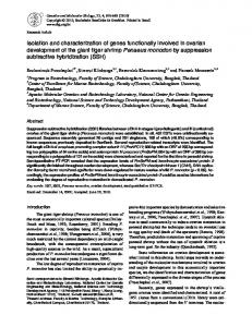
Fig. 1. Ball-and-stick representation of Mb. (A) Detailed view of residues in the wild-type protein in the region near the heme group where the metal ion binding site was designed. (B) The conformation of bound Mn2⫹ and its associated ligand interactions in the new metal ion binding site of the K45E兾K63E variant of Mb. Note that H113 is from a related molecule in the crystalline lattice and is expected to be replaced by a water molecule in solution. (C) The conformation of bound Cd2⫹ in the new metal ion binding site of the K45E兾K63E Mb variant. (D) A view of the natural metal binding site observed in Mb in structural studies of the Cd2⫹-bound form of K45E兾K63E Mb. In related Mn2⫹ studies this site remained unoccupied.
The dependence of peroxidase activity on pH was evaluated at 25°C between pH 5.0 and 8.0 with a mixed buffer system (5 mM Mes兾5 mM Mops兾5 mM TAPS兾95 mM NaCl) [TAPS, N-tris(hydroxymethyl)methyl-3-aminopropanesulfonic acid] with H2O2 (0.98 mM) and ABTS (0.19 mM) concentrations as indicated. Manganese peroxidase activities were determined by monitoring the rate of formation of the Mn(III) malonate complex at 270 nm in 0.1 M sodium malonate buffer, pH 6.2, at 25°C (10 mM MnSO4兾0.1 mM H2O2兾0.2 M Mb) (32). A unit of activity is defined as the amount of protein required to achieve an increase in A270 of 1 per min. Hunter et al.
Results Structures of the K45E and K45E兾K63E Variants. The global folds
of the polypeptide chains of these two variants are very similar to the fold found for wild-type horse heart Mb. For the K45E variant, the overall average deviation for all main-chain atoms is 0.13 Å. The largest structural differences are isolated to the disordered N and C termini of the polypeptide chain (G1, Q152, and G153) and the side chains of residues highly exposed to solution (D4, Q9, K56, L86, and T95). Furthermore, despite the loss of the hydrogen bond interaction between K45 and heme propionate-6 in the K45E variant, the positioning and conformation of the heme group is retained, with the average positional PNAS 兩 April 1, 2003 兩 vol. 100 兩 no. 7 兩 3649
Table 2. Equilibrium binding constants K1 pH Wild-type 6.92 K45E兾K63E 6.96 5.99 5.49 5.98
I, mM
M⫺1
17.2
—
17.2 17.2 17.2 100
1.28 (4) ⫻ 104 3.1 (5) ⫻ 103 5.2 (4) ⫻ 102 3.0 (1) ⫻ 102
K2 q
0.233 (5) 0.19 (4) 0.512 (3) 0.66 (2)
M⫺1
q
6.9 (2) ⫻ 102
0.226 (3)
6.2 (2) ⫻ 102 3.8 (8) ⫻ 102 — —
0.780 (7) 0.66 (2)
Equilibrium binding constants were determined from potentiometric titrations at various pH and I values (25°C) fit to either a one-site (wild-type) or a two-site (variant) model. The change in proton binding that is associated with Mn2⫹ binding (q) is also indicated. Uncertainties (standard deviations) are shown in parentheses.
Fig. 2. Potentiometric titration of the K45E兾K63E variant with MnSO4 at pH 7.0 and 17.2 mM KCl. The data were fit to one-site (A) and two-site (B) models, the two-site model giving the better fit.
deviation of all 43 heme atoms being 0.11 Å (Fig. 1). For the K45E兾K63E variant, the overall average value for main-chain differences from wild-type protein is 0.26 Å. As with the K45E variant, the largest structural deviations occur at the disordered polypeptide chain termini. However, in this structure other notable displacements involve residues 48–53 (C–D loop region, average deviation 1.0 Å), A57 (average deviation 0.74 Å), and L89 (average deviation 0.72 Å). The heme group of this variant also retains a conformation very similar to that observed in the wildtype structure. Heme pKa. The pKa of the distally coordinated H2O ligand of the
wild-type protein agrees well with the value reported previously (8.93 at 20°C; ref. 33). The corresponding values obtained for the K45E (9.6) and K45E兾K63E (10.1) variants indicate that the substitutions introduced in the current work stabilize the neutral (protonated) form of this ligand significantly.
Thermal Stability Measurements. Comparison of the far-UV CD spectra of the Mb variants with the spectrum of the wild-type protein indicated that no apparent change in secondary structure resulted from the amino acid substitutions introduced in the variants studied in the current work. The thermal stability of the K45E (Tm ⫽ 74.7 ⫾ 0.5°C) and K45E兾K63E (Tm ⫽ 73 ⫾ 1°C) variants is slightly lower than that of the wild-type protein (Tm ⫽ 79 ⫾ 1°C). Electrochemical Measurements. The midpoint potentials of the K45E and K45E兾K63E variants obtained by spectroelectrochemistry were 26 ⫾ 3 mV and 0 ⫾ 3 mV vs. the normal hydrogen electrode (NHE), respectively. The reduction potential of wild-type Mb under these conditions is 52 ⫾ 2 mV vs. NHE (all Nernst slopes were 59 ⫾ 2 mV). Potentiometric Analysis of Mn2ⴙ Binding. The binding of Mn2⫹
ions to wild-type Mb and the variant that exhibited the greatest 3650 兩 www.pnas.org兾cgi兾doi兾10.1073兾pnas.0636702100
affinity, K45E兾K63E, was quantified by potentiometric titration. The resulting titration curves were fit to either a one- or a twosite model (Fig. 2). The results obtained for wild-type Mb were best fit by a model that assumes the presence of a single binding site for Mn2⫹, and the association constant obtained from this analysis indicated that the affinity of this site for metal ions is relatively low. On the other hand, at pH 7.0 and low I (17.2 mM KCl), the results obtained for the double variant were best with a two-site model. The association constants obtained from this analysis (Table 2) indicated that the variant possesses the same low-affinity, endogenous site (defined by K2) as wild-type Mb and an additional, higher-affinity site (defined by K1) that exhibits a 20-fold greater affinity for Mn2⫹ than does the low-affinity site. Both binding sites exhibit strong pH and I dependences, with affinity decreasing as the pH is lowered and decreasing as I is raised. At sufficiently low pH and high I, the low-affinity site (K2) is not detectable by this method. Functional Linkages of Metal Ion Binding to the Variant. Because
the new binding site for metal ions is at the heme edge, the effect of Mn2⫹ binding on the midpoint potential and azide binding affinity of the K45E兾K63E variant was compared with their effect on these properties of wild-type Mb. Notably, addition of 50 mM MnSO4 (50 mM Hepes buffer, pH 7.0) had no influence on wild-type Mb (45 ⫾ 2 mV without Mn2⫹, 50 ⫾ 4 mV with Mn2⫹ added) but increased the potential of the variant from 9 ⫾ 2 mV to 40 ⫾ 5 mV, a value approaching that of the wildtype protein. In these experiments, the influence of MnSO4 on I was accounted for by making solutions of 50 mM in NaCl in the presence of Mn2⫹ and 200 mM in NaCl in its absence. The effect of metal ion binding on the affinity of the K45E兾 K63E variant for azide is similarly substantial. In this case, Kd for azide binding to wild-type Mb in the absence and presence of Mn2⫹ was found to be 28 ⫾ 1 and 26 ⫾ 1 M, respectively (50 mM Hepes buffer, pH 7.0). For the K45E兾K63E variant, Kd changed from 277 ⫾ 9 mM to 32 ⫾ 3 mM with the addition of azide. The contribution of MnSO4 to I was accounted for as in the electrochemical studies above. 1H NMR Analysis of Mn2ⴙ Binding. The paramagnetically shifted heme resonances of native metMb have been assigned for horse heart Mb (34) as indicated in Fig. 3. The 1H NMR spectra of both variants are highly similar to the spectrum of wild-type Mb except for small shifts in the resonances of the protons near the residues that differ in the sequences of the two proteins. Specifically, the 5-CH3, 6-␣, and 7- proton resonances shift downfield ⬇1 ppm and the 6-␣⬘ resonance shifts downfield ⬇2 ppm. The paramagnetic Mn2⫹ ion induces broadening of the resonances near the metal ion binding site through dipolar coupling be-
Hunter et al.
SPECIAL FEATURE
Table 3. Rates of Mn2ⴙ turnover and rate constants for Mb compound I formation (k1) for the Mb variants Rate of Mn2⫹ turnover* Variant Wild type K45E K45E兾K63E
Units兾 mol 14(2) 44(7) 51(8)
Reaction with H2O2†
Relative
k1, M⫺1䡠sec⫺1
Relative
1.0 3.1 3.6
4.3(2) ⫻ 1.27(5) ⫻ 103 1.05(7) ⫻ 103
1.0 3.0 2.4
102
Fig. 3. The 200-MHz 1H NMR spectra of the 100-ppm downfield region of K45E兾K63E Mb variant with 0 (trace A), 0.2 (trace B), 0.5 (trace C), and 0.8 (trace D) eq of MnSO4 [deuterated sodium phosphate buffer, I ⫽ 0.1 M, pH 7.0 (uncorrected pH meter reading), 20°C]. The proton resonances that were broadened on addition of Mn2⫹ are indicated by the dotted lines.
tween the Mn2⫹ ion and adjacent nuclei (35, 36). The effect of Mn2⫹ binding on the low-field region of the spectrum of the K45E兾K63E Mb variant is shown in Fig. 3. The greatest broadening is observed for the 6-␣ proton of the heme 6-propionate group, consistent with Mn2⫹ binding near this proton. Broadening is also observed for the 6-␣⬘ proton resonance, but it is difficult to quantify the magnitude of the broadening because of the similar chemical shift of the 4-␣ proton resonance of the 4-vinyl heme substituent. The 5-CH3 heme resonance also undergoes minor broadening. The spectrum of wild-type Mb exhibited little or no broadening of any of the heme resonances in the presence of various concentrations of Mn2⫹, consistent with the inability of this protein to bind divalent metal ions near the heme prosthetic group. Structures of the Manganese and Cadmium K45E兾K63E Complexes.
Soaking experiments to produce metal complexes with the K45E variant for structural studies were unsuccessful. However, for the K45E兾K63E variant, both metal ions were found to bind near heme 6-propionate and the substituted E45 side chain. Notably, the overall conformations of the unbound and bound forms of the K45E兾K63E variant were found to be very similar, with an overall average value for main-chain differences of 0.13 Å for each of the bound forms. Beyond the expected structural differences at the disordered N and C termini of the polypeptide chain, the largest positional shifts for both the Mn2⫹ and Cd2⫹ complexes involve the side chain of E45, which substantially reorients to accommodate metal binding. A related repositioning of heme 6-propionate is observed to occur as this group moves to optimize its metal–ligand interaction. As Fig. 1 shows, the ligands involved in binding to a Mn2⫹ or Cd2⫹ ion at the engineered metal binding site adjacent to the heme edge include the heme 6-propionate group, the side chain of the substituted E45, two water molecules, and the side chain Hunter et al.
of H113. This latter group is donated from a symmetry-related molecule in the crystalline lattice and in solution is likely replaced by a water molecule. Interestingly, a second binding site is also found in the Cd2⫹ complex that appears to be related to a metal ion binding site previously observed for sperm whale Mb (37, 38) and corresponds to the low-affinity site observed by potentiometric titration (Fig. 2 and Table 2). This site occurs near the side chains of H119 and D122, both of which take part in metal–ligand interactions (Fig. 1). Here, cadmium binding requires a small reorientation of the D122 side chain. Also taking part in ligand interactions at this site are two water molecules. No evidence of Mn2⫹ binding could be found in this site in structural studies. A summary of metal ligation partners and interaction distances at both metal ion binding sites is provided in Table 4, which is published as supporting information on the PNAS web site, www.pnas.org. Activity Assays. The ability of Mb to oxidize the bound Mn2⫹
ions was assessed by comparing the manganese peroxidase-like activity of wild-type Mb with that of the variant. These assays were carried out in the presence of malonate as described by Wariishi et al. (32) for activity assays of an authentic manganese peroxidase. These authors have proposed that malonate facilitates Mn3⫹ dissociation from the protein and stabilizes Mn(III) in aqueous solution through formation of the Mn3⫹–malonate complex (32). The rate constants for compound I formation determined as described above are shown in Table 3. It is apparent from these results that wild-type Mb oxidizes Mn2⫹ slowly and that the variant exhibits a 3-fold increase in the rate constant for reaction with hydrogen peroxide that accounts for the 3-fold increased activity in oxidation of Mn2⫹ exhibited by the variant. The pH dependence of the rate of ABTS oxidation by H2O2 catalyzed by wild-type Mb and the K45E and K45E兾 K63E variants was also determined over the pH range 4.5– 8.0. The extinction coefficient of the ABTS radical cation is not dependent on pH over this range. For both proteins, the rate of ABTS oxidation increased as the pH was decreased. Notably, the pH dependence of peroxidase activity is the opposite of the pH dependence of Mn2⫹ binding affinity. Discussion The present results establish that replacement of two surface lysyl residues located near the heme 6-propionate group of horseheart Mb with glutamyl residues introduces a functionally linked metal ion binding site into this protein with minimal structural perturbation. Whereas addition of Mn2⫹ to wild-type Mb has little effect on the reduction potential of the heme iron or on the affinity of the protein for azide, these properties of the K45E兾K63E variant exhibit an unambiguous dependence on Mn2⫹ binding. Both of these effects can be understood in terms PNAS 兩 April 1, 2003 兩 vol. 100 兩 no. 7 兩 3651
CHEMISTRY
Steady-state rate enhancements relative to wild-type are shown. *In 0.1 M malonate buffer, pH 6.2, 25°C, 10 mM MnSO4, 0.1 mM H2O2, 2 M Mb. †In Mes buffer (I ⫽ 0.1 M, pH 6.0, 25°C), 0.24 mM ABTS, 0.25– 4.0 mM H O , 2 2 2 M Mb.
of the electrostatic effects of the amino acid substitutions involved and the resulting charge neutralization that occurs on Mn2⫹ binding. In the case of the midpoint potential, the introduction of negative charges near the heme edge stabilizes metMb and thereby lowers the potential. Binding of Mn2⫹ neutralizes these negative charges and returns the potential to that of wild-type Mb. Similarly, the more negatively charged entrance to the heme pocket of the K45E兾K63E variant destabilizes the binding of the anionic ligand azide. Charge neutralization upon binding of Mn2⫹ restores affinity for azide to that of wild-type Mb. These observations demonstrate that introduction of a site for metal ion binding near a protein active site can be an effective means of creating new functional linkages in proteins through modulation of electrostatic properties of the active site. Electrostatic effects can also account for other functional changes observed in the variants studied here. The increased pKa of the distally coordinated water molecule in the active site of the variant presumably arises from electrostatic destabilization of a coordinated hydroxyl group that arises from the proximity of two carboxylate groups to the heme pocket in this protein. This interpretation is supported by the observation of a greater increase in this pKa for the double variant than for the single variant. The small decrease in thermal stability observed for the variants can be understood in terms of the loss of the hydrogen bond to the heme propionate upon replacement of K45 and a small destabilizing electrostatic interaction between the heme propionate group and E63. The role of hydrogen bonding interactions between the heme propionates and surface residues of Mb has been studied more extensively in previous studies (39, 40). While the new binding site captures some of the characteristics of the corresponding site in manganese peroxidase, notable differences are also apparent. For example, although the affinity of Mn2⫹ binding to the Mb double variant is ⬇20-fold greater at the newly created site than at the endogenous metal ion binding site, the affinity of the peroxidase for Mn2⫹ is about two orders of magnitude greater (29). Interestingly, however, the affinity of the peroxidase for Mn2⫹ at acidic pH, where catalytic activity of the enzyme is greatest, is the same order of magnitude as that for the variant studied here. This fact presumably reflects the balance that must be struck between a high affinity for metal ion binding and the need for facile metal ion release to achieve efficient catalytic turnover. Structurally, the site we have introduced into Mb provides only two metal ligands as opposed to
the four protein ligands provided in the corresponding site in manganese peroxidase. In the wild-type peroxidase enzyme, three carboxylate groups and the heme propionate group provide ligands to the bound metal ion (9). Removal of one of the ligands (D179) in manganese peroxidase significantly decreases the affinity for Mn2⫹ (41). The crystal structures of the metalbound forms of K45E兾K63E Mb identify only the heme 6-propionate and E45 as providing ligands to the metal ion; however, the additional negative charge at position 63 may help stabilize metal ion binding in that it helps to create a small negative patch near the heme 6-propionate group. The Mn2⫹-induced line-broadening observed in the NMR experiments indicates that the mechanism of metal ion binding to the variant in solution is similar if not identical to that observed in the crystal. The peroxidase activity of Mb has been recognized since at least the 1950s (reviewed in ref. 42), and considerable interest has arisen in recent years in increasing this activity through the application of protein engineering technology (14, 42, 43). Although this objective was not a goal of the current work, it is noteworthy that amino acid substitutions introduced in the two variants studied here increase the peroxidase activity ⬇3-fold and that this increase arises primarily through an increased rate of formation of the presumed compound I intermediate (k1, Table 3). Because the metal ion binding site introduced in the current work is in many ways analogous to that present in manganese peroxidase, the possible manganese peroxidase activity of these variants was also of interest. As far as we are aware, wildtype Mb has not been reported previously to possess manganese peroxidase activity, so although the activity of the wild-type protein is quite limited, it is interesting to note that this activity is detectable. Significantly, these variants also exhibited a 3-fold increase in manganese peroxidase activity. This observation suggests that the peroxidase and manganese peroxidase activities of Mb are closely linked and that incorporation of the current amino acid substitutions into a variant with significantly enhanced peroxidase activity with ABTS or other organic substrates may reasonably be expected to exhibit a significant enhancement in manganese peroxidase activity.
1. Todd, R. J., Van Dam, M. E., Casimiro, D., Haymore, B. L. & Arnold, F. H. (1991) Proteins 10, 156–161. 2. Suh, S. S., Haymore, B. L. & Arnold, F. H. (1991) Protein Eng. 4, 301–305. 3. Hellinga, H. W. & Richards, F. M. (1991) J. Mol. Biol. 222, 763–785. 4. Hellinga, H. W. (1998) Folding Des. 3, R1–R8. 5. Benson, D. E., Wisz, M. S. & Hellinga, H. W. (2000) Proc. Natl. Acad. Sci. USA 97, 6292–6297. 6. Hellinga, H. W., Caradonna, J. P. & Richards, F. M. (1991) J. Mol. Biol. 222, 787–803. 7. Marvin, J. S. & Hellinga, H. W. (2001) Proc. Natl. Acad. Sci. USA 98, 4955–4960. 8. Benson, D. E., Haddy, A. E. & Hellinga, H. W. (2002) Biochemistry 41, 3262–3269. 9. Sundaramoorthy, M., Kishi, K., Gold, M. H. & Poulos, T. L. (1994) J. Biol. Chem. 269, 32759–32767. 10. Yeung, B. K., Wang, X., Sigman, J. A., Petillo, P. A. & Lu, Y. (1997) Chem. Biol. 4, 215–221. 11. Wilcox, S. K., Putnam, C. D., Sastry, M., Blankenship, J., Chazin, W. J., McRee, D. E. & Goodin, D. B. (1998) Biochemistry 37, 16853–16862. 12. Mester, T. & Tien, M. (2001) Biochem. Biophys. Res. Commun. 284, 723–728. 13. Bonagura, C. A., Sundaramoorthy, M., Bhaskar, B. & Poulos, T. L. (1999) Biochemistry 38, 5538–5545. 14. Wan, L., Twitchett, M. B., Eltis, L. D., Mauk, A. G. & Smith, M. (1998) Proc. Natl. Acad. Sci. USA 95, 12825–12831. 15. Hildebrand, D. P., Lim, K. T., Rosell, F. I., Twitchett, M. B., Wan, L. & Mauk, A. G. (1998) J. Inorg. Biochem. 70, 11–16. 16. Guillemette, J. G., Matsushima-Hibiya, Y., Atkinson, T. & Smith, M. (1991) Protein Eng. 4, 585–592. 17. Lloyd, E. & Mauk, A. G. (1994) FEBS Lett. 340, 281–286. 18. Zoller, M. J. & Smith, M. (1987) Methods Enzymol. 154, 329–350. 19. Rafferty, S. P., Guillemette, J. G., Smith, M. & Mauk, A. G. (1996) Inorg. Chim. Acta 242, 171–177. 20. Reid, L. S., Lim, A. R. & Mauk, A. G. (1986) J. Am. Chem. Soc. 108, 8197–8201. 21. Pladziewicz, J. R., Meyer, T. J., Broomhead, J. A. & Taube, H. (1973) Inorg. Chem. 12, 639.
22. Maurus, R., Overall, C. M., Bogumil, R., Luo, Y., Mauk, A. G., Smith, M. & Brayer, G. D. (1997) Biochim. Biophys. Acta 1341, 1–13. 23. Evans, S. V. & Brayer, G. D. (1988) J. Biol. Chem. 263, 4263–4268. 24. Higashi, T. (1990) J. Appl. Crystallogr. 23, 253–257. 25. Sato, M., Yamamoto, M., Imada, Y., Katsube, Y., Tanaka, N. & Higashi, T. (1992) J. Appl. Crystallogr. 25, 348–356. 26. Bru ¨nger, A. T. (1990) X-PLOR, A System for Crystallography and NMR (Yale Univ., New Haven, CT), Version 2.1. 27. Mauk, M. R., Barker, P. D. & Mauk, A. G. (1991) Biochemistry 30, 9873–9881. 28. Mauk, M. R., Kishi, K., Gold, M. H. & Mauk, A. G. (1998) Biochemistry 37, 6767–6771. 29. Mauk, M. R., Ferrer, J. C. & Mauk, A. G. (1994) Biochemistry 33, 12609–12614. 30. Inubushi, T. & Becher, E. D. (1983) J. Magn. Reson. 51, 128–133. 31. Dunford, H. B. (1991) in Peroxidases in Chemistry and Biology, eds. Everse, J., Everse, K. E. & Grisham, M. B. (CRC Press, Boca Raton, FL), pp. 1–24. 32. Wariishi, J., Valli, K. & Gold, M. H. (1992) J. Biol. Chem. 267, 23688–23695. 33. Antonini, E. & Brunori, M. (1971) Hemoglobin and Myoglobin in Their Reactions with Ligands, Frontiers of Biology (Elsevier, New York), Vol. 21, p. 47. 34. Luo, Y., Peyton, D. H. & Yee, S. (1998) Biochim. Biophys. Acta 1388, 66–76. 35. Banci, L., Bertini, I., Bini, T., Tien, M. & Turano, P. (1993) Biochemistry 32, 5825–5831. 36. Bertini, I. & Lucninat, C. (1986) NMR of Paramagnetic Molecules in Biological Systems (Benjamin兾Cummings, Menlo Park, CA). 37. Banaszak, L. J., Watson, H. C., Kendrew, J. C. (1965) J. Mol. Biol. 12, 130–137. 38. Van Dyke, B. R., Bakan, D. A., Glover, K. A., Hegenauer, J. C., Saltman, P., Springer, B. A. & Sligar, S. G. (1992) Proc. Natl. Acad. Sci. USA. 89, 8016–8019. 39. Hunter, C. L., Lloyd, E., Eltis, L. D., Rafferty, S. P., Lee, H., Smith, M. & Mauk, A. G. (1997) Biochemistry 36, 1010–1017. 40. Hunter, C. L., Mauk, A. G. & Douglas, D. J. (1997) Biochemistry 36, 1018–1025. 41. Kusters-van Someron, M., Kishi, K., Lundell, T. & Gold, M. H. (1995) Biochemistry 34, 10620–10627. 42. Raven, E. L. & Mauk, A. G. (2001) Adv. Inorg. Chem. 51, 1–49. 43. Ozaki, S., Roach, M. P., Matsui, T. & Watanabe, Y. (2001) Acc. Chem. Res. 34, 818–825.
3652 兩 www.pnas.org兾cgi兾doi兾10.1073兾pnas.0636702100
This work was supported by the Protein Engineering Networks of Centres of Excellence (G.D.B. and A.G.M.), Canadian Institutes of Health Research Grant MT-14021 (to A.G.M.), a Medical Research Council of Canada Studentship (to R.M.), and a Canada Research Chair (to A.G.M.).
Hunter et al.