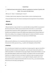
Neodymium magnet. (b) Magnetic strength contour (A/m) under external magnetic source with the surface flux of 0.4 T. Ref
A microfluidic system for capturing malaria-infected bloodcells using an array of nickel structures P. Noosawad, A. Pimpin and W. Srituravanich Department of Mechanical Engineering, Faculty of Engineering, Chulalongkorn University
Introduction & Background Treatment and prevention of malaria, a disease having an anopheles mosquito as a carrier, have become a severe medical issue to many countries in a tropical zone. A diagnosis technology plays an important role to achieve the treatment and prevention including the elimination of malaria. Among various techniques, an employment of a magnetic force for quick diagnosis was extensively studied due to malaria-infected red blood cells exhibit a unique magnetic property that is different from healthy blood cells. However, the magnitude of the exerted magnetic force is very small and typically effective within a small area close to a magnetic source. For few years, our research group has studied and developed a lab-on-a-chip technology to tackle this issue[1].
(a) Velocity contour (m/s) and streamlines at the flow rate of 0.2 µl/min
Objective This study aims to improve a system design to efficiently capture infected red blood cells by proposing the additional nickel microstructures inside the microchannel. The system design was conducted using COMSOL MULTIPHYSICS®.
Computational Methods Figure 1 shows a 2D computation domain consisting of two magnets and an array of 200 x 200 µm2 nickel microstructures inside a microchannel. An array of nickel structures was aligned in a zigzag pattern, and two magnets were placed in a counterpole manner. Magnets Nickel microstructures N S S Microchannel N Fig.1 An array of nickel microstructures sandwiched between two magnets, whose dimensions are 20 x10x10 mm2. A magnetic type is Neodymium magnet.
Results Microfluidics and AC/DC module with 2-D simulation were employed in order to compare the magnitude of both hydrodynamic-drag and magnetic force at a certain position around the structures for different shapes of nickel structures. Figure 2a and b exhibit the velocity of fluid flow and magnetic strength around the square, triangular, circular and modifiedsquare microstructure. In general, at the corners of microstructures, they tend to be locations where the magnitude of magnetic force as well as hydrodynamic-drag force is large. Therefore, the effects of magnetic force on the capturing efficacy are not strong. For the modified-square structures, the locations where the two forces were largely induced could be slightly relocated and separated apart from each other.
(b) Magnetic strength contour (A/m) under external magnetic source with the surface flux of 0.4 T. Fig. 2 Computational results with different shapes of nickel microstructures
Conclusions The additional nickel microstructures inside the microchannel were proposed so that the opportunity to capture the infected blood cells under the influence of magnetic forces increases. One important issue is that the placement of microstructures should be at a proper and right position in order to maximize the magnitude of magnetic force and minimize the magnitude of hydrodynamic-drag force exerting on the targeted cells. For this reasons, this study aims to examine the effects of microstructure shape and alignment that could provide the desired condition.
Acknowledgements This work was funded by Chulalongkorn University through the Chulalongkorn Academic Advancement into Its 2nd Century Project (Smart Medical Device) from Chulalongkorn University.
References [1] Kasetsirikul S., Srituravanich W., Piyaviriyakul P., Pimpin A., (2017), “Separation of Magnetic Particles Using an Array of Magnets —A Model of a Separation Device for Malaria-Infected Blood Cells,” Sensors and Materials, Vol. 29, No. 3, pp. 281–291.