recognition sites from TUL, PH, HTN, DOB and SEO hantaviruses. However, this panel of MAbs was unable to distinguish the PUU serotype from KBR, ...
Journal of General Virology (1998), 79, 2603–2614. Printed in Great Britain ...................................................................................................................................................................................................................................................................................
Isolation and characterization of Puumala hantavirus from Norway : evidence for a distinct phylogenetic sublineage AI ke Lundkvist,1, 2 Donna Wiger,3 Jan Ho$ rling,2, 4 Katarina Brus Sjo$ lander,1 Angelina Plyusnina,5 Reidar Mehl,6 Antti Vaheri5 and Alexander Plyusnin5 1
Swedish Institute for Infectious Disease Control, S-105 21 Stockholm, Sweden Microbiology and Tumour Biology Centre, Karolinska Institute, S-171 77 Stockholm, Sweden 3 Norwegian Medicines Control Authority, Sven Oftedalsvei 6, N-0950 Oslo, Norway 4 National Institute of Public Health, PO Box 4404 Torshov, N-0403 Oslo, Norway 5 Department of Virology, Haartman Institute, PO Box 21, FIN-00014 University of Helsinki, Finland 6 Pharmacia and Upjohn, S-112 87 Stockholm, Sweden 2
Puumala (PUU) hantavirus is the aetiological agent of nephropathia epidemica (NE), a mild form of haemorrhagic fever with renal syndrome, which occurs in Fennoscandia, central Europe and Russia. In Norway, NE-like disease has been reported since 1946 and about 50 cases are diagnosed annually ; however, the causative agent has not been characterized. In this study, a virus originating from bank voles (Clethrionomys glareolus) trapped near the town of Eidsvoll (Akershus county) was isolated and passaged in laboratory-bred bank voles. The bank vole strain was identified as a PUU virus by serological typing and by sequence analysis of the S and M gene segments. For comparison, complete or partial S sequences were determined for wild-type
Introduction Hantaviruses, members of the family Bunyaviridae, are known to cause two serious and often fatal human diseases : haemorrhagic fever with renal syndrome (HFRS) and hantavirus pulmonary syndrome (HPS). Small mammals, mainly rodents, are the natural reservoirs of hantaviruses and transmission to humans occurs via aerosolized animal excreta (Plyusnin et al., 1996 a). Hantaan (HTN), Seoul (SEO), Dobrava (DOB) and Puumala (PUU) viruses are known to cause HFRS, Author for correspondence : AI ke Lundkvist (mail to address 1). Fax 46 8 7351424. e-mail Ake.Lundkvist!smi.ki.se The nucleotide sequence data (consensus sequences) reported in this paper have been deposited in the EMBL, GenBank and DDBJ nucleotide sequence databases under the accession numbers AJ223367–AJ223383.
0001-5628 # 1998 SGM
PUU strains from five locations in Sweden, two inhabited by the southern variant of bank vole present in Fennoscandia, and three by the northern variant. Phylogenetic analysis showed that Norwegian PUU strains are clustered together with Swedish strains from the first group forming a wellsupported sublineage within the PUU genotype, distinct from other sublineages from northern Sweden, Finland, Russia and France. The results are consistent with the view of a complex evolutionary history of PUU strains in post-glacial Fennoscandia. Analyses of the current collection of nucleotide sequences suggest that PUU is the most variable genotype of the known hantaviruses.
characterized by fever, renal failure and, in severe cases, haemorrhagic manifestations (Antoniadis et al., 1996 ; Kanerva et al., 1998). Sin Nombre (SN) and related viruses cause HPS in the Americas (Nichol et al., 1996 ; Padula et al., 1998). HPS is characterized by acute respiratory dysfunction with a mortality of approximately 50 %. In addition to the pathogenic hantaviruses, there are at least six distinct hantaviruses at present not associated with human disease : Prospect Hill (PH), Thailand (THAI), Thottapalayam, Khabarovsk (KBR), Tula (TUL) and Topografov (TOP) viruses (Plyusnin et al., 1996 a, b). Several other hantaviruses have been characterized, mainly genetically from rodent samples, but have not yet been isolated in cell culture or animals (Nichol et al., 1996 ; Plyusnin et al., 1996 a). In common with other members of the Bunyaviridae, hantaviruses have a single-stranded, tripartite RNA genome, packed in enveloped helical nucleocapsids. The negative-
CGAD
AI . Lundkvist and others
stranded genome of approximately 12 kb encodes four structural proteins : the RNA polymerase, a glycoprotein precursor (GPC), cotranslationally processed into two envelope proteins (G1 and G2), and a nucleocapsid protein (N), respectively (Schmaljohn, 1996). Geographically isolated rodent populations allow comparison of the genetic properties of hantavirus strains which have been separated long ago and have been evolving independently. Fennoscandia may constitute an optimal region to study both faunal history (Siivonen, 1982) and the evolution of hantaviruses co-evolving with their natural hosts. The contact zone between the two major phylogeographical groups of the bank vole (Clethrionomys glareolus), the natural host of PUU, which recolonized Sweden from two directions after the last glacial period (approx. 10 000 years ago), is still only about 50 km wide (Jaarola & Tegelstro$ m, 1995). Notably, PUU strains circulating on the two sides of the Swedish bank vole population-border belong to two different genetic lineages (Ho$ rling et al., 1996 b). Similarly, geographically separated PUU strains from Finland and from pre-Ural Russia form two lineages (Plyusnin et al., 1995 a) which most probably originate from different ancestors presented in separate glacial refugia of bank voles. Nephropathia epidemica (NE)-like disease, a mild form of HFRS, has been known to occur in Norway for several decades and serological confirmation of human cases using hantavirus antigen has been routinely used since 1981 (Traavik et al., 1984). Previous studies have identified hantavirus-reactive antibodies and}or antigen in four rodent species : Clethrionomys glareolus, C. rutilus, C. rufocanus and Apodemus sylvaticus, and suggested C. glareolus as the main NE virus reservoir (Traavik et al., 1984 ; Sommer & Traavik, 1985). However, the causative agent of NE in Norway has not been identified. Like other hantaviruses, PUU virus has proven difficult to isolate and to date only a few isolates have been reported from Western Europe or Fennoscandia. This report describes the first serological and genetic characterization of PUU virus from Norway.
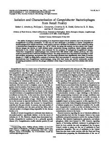
Methods + Trapping of small rodents. Bank voles were collected with live traps at three locations in Norway. Trapping locations, selected by the previous occurrence of serologically confirmed NE cases, were numbered from north to south (Fig. 1). Animal 1109 was trapped in November 1986 near Kvikne, about 10–15 km into the mountains west of the town of Vinstra (locality 7) in the county of Oppland. Animals 1124, 1138 and 1192 were trapped in November 1987 north of Eidsvoll (locality 8) in the county of Akershus. The animals designated C11 and C12 were trapped in October 1995 near Tangvall about 1 km south-east from the town of Søgne (locality 9) in Vest Agder county. In addition, samples from bank voles trapped in Sweden in 1994–95 (Ho$ rling et al., 1996 b) were analysed (Fig. 1, localities 1–6). + Virus strains. Hantaviruses, PUU strains Sotkamo (Vapalahti et al., 1992), 83-L20 (Ho$ rling et al., 1995) and Kazan-E6-I (Lundkvist et al., 1997 a), KBR virus (Ho$ rling et al., 1996 a), TUL virus (Vapalahti et al.,
CGAE
1996) and HTN virus strain 76-118 (Lee et al., 1978), were passaged on monolayers of Vero E6 cells in Eagle’s MEM supplemented with 2 % foetal calf serum (FCS), 2 mM -glutamine and antibiotics. Virus stocks were kept at ®70 °C until used. + Antibodies. Generation and characterization of PUU and TUL virus-specific monoclonal antibodies (MAbs) have been described elsewhere (Lundkvist et al., 1991, 1993, 1996 b ; Lundkvist & Niklasson, 1992). Large scale MAb production was performed by culturing hybridomas in roller bottles followed by purification on protein G–Sepharose as previously described (Lundkvist et al., 1991). + Virus isolation. Four- to ten-week-old hantavirus-free bank voles, from a laboratory colony established from animals trapped in Sweden (Lundkvist et al., 1996 a), were inoculated subcutaneously with hantavirus antigen}RNA-containing lung suspensions from trapped bank voles. The rodents were sacrificed 21 days later and analysed for the presence of hantavirus antigen, RNA, G2-reactive and neutralizing antibodies. + Immunoassays. Bank vole serum samples were examined for the presence of PUU-reactive antibodies by an immunofluorescence assay (IFA) and for PUU G2-reactive antibodies by an ELISA as described previously (Wiger et al., 1991 ; Lundkvist et al., 1997 a). Neutralizing antibody responses against hantavirus were analysed by focus reduction neutralization test (FRNT) (Niklasson et al., 1991 ; Lundkvist et al., 1997 b). Lung samples from naturally or experimentally infected bank voles were examined for the presence of PUU virus N by hantavirus-antigen ELISA and by immunoblotting (WB) as previously described (Lundkvist et al., 1995). The isolated virus strain was analysed antigenically and compared to other hantaviruses by a panel of six MAbs by an ELISA as previously described (Lundkvist et al., 1996 b). + RT–PCR, cloning and sequencing. RT–PCR of the entire S segment and the proximal part (nt 2775–3657) of the M segment was performed as described previously (Plyusnin et al., 1994) with one modification : RT for the M-segment sequences was performed in the presence of amplification primers. The PCR products were purified, cloned, and sequenced manually or automatically. RT–PCR of partial S (nt 799–1106) and M (nt 2147–2632) segments was performed as described earlier (Plyusnin et al., 1997). Amplicons were purified and sequenced automatically. Each sequence was obtained either from at least three individual cDNA clones, or two independent RT–PCR reactions. The following PUU strains were not isolated and were characterized by sequencing only : PUU}Eidsvoll}1138Cg}87 (Eids1138), PUU} Eidsvoll}1124Cg}87 (Eids1124), PUU}Vinstra}1109Cg}86 (Vins1109), PUU}Gra$ smark}76Cg}95 (Gra$ s67), PUU}Hundberget}36Cg}94 (Hund36), PUU}Hundberget}48Cg}94 (Hund48), PUU}Lungvik} Cg72}95 (Lung72), PUU}Mellansel}Cg47}94 (Mell-47), PUU} Mellansel}49Cg}94 (Mell-49), PUU}Solleftea/ }3Cg}95 (Soll-3), PUU} Solleftea/ }6Cg}95 (Soll-6), PUU}Sundsvall}126Cg}95 (Sund126), PUU}Sundsvall}255Cg}95 (Sund255), PUU}Tavelsjo$ }81Cg}94 (Tav81), PUU}Vindeln}4Cg}94 (Vind4) and PUU}Vindeln}8Cg}94 (Vind8). + Phylogenetic analysis. The PHYLIP program package (Felsenstein, 1993) was used to generate 200 bootstrap replicates of the sequence data (Seqboot). Distance matrices were calculated using Kimura’s two-parameter model (Dnadist) and analysed by the Fitch– Margoliash tree fitting algorithm (Fitch). The bootstrap support percentages of particular branching points were calculated from these trees (Consense).
PUU virus in Norway
Results Screening of bank voles
Five bank voles, collected at three different locations in Norway in 1986, 1987 and 1995, respectively (Fig. 1), were hantavirus-antibody-positive when analysed by IFA. All three trapping locations were situated south of the estimated bank vole population border. Lung samples were analysed for the presence of viral N antigen by WB and for viral RNA by RT–PCR (data summarized in Table 1). N antigen was detected in all five antibody-positive animals, which were also positive for viral RNA when analysed by a highly sensitive nested RT–PCR, amplifying nt 799–1106 of the S segment. The entire S segment could only be amplified from two animals, while amplification of the partial M segment (nt 2147–2632) was unsuccessful in all cases. Isolation of Norwegian PUU virus from laboratory-bred bank voles
Pairs of hantavirus-free laboratory-bred voles were inoculated with lung suspensions from five animals positive for hantavirus antibody}antigen}RNA. Two pairs of the animals (injected with g1109 and g1124, respectively) developed high levels of PUU G2-reactive antibodies while only one of the pairs, injected with g1124, developed detectable levels of N antigen as determined by ELISA and WB. Lung samples from the PUU G2-antibody-positive animals were further passaged to new animals ; only the g1124 (passage 1)-injected animals were G2-antibody}N-antigen-positive when analysed 21 days after inoculation. This strain was designated PUU}Eidsvoll} 1124v (Eid1124v). All attempts to isolate virus in Vero E6 cell culture, either directly from the original lungs or from experimentally infected animals, were unsuccessful. The authenticity of the bank vole strain (Eid1124v) was later confirmed by comparison of its S segment sequence (nt 799–1106) with the one obtained directly from lung tissue of trapped bank vole g1124 (wild-type strain PUU} Eidsvoll}1124Cg}87) ; the sequences were identical. Serological and antigenic characterization
The N antigens of the Eid1124v strain and hantavirus prototype strains were compared by an antigen-capture ELISA using a selected panel of MAbs (Table 2). The results revealed an identical epitope pattern for Eid1124v and PUU Sotkamo. The pattern differed in at least two recognition sites from TUL, PH, HTN, DOB and SEO hantaviruses. However, this panel of MAbs was unable to distinguish the PUU serotype from KBR, a serotype previously shown to be closely related to PUU (Ho$ rling et al., 1996 a). An FRNT comparison with antisera from the experimentally infected bank voles showed similar high neutralizing titres to the Finnish Sotkamo, Swedish Vindeln83-L20 and Russian
Fig. 1. Map of Fennoscandia and Denmark. The contact zone between the two bank vole populations with different recolonization histories and the trapping localities (1–9) are marked. 1, Hundberget ; 2, Tavelsjo$ /Vindeln ; 3, Mellansel ; 4, Solleftea/ /Lungvik ; 5, Sundsvall ; 6, Gra$ smark ; 7, Vinstra ; 8, Eidsvoll ; 9, Søgne.
Kazan-E6-I PUU strains, while the reactivities to other hantaviruses, including KBR, TUL and HTN, were lower or absent (Table 3).
Genetic analysis
For genetic characterization of the bank vole strain Eid1124v, PCR products which contained the full-length cDNA copy of the S segment and the proximal part (nt 2775–3657) of the M segment were cloned and sequenced. The S segment sequence of Eid1124v (including the 22 terminal nucleotides that were involved in the primer’s annealing, and, therefore, not determined directly) contained a 5«NCR of 42 nt, an ORF for the N protein (433 aa), followed by a 3«NCR of 504 nt. Also, in the 1 reading frame, there was an additional ORF encoding a putative NSs protein of 90 aa, as described earlier for PUU and other hantaviruses (Plyusnin et al., 1996 a). The deduced sequence of the N protein contained five cysteine residues. Three of them (aa 203, 244 and 319) are common to all hantaviruses, the fourth (aa 314) is present in all but HTN-like viruses, and the fifth (aa 323) can be seen in all but SN-like viruses. The M sequence contained nt 2775–3462 of the coding region followed by 195 nt of the 3«NCR (in the plus-sense strand). This part of the hantaviral M segment has previously
CGAF
AI . Lundkvist and others
Table 1. Screening of trapped Clethrionomys glareolus RNA (RT–PCR) Locality/animal (year of collection)
Antibody (IFA)*
Antigen (WB)
Partial S (nt 799–1106)
Complete S
Partial M (nt 2147–2632)
" 20
®
®
" 20 " 20 " 20
®
® ® ®
" 20 ! 20
®
®
® ®
® ®
Vinstra ‘ locality 7 ’ 1109 (86) Eidsvoll ‘ locality 8 ’ 1124 (87) 1138 (87) 1192 (87) Søgne ‘ locality 9 ’ C11 (95) C12 (95)
* Reciprocal titre : , positive ; ®, negative.
Table 2. Antigenic comparison of hantavirus N in a MAb antigen-capture assay
Table 3. Serological characterization of Norwegian PUU Reciprocal FRNT titre
Capture antibody* Antigen Eid1124v PUU}Sotk KBR TUL PH HTN DOB SEO
3H9
5E1
2E12
6A6
3D3
® ® ® ®
® ® ® ®
® ® ® ® ® ®
® ® ® ® ® ® ®
* , Absorbance obtained with antigen-capture by MAb " 3¬ the background absorbance obtained with negative IgG.
been shown to be representative for genetic analysis (Plyusnin et al., 1994, 1995 b). The deduced sequence of the G2 protein (aa 931–1148) contained all 11 cysteine residues common to all hantaviruses as well as an additional cysteine present in all PUU strains. Also, it contained the putative N-glycosylation site (Asn-Val-Thr), aa 947–949, that is conserved in all hantaviruses. The 3«NCR of the M segment was 195 nt in length, the same as in almost all sequenced PUU strains. Data on the pairwise comparison of the Eid1124v S}N and M}G2 sequences with those of other hantaviruses (Table 4) correlated well with the results of the serological analysis. The highest identity was observed with other PUU strains (about 80 % or more at the nucleotide level, and 85 % or more at the amino acid level). For other hantavirus genotypes, the percentage identities were substantially lower : the best scores
CGAG
Virus PUU Vindeln83-L20 PUU Sotkamo PUU Kazan-E6-I KBR TUL HTN
Serum : 1109-p1-b 1124-p1-b 1124-p2-b 320 320 1280 ! 80 80 ! 80
1280 2560 1280 160 160 ! 80
80 80 80 ! 80 ! 80 ! 80
were observed for KBR, while TUL and related viruses (data on PH and ILV are not shown) were less similar ; SN and related viruses (NY, BCC, BAY and ELMC – not shown) gave even lower identities, and HTN, DOB and SEO were the most distantly related. As PUU strains from Sweden were expected to be the closest relatives of the Norwegian strain, the complete S segment sequences were also determined for two strains from Solleftea/ , (PUU}Solleftea/ }3Cg}95 and PUU}Solleftea/ } 6Cg}95), located immediately south of the bank vole population border, and for two strains from Mellansel (PUU} Mellansel}47Cg}94 and PUU}Mellansel}49Cg}94), as well as for strains from Hundberget (PUU}Hundberget}Cg36}94) and Tavelsjo$ (PUU}Tavelsjo$ }81Cg}94), all three locations situated north of the population border (Fig. 1). Of these strains, the ones from Solleftea/ showed the highest level of identity with the sequences of the Norwegian PUU strain (87–88 % at the nucleotide level and 98 % at the amino acid level ; Table 4). Comparison of the S segment 3«NCR sequences
PUU virus in Norway
Table 4. Percentage identities between sequences of the strain PUU/Eidsvoll/1124v and those of other hantaviruses Hantavirus type* PUU KBR TUL SN DOB HTN SEO Sotk B-1820 Vind Hund36 Mell-47 Soll-3 Soll-6 Paris Mf43 5302v H10 Slov 76-118 SR-11 S segment† N protein (aa1– 433) M segment‡ G2 protein (aa 931–1148)
85±4 96±1 80±0 86±5
82±9 96±1 79±8 87±3
84±5 96±5 79±5 85±6
84±4 96±8
84±7 96±3
88±1 98±0
87±1 98±0
83±3 97±2 81±2 91±3
77±9 87±3
72±4 79±7 74±2 79±0
68±2 71±8 68±7 69±9
61±2 61±0 60±1 58±1
62±5 61±9 59±4 57±2
62±7 63±3 61±9 58±1
* For abbreviations, see legend to Fig. 3. † Coding region (complete), nt 43–1344. ‡ Coding region (part), nt 2775–3462. All numbering is according to the sequences of the prototype PUU Sotkamo strain. , Not determined.
revealed substantially lower percentage identities but showed the same pattern : Solleftea/ strains were the closest relatives (identity 84 %) while the others were more distantly related (71–78 %). Multiple alignment of the NCRs located downstream of the main ORFs in the M and S segments (Fig. 2) clearly showed that these regions represent the most variable parts of the PUU genome. Sequence differences within the S-3«NCR of different PUU strains can reach 30 %, and within the M-3«NCR 37 %, which is the highest level of intra-genotype diversity registered for all hantaviruses. Genetic variations within Norwegian PUU strains
To estimate the range of genetic diversity within Norwegian PUU strains, the complete S sequence was determined for the RT–PCR product obtained directly from the lung tissue of bank vole g1138 (strain Eidsvoll} 1138Cg}87), and partial S sequences (nt 799–1106) were determined for strains Vinstra}1109Cg}86 and Eidsvoll} 1124Cg}87. Sequences of the partial S PCR-amplicons from bank voles g1192, locality Eidsvoll, and gC11, locality Søgne, could not be recovered. The complete S sequences from strains Eid1124v and Eid1138Cg, which originated from the same population of bank voles, differed only at nine positions (identity 99±5 %). All seven mutations in the coding region were silent ; within the 3«NCR two deletions of single bases were found in the Eid1124v strain. The partial nucleotide sequence of strain Vinstra}1109Cg}86, which originated from another locality, 160 km north-west from Eidsvoll, differed from those of the Eidsvoll strains in 30–32 positions (identity 87±9–88±6 %). Deduced amino acid sequences of the N protein differed at four positions (identity of 95±4 %), and all mutations were conservative (K#'& ! R, V#') ! I, M#(" ! I and S$!# ! N). In short, the closer the geographical origins of the Norwegian PUU strains, the higher were their sequence identities.
Phylogenetic analysis of Norwegian PUU
On a phylogenetic tree based on the complete coding region of the S segment (Fig. 3 a), strains Eid1124v and Eid1138Cg were located together with other strains belonging to the PUU clade sharing with all of them a common ancient ancestor. Within the PUU clade, the Norwegian strains, together with strains from Solleftea/ in Sweden, formed a distinct sublineage, separated from the other four sublineages described so far from Finland (strains Sotkamo, Puumala, Evo12–15 and Virrat), Sweden, north of the bank vole population border (NPB) (strains Mellansel-47 and -49, Vindeln-L20, Tavelsjo$ -81, Hundberget-36 and ‘ Vranica ’} Ha$ llna$ s), Russia (strains Bashkiria}Cg1820 and P360, and Udmurtia 338, 458, 444 and 894), and France (strain Paris 9013). While the monophyletic origin of strains belonging to each of the sublineages was well supported, the relationships between sister PUU sublineages were poorly resolved as shown by the weak bootstrap probabilities (34 and 60 %). The phylogenetic tree calculated for the partial sequences of the S segment (Fig. 3 b), which included additional strains,† showed a similar branching order : Norwegian PUU strains from Eidsvoll and Vinstra are clustered together with the Swedish strains from Solleftea/ , Sundsvall, Gra$ smark and Lungvik, all localities inhabited by the southern-type bank voles (Ho$ rling et al., 1996 b). As in the previous tree, wellsupported sister sublineages from Finland, Russia and
† Partial S segment sequences (nt 799–1106) were determined for the following additional Swedish PUU strains : PUU}Vindeln}4Cg}94, PUU}Vindeln}8Cg}94 and PUU}Gra$ smark}76Cg}95 (localities shown in Fig. 1). Partial S segment sequences for the following strains have been determined previously : PUU}Hundberget}48Cg}94, PUU}Sundsvall}126Cg}95, PUU}Sundsvall}255Cg}95 and PUU}Lungvik}72Cg}95 (Ho$ rling et al., 1996 b).
CGAH
CGAI
(b)
(a)
AI . Lundkvist and others
PUU virus in Norway
Sweden (NPB), showed an uncertain pattern of interrelationships (bootstrap probabilities from 26–53 % were observed). On the tree calculated for the M segment sequences (Fig. 3 c), the Norwegian variant of PUU again represented a sublineage distinct from other sister sublineages from France (strain Paris 90-13), Sweden}NPB (strains Ha$ llna$ s and Vindeln-L20), Finland (strain Sotkamo) and Russia (strains from Udmurtia and Bashkiria). The results suggested an absence of reassortant events such as an import of S or M segments from another PUU sublineage in the evolutionary history of strain Eid1124v. Taken together, the results of the phylogenetic analysis support a monophyletic origin of the Norwegian PUU strains from Eidsvoll and Vinstra together with the Swedish PUU strains originating south of the population border (SPB).
Discussion Isolation and serological characterization of PUU from Norway
NE, caused by PUU virus, has been known to be endemic in Norway since 1946 and approximately 50 cases are reported annually (Knutrud, 1946 ; Traavik et al., 1983 ; D. Wiger and others, unpublished). Epidemiological studies have suggested C. glareolus as the main reservoir of the hantavirus causing NE in Norway, although hantavirus antibodies}antigen have also been detected in C. rutilus, C. rufocanus and A. sylvaticus (Sommer & Traavik, 1985). Isolation attempts have not been successful (D. Wiger and others, unpublished data) and no data on the virus have been available. Thus, the virus reported here is the first PUU strain from Norway. The analysis of hantavirus antigen}antibodies}RNA revealed five positive animals. It should be noted that significant differences in sensitivities of the various RT–PCR protocols were observed (Table 1). The most sensitive PCR on this material, amplifying nt 799–1106 of the S gene, detected PUU RNA in all five animals, while the M segment PCR was negative for all samples. These results clearly emphasize the need for optimized and standardized protocols in analysis and interpretation of data on, for example, the ratio between antibody}antigen}RNA positivity in wild-trapped animals. Attempts to isolate virus from the naturally infected animals resulted in one strain which could be further passaged to new bank voles. Hantaviruses, especially PUU, are usually most difficult to isolate in cell culture (McKee et al., 1991 ; Kanerva et al., 1998). Although initial passage in the specific natural rodent reservoir has enhanced the success rate in subsequent
cell culture isolation on several occasions (e.g. SN, TOP hantaviruses ; Elliott et al., 1994 ; O. Vapalahti and others, unpublished), our repeated trials to isolate these PUU strains in Vero E6 cells were not successful. However, by amplifying the virus in laboratory-bred bank voles, we were able to collect enough viral RNA for sequencing of the S and M genes of one strain, as well as viral antigen for MAb characterization, and also to raise antibodies for serological typing. It should be noted that the other virus (from g1109), although most likely replicating as indicated by the induction of high titres of neutralizing antibodies when inoculated to bank voles, could not be isolated by this method. Why such differences in infectivity occur, i.e. some wild-type PUU strains cause only abortive infection in bank voles, while others can be efficiently passaged (Brummer-Korvenkontio et al., 1982 ; our unpublished observation) is not known. One explanation may be that the infectivity of the virus changes over time, and thereby, viruses from animals that have been infected for longer periods of time could be attenuated to various degrees. PUU virus has previously been defined as a distinct hantavirus serotype (Schmaljohn et al., 1985 ; Chu et al., 1994 ; Ho$ rling et al., 1996 a ; Vapalahti et al., 1996). In earlier studies only minor antigenic differences between PUU strains from distantly located geographical regions were found using MAbs (Lundkvist et al., 1991), although nucleotide sequence differences up to 20 % within the coding regions of the structural proteins have been reported (Bowen et al., 1995 ; Ho$ rling et al., 1995 ; Plyusnin et al., 1995 a). To allow for a serological typing of Eid1124v, sera from experimentally infected animals were analysed by FRNT. The reciprocal titres, ranging from 80 to 1280, are similar to the homologous titres usually found for bank voles naturally or experimentally infected with PUU (Lundkvist et al., 1996 a). In combination with the MAb data, the high titres of neutralizing antibodies to PUU strains originating from Sweden, Finland and Russia, but low or absent titres to the other hantaviruses, including KBR, clearly placed the bank vole strain within the PUU serotype. Genetic properties and evolution of PUU from Norway
The general features of the nucleotide sequences of S and M gene segments of the bank vole strain from Eidsvoll are those of a typical PUU strain : thus, the genetic analysis data confirmed the serological results. The data on sequence variation within Norwegian PUU strains from two localities were in line with the previous observations on geographical clustering of hantavirus genetic variants in general, and PUU in particular (Plyusnin et al., 1996 a). Phylogenetic analysis
Fig. 2. Multiple alignment of nucleotide sequences of the 3«NCRs (plus-sense) : (a) S segment ; (b) M segment. Numbering starts with the termination codons. The terminal panhandle-forming nucleotides are underlined ; the putative transcription termination signal on the M-segment is boxed ; the long inverted repeat with two ‘ TATATATA ’-octets in the S segment is marked by double-underlining. Dots (.) indicate nucleotides identical to the uppermost sequence ; dashes (–) indicate gaps introduced to align the sequences ; colons ( :) indicate positions to which consensus sequences could not be judged based on the 50 % majority rule. As nt 9 and 10 from the right termini of both segments were determined directly for the French strain 90-13 only, the corresponding positions in other sequences are shown by small black circles.
CGAJ
(a)
Fig. 3. For legend see facing page.
(b)
AI . Lundkvist and others
CGBA
(c) Fig. 3. Phylogenetic trees of hantaviruses based on: (a) complete coding region sequences of the S segment, (b) partial sequences of the S segment (nt 799–1106) and (c) partial sequences of the M segment (nt 2147–2632). Abbreviations for Puumala virus strains: Eid1124, Eidsvoll/1124v; Eid1138, Eidsvoll/1138Cg/87; Vins1109, Vinstra/1109Cg/86; Hund36, Hundberget/36Cg/94; Hund 48, Hundberget/48Cg/94; Vind4, Vindeln/4Cg/94; Vind8, Vindeln/8Cg/94; Tav81, Tabelsjö/81Cg/94; Mell-47, Mellansel/47Cg/94; Mell-49, Mellansel/49Cg/94; Soll-3, Sollefteå/3Cg/94; Soll-6, Sollefteå/6Cg/95; Lung, Lungvik/72Cg/95; Sund126, Sundsvall/126Cg/95; Sund255, Sundsvall/255Cg/95; Gräs47, Gräsmark/47Cg/95; Sotk, Sotkamo; Puu, Puumala/1324Cg/79; Evo12–15, Evo/12Cg/93 to 15Cg/93; Virrat, Virrat/25Cg/95; B-1820, Bashkiria/CG1820; B-P360, Bashkiria/P360; B-K27, Bashkiria/K27; Udm338, Udmurtia/338Cg/92; Udm444, Udmurtia/444Cg/88 Udm458, Udmurtia/458Cg/88; Udm894, Udmurtia/894Cg/91; Paris, Paris 90-13; Berkel, Berkel; VindL20, Vindeln/L20Cg/83; Vran/Häll, ‘Vranica’ (presumably Hällnäss B1). Abbreviations for other handtaviruses: TULv, Tula, strain Moravia/5302v/95; TUL76, Tula strain Tula/76Ma/87; TUL86, Tula, strain Moravia/5286/95; KBR, Khabarovsk virus, strain MF43; PH, Prospect Hill virus, strain PH-1; ILV, Isla Vista, strain MC-SB-1; SN, Sin Nombre virus, strain H10; NY, New York virus, strain RI-1; ELMC, El Moro Canyon virus, strain RM-97; RIOS, Rio Segundo Virus, strain RMx-Costa-1; BAY, Bayou virus, strain Louisiana; BCC, Black Creek Canal virus; SEO, Seoul virus, strain SR-11; HTN, Hantaan virus, strain 76-118; DOB, Dobrava virus; THAI, Thailand virus, strain 749. GenBank accession numbers. The S segment includes PUU, strain Sotkamo (X61035); PUU, strain Puumala/1324Cg/79 (Z46942); PUU, strain Evo/12Cg/93–Evo/15Cg/93 (Z30702–Z30705); PUU, strain K27 (L08804): PUU, strain P360 (L11347): PUU, strain Bashkiria/CG1820 (M32750); PUU, strain Udmurtia/894Cg/91 (Z21497); PUU, strain Udmurtia/444Cg/88 (Z30706); PUU, strain Udmurtia/458Cg/88 (Z30707); PUU, strain Udmurtia/338Cg/92 (Z30708); PUU, strain Vindeln/83L20 (Z48586); PUU, Paris strain 90-13 (U22423); PUU, strain ‘Vranica’/Hällnäs (U14137); PUU, strain Berkel (L36944); KBR, strain MF43 (U35254); TUL, strain Moravia/5286Ma/94 (Z48573); TUL, strain Tula/76Ma/87 (Z30941); PH, strain PH-1 (Z49098); Isla Vista (ILV), strain MC-SB-1 (U31534); SN, strain H10 (L25784); New York (NY), strain RI-1 (U09488); El Moro Canyon (ELMC), strain RM-97 (U11427); Bayou (BAY), strain Louisiana (L36929); Black Creek Canal (BCC) (L39949); SEO, strain SR-11 (M34882); HTN, strain 76118 (M146271); and DOB (L41916). The M segment sequences included PUU, strain Sotkamo (X61034); PUU, strain Vindeln/83-L20 (Z49214); PUU, strain Bashkiria/CG1820 (M29979); PUU, strain Udmurtia/894Cg/91 (Z21509); PUU, strain P360 (L08755); PUU, strain K27 (L08754); PUU, Paris strain 90-13; PUU, strain ‘Vranica’/Hällnäs (U14136); TUL, strain Moravia/5302v/95 (Z69993); TUL, strain Moravia/5286Ma/94 (Z65538); PH, strain PH-1 (Z55129); SN, strain H10 (L25783); NY, strain RI-1 (U36801); ELMC, strain RM-97 (U26828); BAY, strain Louisiana (L36930); BCC (L39950); SEO, strain SR-11 (M34881); HTN, strain 76118 (M14627); DOB (L33685); and THAI, strain 749 (L08756).
PUU virus in Norway
CGBB
AI . Lundkvist and others
revealed a new genetic sublineage in PUU, formed by the Norwegian strains from Eidsvoll and Vinstra, together with the Swedish strains from Solleftea/ , Lungvik, Sundsvall and Gra$ smark, all located south of the bank vole population border. The data support the previously proposed scenario of a postglacial spreading of PUU through Scandinavia with separate glacial refugia of infected bank voles (Ho$ rling et al., 1996 b). Taking our findings into consideration, together with the recently described new sublineage of PUU from Austria (Bowen et al., 1997), one can conclude that there are at least six distinct genetic sublineages of European PUU. Interrelationships between the sublineages are still poorly resolved, indicating that more sequence data are needed. Although the history of PUU co-evolution with its natural host in Europe still awaits careful reconstruction, some features of the virus suggest that this history was complex. For instance, the ranges of genetic diversity for PUU S-3«NCR (30 %) and M-3«NCR (37 %) are substantially higher than the corresponding values (13 % and 30 %, respectively) for another hantavirus, TUL, carried by the European common vole (Plyusnin, 1995 b ; C. Sibold and others, unpublished). This may reflect differences in the patterns of vole migrations during the recolonization process which took place in northern and central Europe after the retreat of the Weichselian ice sheet. Data obtained in this study allow, for the first time, comparison of sequences of the 3«NCRs of M and S segments for five genetic lineages of PUU. Multiple alignment of these sequences showed a similar distribution of variability (Fig. 2). About 100 bases from the 3« termini of both regions are highly conserved while the rest of the sequence is more variable. Within the conserved areas of both 3«NCRs there are nucleotide sequences that can be found in all other hantaviruses. In the M segment, they are : (1) the terminal panhandleforming stretch ATCATCATACGAGGCGTTCT (Chizhikov et al., 1995) and (2) the putative transcription termination– polyadenylation signal TATATAG(A) CTAGCACTT (nt ) 3572–3595) (Hutchinson et al., 1996). In the S segment, they are : (1) the terminal panhandle-forming sequence ATCATCATACGAGGAACT and (2) the long inverted repeat, which includes two ‘ TATATATA ’-octets (Fig. 2 a, doubleunderlined). While the variable areas of the 3«NCRs are conserved within a specific genetic lineage of PUU, the only parameter which remains conserved for all lineages of PUU is the length of the sequence and, therefore, the total length of the S RNA molecules. This may indicate the requirement of constant lengths of the S and M segments for proper packaging. Our observations on, so far, the largest collection of PUU nucleotide sequences suggest that PUU is the most variable of the known hantavirus types. The range of diversity of the coding region of the S segment is about 20 % ; for the NCR the range is about 30 % – and that of the M-3«NCR is 37 %. All the values for PUU are much higher than those for other hantavirus types, i.e. HTN (Schmaljohn et al., 1988), SEO (Kariwa et al.,
CGBC
1994), SN (Spiropoulou et al., 1994 ; Li et al., 1995) and TUL (Plyusnin et al., 1995 b ; C. Sibold and others, unpublished). This high level of genetic variability correlates with the sometimes high degree of antigenic variability observed among various PUU strains : neutralizing antibody titres differ to a large extent, in several cases up to 16-fold, between homologous and heterologous strains (Niklasson et al., 1991 ; Lundkvist et al., 1997 a). These features of PUU virus should be taken into consideration, for example when selecting quantitative parameters for taxonomic purposes. We thank Mari Gilljam for excellent technical support. This project was supported by the Joint Committee of the Nordic Medical Research Councils, the Swedish Medical Research Council (Projects no. 12177 and 12642), the Swedish Society of Medicine, The European Community (Biomed-2), and by grants from the Medical Research Council of the Academy of Finland and The Sigrid Juse! lius Foundation, Helsinki.
References Antoniadis, A., Stylianakis, A., Papa, A., Alexiou-Daniel, S., Lampropoulos, A., Nichol, S. T., Peters, C. J. & Spiropoulou, C. F. (1996).
Direct genetic detection of Dobrava virus in Greek and Albanian haemorrhagic fever with renal syndrome (HFRS) patients. Journal of Infectious Diseases 174, 407–410. Bowen, M. D., Kariwa, H., Rollin, P. E., Peters, C. J. & Nichol, S. T. (1995). Genetic characterization of a human isolate of Puumala
hantavirus from France. Virus Research 38, 279–289. Bowen, M. D., Gelbmann, W., Ksiasek, T., Nichol, S. T. & Nowotny, N. (1997). Puumala virus and two genetic variants of Tula virus are present
in Austrian rodents. Journal of Medical Virology 53, 174–181. Brummer-Korvenkontio, M., Henttonen, H. & Vaheri, A. (1982).
Hemorrhagic fever with renal syndrome in Finland : ecology and virology of nephropathia epidemica. Scandinavian Journal of Infectious Diseases (Suppl.) 36, 88–91. Chizhikov, V. E., Spiropoulou, C., Morzunov, S. P., Monroe, M. C., Peters, C. J. & Nichol, S. T. (1995). Complete genetic characterization
and analysis of isolation of Sin Nombre virus. Journal of Virology 69, 8132–8136. Chu, Y.-K., Rossi, C., LeDuc, J. W., Lee, H. W., Schmaljohn, C. S. & Dalrymple, J. M. (1994). Serological relationships among viruses in the
Hantavirus genus, family Bunyaviridae. Virology 198, 196–204. Elliott, L. H., Ksiazek, T. G., Rollin, P. E., Spiropoulou, C. F., Morzunov, S., Monroe, M., Goldsmith, C. S., Humphrey, C. D., Zaki, S. R., Krebs, J. W. and others (1994). Isolation of the causative agent of hantavirus
pulmonary syndrome. American Journal of Tropical Medicine and Hygiene 51, 102–108. Felsenstein, J. (1993). PHYLIP (Phylogeny Inference Package) version 3.5c. Ho$ rling, J., Cheng, Y., Plyusnin, A., Persson, K., Lehva$ slaiho, H., Vaheri, A., Niklasson, B. & Lundkvist, AI . (1995). Nucleotide and
deduced amino acid sequences of the M and S genome segments of a Swedish Puumala virus isolate. Virus Research 39, 321–330. Ho$ rling, J., Chizhikov, V., Lundkvist, AI ., Jonsson, M., Ivanov, L., Dekonenko, A., Niklasson, B., Dzagurova, T., Peters, C. J., Tkachenko, E. & Nichol, S. (1996 a). Khabarovsk virus, a phylogenetically and
serologically distinct hantavirus isolated from Microtus fortis trapped in far-east Russia. Journal of General Virology 77, 687–694.
PUU virus in Norway Ho$ rling, J., Lundkvist, AI ., Jaarola, M., Plyusnin, A., Tegelstro$ m, H., Persson, K., Lehva$ slaiho, H., Ho$ rnfeldt, B., Vaheri, A. & Niklasson, B. (1996 b). Distribution and genetic heterogeneity of Puumala virus in
Sweden. Journal of General Virology 77, 2555–2562. Hutchinson, K. L., Peters, C. J. & Nichol, S. T. (1996). Sin Nombre virus
mRNA synthesis. Virology 224, 139–149. Jaarola, M. & Tegelstro$ m, H. (1995). Colonization history of north European field voles (Microtus agrestis) revealed by mitochondrial DNA. Molecular Ecology 4, 299–310. Kanerva, M., Mustonen, J. & Vaheri, A. (1998). Pathogenesis of Puumala and other hantavirus infections. Reviews in Medical Virology 81, 67–86. Kariwa, H., Isegawa, Y., Arikawa, J., Takashima, I., Ueda, S., Yamanishi, K. & Hashimoto, N. (1994). Comparison of nucleotide sequences of M
genome segments among Seoul virus strains isolated from Eastern Asia. Virus Research 33, 27–38. Knutrud, O. (1946). Nephropathia epidemica. Tidsskrift Norske Lægeforen 69, 259–261. Lee, H. W., Lee, P. W. & Johnson, K. M. (1978). Isolation of the etiologic agent of Korean hemorrhagic fever. Journal of Infectious Diseases 137, 298–308. Li, D., Schmaljohn, A. L., Anderson, K. & Schmaljohn, C. S. (1995).
Complete nucleotide sequences of the M and S segments of two hantavirus isolates from California : evidence for reassortment in nature among viruses related to hantavirus pulmonary syndrome. Virology 206, 973–983. Lundkvist, AI . & Niklasson, B. (1992). Bank vole monoclonal antibodies against Puumala virus envelope glycoproteins ; identification of epitopes involved in neutralization. Archives of Virology 126, 93–105. Lundkvist, AI ., Fatouros, A. & Niklasson, B. (1991). Antigenic variation of European haemorrhagic fever with renal syndrome virus strains characterized using bank vole monoclonal antibodies. Journal of General Virology 72, 2097–2103. Lundkvist, AI ., Ho$ rling, J., Athlin, L., Rose! n, A. & Niklasson, B. (1993). Neutralizing human monoclonal antibodies against Puumala virus, causative agent of nephropathia epidemica : a novel method using antigen-coated magnetic beads for specific B cell isolation. Journal of General Virology 74, 1303–1310. Lundkvist, AI ., Ho$ rling, J., Bjo$ rsten, S. & Niklasson, B. (1995). Sensitive detection of hantaviruses by biotin-streptavidin enhanced immunoassays based on bank vole monoclonal antibodies. Journal of Virological Methods 52, 75–86. Lundkvist, AI ., Kallio-Kokko, H., Brus Sjo$ lander, K., Lankinen, H., Niklasson, B., Vaheri, A. & Vapalahti, O. (1996 a). Characterization of Puumala virus nucleocapsid protein : identification of B-cell epitopes and domains involved in protective immunity. Virology 216, 397–406. Lundkvist, AI ., Vapalahti, O., Plyusnin, A., Brus Sjo$ lander, K., Niklasson, B. & Vaheri, A. (1996 b). Characterization of Tula virus antigenic determinants defined by monoclonal antibodies raised against baculovirus-expressed nucleocapsid protein. Virus Research 45, 29–44. Lundkvist, AI ., Cheng, Y., Brus Sjo$ lander, K., Niklasson, B., Vaheri, A. & Plyusnin, A. (1997 a). Cell culture adaptation of Puumala hantavirus changes the infectivity for its natural reservoir, Clethrionomys glareolus, and leads to accumulation of mutants with altered genomic RNA S segment. Journal of Virology 71, 9515–9523. Lundkvist, AI ., Hukic, M., Ho$ rling, J., Gilljam, M., Nichol, S. & Niklasson, B. (1997 b). Puumala and Dobrava viruses cause hemorrhagic fever with renal syndrome in Bosnia–Herzegovina : evidence of highly crossneutralizing antibody responses in early patient sera. Journal of Medical Virology 53, 51–59.
McKee, K. T., Jr, LeDuc, J. W. & Peters, C. J. (1991). Hantaviruses. In
Textbook of Human Virology, vol. 2, pp. 615–632. Edited by R. B. Belshe. St Louis, MO : Mosby. Nichol, S. T., Ksiazek, T. G., Rollin, P. E. & Peters, C. J. (1996).
Hantavirus pulmonary syndrome and newly described hantaviruses in the United States. In The Bunyaviridae, pp. 269–280. Edited by R. M. Elliott. New York : Plenum Press. Niklasson, B., Jonsson, M., Lundkvist, AI ., Ho$ rling, J. & Tkachenko, E. (1991). European isolates of viruses causing hemorrhagic fever with
renal syndrome compared by neutralization test. American Journal of Tropical Medicine and Hygiene 45, 660–665. Padula, P. J., Edelstein, A., Miguel, S. D. L., Lopez, N. M., Rossi, C. M. & Rabinovich, R. D. (1998). Hantavirus pulmonary syndrome outbreak
in Argentina : molecular evidence for person-to-person transmission of Andes virus. Virology 241, 323–330. Plyusnin, A., Vapalahti, O., Ulfves, K., Lehva$ slaiho, H., Apekina, N., Gavrilovskaya, I., Blinov, V. & Vaheri, A. (1994). Sequences of wild
Puumala virus genes show a correlation of genetic variation with geographic origin of the strains. Journal of General Virology 75, 405–409. Plyusnin, A., Vapalahti, O., Lehva$ slaiho, H., Apekina, N., Mikhailova, T., Gavrilovskaya, I., Laakkonen, J., Niemimaa, J., Henttonen, H., Brummer-Korvenkontio, M. & Vaheri, A. (1995 a). Genetic variation of
wild Puumala viruses within the serotype, local rodent populations and individual animal. Virus Research 38, 25–41. Plyusnin, A., Cheng, Y., Vapalahti, O., Pejcoch, M., Unar, J., Jelinkova, Z., Lehva$ slaiho, H., Lundkvist, AI . & Vaheri, A. (1995 b). Genetic
variation in Tula hantaviruses : sequence analysis of the S and M segments of strains from central Europe. Virus Research 39, 237–250. Plyusnin, A., Vapalahti, O. & Vaheri, A. (1996 a). Hantaviruses : genome structure, expression and evolution. Journal of General Virology 77, 2677–2687.
Plyusnin, A., Vapalahti, O., Lundkvist, AI ., Henttonen, H. & Vaheri, A. (1996 b). Newly recognized hantavirus in Siberian lemmings. Lancet
347, 1835–1836. Plyusnin, A., Ho$ rling, J., Kanerva, M., Mustonen, J., Cheng, Y., Partanen, J., Vapalahti, O., Kukkonen, S. K. J., Niemimaa, J., Henttonen, H., Niklasson, B., Lundkvist, AI . & Vaheri, A. (1997).
Puumala hantavirus genome in patients with nephropathia epidemica : correlation of PCR positivity with HLA haplotype and link to viral sequences in local rodents. Journal of Clinical Microbiology 35, 1090–1096. Schmaljohn, C. S. (1996). Molecular biology of hantaviruses. In The Bunyaviridae, pp. 63–90. Edited by R. M. Elliott. New York : Plenum Press. Schmaljohn, C. S., Hasty, S. E., Dalrymple, J. M., LeDuc, J. W., Lee, H. W., von Bonsdorff, C.-H., Brummer-Korvenkontio, M., Vaheri, A., Tsai, T. F., Regnery, H. L., Goldgaber, D. & Lee, P. W. (1985). Antigenic and
genetic properties of viruses linked to hemorrhagic fever with renal syndrome. Science 227, 1041–1044. Schmaljohn, C. S., Arikawa, J., Hasty, S. E., Rasmussen, L., Lee, H. W., Lee, P. W. & Dalrymple, J. M. (1988). Conservation of antigenic
properties and sequences encoding the envelope proteins of prototype Hantaan virus and two virus isolates from Korean haemorrhagic fever patients. Journal of General Virology 69, 1949–1955. Siivonen, L. (1982). The history of the Fennoscandian mammal fauna. Acta Zoologica Fennica 169, 7–10. Sommer, A.-I. & Traavik, T. (1985). Reservoir animals for nephropathia epidemica in Norway : indications of a major role for the bank vole (C. glareolus) in comparison with the woodmouse (A. sylvaticus). Journal of Hygiene 94, 123–127.
CGBD
AI . Lundkvist and others Spiropoulou, C. F., Morzunov, S., Feldmann, H., Sanchez, A., Peters, C. J. & Nichol, S. T. (1994). Genome structure and variability of a virus
strain variation in hantaviruses and expression of the nucleocapsid protein. Journal of General Virology 73, 829–838.
causing hantavirus pulmonary syndrome. Virology 200, 715–723.
Vapalahti, O., Lundkvist, AI ., Kukkonen, S. K. J., Cheng, Y., Gilljam, M., Kanerva, M., Manni, T., Pejcoch, M., von Bonsdorff, C.-H., Niemimaa, J., Kaikusalo, A., Henttonen, H., Vaheri, A. & Plyusnin, A. (1996).
Traavik, T., Mehl, R., Berdal, B. P., Lund, S. & Dalrymple, J. M. (1983).
Nephropathia epidemica in Norway : description of serological response in human disease and implication of rodent reservoirs. Scandinavian Journal of Infectious Diseases 15, 11–16. Traavik, T., Sommer, A.-I., Mehl, R., Berdal, B. P., Stavem, K., Hunder, O. H. & Dalrymple, J. M. (1984). Nephropathia epidemica in Norway :
antigen and antibodies in rodent reservoirs and antibodies in selected human populations. Journal of Hygiene 93, 139–146.
Isolation and characterization of Tula virus, a distinct serotype in the genus Hantavirus, family Bunyaviridae. Journal of General Virology 77, 3063–3067. Wiger, D., Børsting, I. E., Mehl, R., Niklasson, B. & Berdal, B. P. (1991). Early serological diagnosis of nephropathia epidemica. Tidsskrift
Norske Lægeforen 111, 3279–3282.
Vapalahti, O., Kallio-Kokko, H., Salonen, E.-M., BrummerKorvenkontio, M. & Vaheri, A. (1992). Cloning and sequencing of
Puumala virus Sotkamo strain S and M RNA segments : evidence for
CGBE
Received 16 April 1998 ; Accepted 15 July 1998