Isolation and Validation of an Endogenous Fluorescent Nucleoid Reporter in Salmonella Typhimurium Ioannis Passaris1, Anirban Ghosh1, William Cenens1, Chris W. Michiels1, Jeroen Lammertyn2, Abram Aertsen1* 1 Laboratory of Food Micobiology, Department of Microbial and Molecular Systems (M2S), Faculty of Bioscience Engineering, KU Leuven, University of Leuven, Belgium, 2 BIOSYST-MeBios, Faculty of Bioscience Engineering, KU Leuven, University of Leuven, Belgium
Abstract In this study we adapted a Mud-based delivery system to construct a random yfp reporter gene (encoding the yellow fluorescent protein) insertion library in the genome of Salmonella Typhimurium LT2, and used fluorescence activated cell sorting and fluorescence microscopy to screen for translational fusions that were able to clearly and specifically label the bacterial nucleoid. Two such fusions were obtained, corresponding to a translational yfp insertion in iscR and iolR, respectively. Both fusions were further validated, and the IscR::YFP fluorescent nucleoid reporter together with time-lapse fluorescence microscopy was subsequently used to monitor nucleoid dynamics in response to the filamentation imposed by growth of LT2 at high hydrostatic pressure (40–45 MPa). As such, we were able to reveal that upon decompression the apparently entangled LT2 chromosomes in filamentous cells rapidly and efficiently segregate, after which septation of the filament occurs. In the course of the latter process, however, cells with a ‘‘trilobed’’ nucleoid were regularly observed, indicative for an imbalance between septum formation and chromosome segregation. Citation: Passaris I, Ghosh A, Cenens W, Michiels CW, Lammertyn J, et al. (2014) Isolation and Validation of an Endogenous Fluorescent Nucleoid Reporter in Salmonella Typhimurium. PLoS ONE 9(4): e93785. doi:10.1371/journal.pone.0093785 Editor: Dipshikha Chakravortty, Indian Institute of Science, India Received December 22, 2013; Accepted March 10, 2014; Published April 2, 2014 Copyright: ß 2014 Passaris et al. This is an open-access article distributed under the terms of the Creative Commons Attribution License, which permits unrestricted use, distribution, and reproduction in any medium, provided the original author and source are credited. Funding: This work was supported by doctoral fellowships from the Flemish Agency for Innovation by Science and Technology (IWT-Vlaanderen; to IP) and the Erasmus Mundus External Cooperation Window (EMECW13-Eurindia; to AG), and grants from the KU Leuven Research Fund (STRT1/10/036, IDO/10/012 and DBOF/12/035) and the Research Foundation of Flanders (FWO-Vlaanderen; G.0580.11). The funders had no role in study design, data collection and analysis, decision to publish, or preparation of the manuscript. Competing Interests: The authors have declared that no competing interests exist. * E-mail:
[email protected]
are often genotoxic and prone to produce artefacts. Such approaches are therefore not suitable to properly follow-up live nucleoid dynamics in cells, especially if the latter are already suffering DNA damage and/or affected in their DNA repairing capabilities. In this study, we screened a random translational YFP library in Salmonella Typhimurium LT2 for chromosome based nucleoid reporters, using a customized transposon. Two nucleoid reporters were found of which one was further validated and subsequently used to track the nucleoid dynamics of LT2 after stressful growth under high hydrostatic pressure (HP), a cryptic but environmentally relevant stress that causes excessive filamentation in mesophilic bacteria [10].
Introduction The intracellular organization of prokaryotes is proving to be increasingly complex, often constituting a structural and functional prelude to eukaryotic counterparts and emphasizing that bacteria are useful models for studying universal cellular mechanisms. The nucleoid is one of the most important structures inside the bacterial cell, and although it has been first described 50 years ago, only very recently more insight was gained about its organization and mechanism of compaction [1,2]. The chromosome is mainly compacted by negative DNA supercoiling which gives rise to supercoiled domains that are topologically insulated from each other [3]. The DNA rotation of these topological domains is restricted by so-called domainins, which have been proposed to include small nucleoid associated proteins (NAPs) [4], structural maintenance of chromosome (SMC) condensing complexes [5], topoisomerases [6], RNA polymerase and even RNA [7,8]. Additional organization of the E. coli nucleoid is accomplished by large DNA regions (around 1 Mb in size), termed macrodomains, which restrict certain rearrangements in the linear-order sequence of the chromosome [9]. Although it is clear that more and more valuable information is obtained about the standing architecture of the nucleoid, studies involving global chromosome dynamics in response to environmental stress are generally lacking. Moreover, the use of specific DNA binding dyes (DAPI, bis-benzimides, …) and fixing procedures to fluorescently stain and image the bacterial nucleoid PLOS ONE | www.plosone.org
Materials and Methods Strains and growth conditions Bacterial strains, phages and plasmids used throughout this study are listed in Table 1. For culturing bacteria, Lysogeny Broth (LB; [11]) medium was used either as broth or as agar plates after the addition of 15% (for spreading plates) or 7% (for soft-agar plates) agar. Cultures were grown in LB broth for 16–20 h at 37uC under well-aerated conditions (200 rpm on an orbital shaker) to reach stationary phase. Exponential phase cultures were in turn prepared by diluting stationary phase cultures 1/100 or 1/1000 in pre-warmed broth, and allowing further incubation at 37uC until an optical density at 600 nm (OD630) of 0.4–0.6 was reached. 1
April 2014 | Volume 9 | Issue 4 | e93785
Fluorescent Nucleoid Reporter in Salmonella
the lack of integrase (Int) activity. To discriminate phage infected from uninfected colonies, mint green plates (MG; [14] and http:// rothlab.ucdavis.edu/Recipes/mintgreen.html) were used to indicate cell lysis. The latter medium contains glucose as a carbon source, and a pH indicator dye that turns dark green at sites where phage infection causes cell lysis and the concomitant release of organic acids.
When appropriate the following chemicals (Applichem, Darmstadt, Germany) were added to the growth medium at the indicated final concentrations: ampicillin (100 mg/ml; Ap100), chloramphenicol (30 mg/ml; Cm30), kanamycin (50 mg/ml; Km50), tetracycline (20 mg/ml; Tc20), oxytetracycline (10 mg/ml; OxyTc10), glucose (0.2%) and L-arabinose (0.2%). For obtaining growth curves, stationary phase cultures were diluted 1/1000 in 300 ml of LB medium, placed in a honeycomb well and incubated in the Bioscreen C system (Thermo Labsystems OY, Helsinki, Finland) for a 24 h period at 37uC, with regular shaking and automatic OD630 nm measurements every 15 min. The resulting OD630 nm values were averaged across three replicate cultures and standard deviations were,11%. Phages were propagated on S. Typhimurium LT2 as plaques in LB soft-agar or as lysates in LB broth as described previously [12]. Phage stocks were filter sterilized with 0.2 mm filters (Fisher Scientific, Aalst, Belgium) and chloroform was added to maintain sterility. Generalized transduction was performed with phage P22 HT105/1 int-201 as described previously [12,13]. This mutant is unable to integrate into the host chromosome as a prophage due to
Construction of MudY Construction of MudY was done by modifying the well characterized MudK transposon [15] using the l red system [16]. The original S. Typhimurium strain (TH2145, Table 1) adapted in this study contains an ampicillin resistance gene in the MudI part, which makes it incompatible with pKD46 antibiotic selection. This prompted us to replace the bla gene in pKD46 with the tetracycline resistance cassette from the mini-Tn10 transposon. For this, phage lNK1323 [17] was mixed with a stationary culture of E. coli K-12 MG1655 pKD46, and plated out on OxyTc10 plates. After overnight incubation, the plates were pooled and subsequently subjected to plasmid DNA extraction (GeneJET
Table 1. Strains, phages and plasmids used in this study.
Name
Relevant characteristic
Source our reference
K-12 DH5a
F2 Q80lacZDM15 D(lacZYA-argF) U169endA1recA1hsdR17deoRthi1supE4412 gyrA96relA1
Laboratory collection
K-12 MG1655
F2 l2 ilvG2 rfb-50 rph-1
[36]
Strains Escherichia coli
Salmonella Typhimurium LT2
Parental strain
[37]
TH2145
LT2 hsiD1284::MudK hisA9944::MudI
Kelly Hughes (University of Utah, USA)
TH2145 MudY
TH2145 in which lacZYA-npt has been replaced by yfp-frt-cat-frt
This work
LT2 iscR::MudY
MudY transposon transposon integrated 433 bp from ATG (+1) of iscR yielding the IscR144::YFP protein
This work This work
LT2 iscR::yfp
C-terminal fusion of yfp to iscR with SGGGG linker, constructed via pAC
LT2 iscR::yfp recA1 srl-202::Tn10
RecA deficient variant of LT2 iscR::yfp
This work
LT2 iscR::yfp-frt-cat-frt
C-terminal fusion of yfp to iscR with SGGGG linker, constructed via pGKBD
This work
TT521
LT2 recA1 srl-202::Tn10
John Roth (University of California at Davis, USA)
LT2 iolR::MudY
MudY transposon transposon integrated 478 bp from ATG (+1) of iolR yielding the IolR159::YFP protein
This work
LT2 iolR::yfp
C-terminal fusion of yfp to iolR with SGGGG linker
This work
LT2K2
SOS-reporter strain of LT2
[31]
P22 HT105/1 int-201
Integration deficient mutant of P22 used for generalized transduction
Kelly Hughes (University of Utah, USA)
lNK1323
l phage used for mini-Tn10 transposon mutagenesis of pKD46
[17]
Phages
Plasmids pKD46
Encodes Lambda red genes under control of arabinose inducible promoter
[16]
pKD46 bla::Tn10
Encodes Lambda red genes under control of arabinose inducible promoter and is OxyTc10 resistant instead of Amp100
This work
pCP20
Encodes Flp for recombining frt sites
[23]
pAc
yfp-frt-cat-frt template for recombineering of yfp. The cat gene is transcribed in the opposite direction compared to the yfp gene.
[18]
pGKBD
yfp-frt-cat-frt template for recombineering of yfp. The cat gene is transcribed in the same direction compared to the yfp gene.
Laboratory collection
doi:10.1371/journal.pone.0093785.t001
PLOS ONE | www.plosone.org
2
April 2014 | Volume 9 | Issue 4 | e93785
Fluorescent Nucleoid Reporter in Salmonella
determined using the protocol described below. YFP fluorescence could be induced in one transformant by adding 0.2% arabinose, indicating the yfp gene became inserted in frame and downstream of the PBAD promoter.
Plasmid Miniprep Kit, ThermoScientific). Next, this pool of plasmid DNA was transformed by electroporation to E. coli DH5a and transformants were first selected on OxyTc10 and then individually screened for loss of ampicillin resistance. As such, a pKD46 derivative could be isolated in which the bla gene was knocked-out by the mini-Tn10 transposon (designated pKD46 bla::Tn10). Subsequently, the MudK donor strain TH2145 was equipped with pKD46 bla::Tn10 and recombineering [16] was performed to exchange the original lacZYA-npt part of MudK with a yfp-frt-cat-frt cassette. For this, the yfp-frt-cat-frt cassette was PCR amplified (Phusion DNA polymerase, ThermoScientific) from plasmid pAC [18] with primers yfp_cat_Fw and yfp_cat_Rev (Table 2). The resulting strain, now harboring the MudY transposon, was purified and then grown in LB broth at 30uC to suppress transposase expression [15] and without OxyTc10 to cure the pKD46 bla::Tn10 plasmid. The resulting strain was named TH2145 MudY (now sensitive to OxyTc10 and Km50, and resistant to Cm30 and Amp100) and was used as the donor strain for constructing a random MudY insertion library.
Construction of random MudY insertion library The P22 HT105/1 int-201 lysate of TH2145 hsiD1284::MudY hisA9944::MudI was mixed with a stationary culture of S. Typhimurium LT2 acceptor strain and incubated for 30 min at 37uC to permit phage adsorption. Next, the mixture was diluted in LB broth complemented with 10 mM EGTA and plated out on LB Cm30 agar plates containing 1 mM EGTA and incubated at 37uC. EGTA was added to block adsorption of P22 and thereby prevent subsequent cycles of infection on the plate [19]. Transposon mutagenesis is accomplished using the method described by Hughes and Roth (1988) [15]. In short, when the MudY transposon, which lacks natural transposition activity, is packed into phage P22 together with the transposase of the adjacent MudI element, it is able to randomly transpose into the bacterial genome. Due to the packaging constraints of P22 (644 kb) MudY and MudI can never transpose simultaneously, as a consequence the transposase is lost by degradation and segregation yielding single insertion mutants with Cm30 resistance. The resulting MudY insertion library constituted of approximately 25.000 mutants and was scraped off the plates using LB broth containing 10 mM EGTA. This large pool was subjected to one round of fluorescence activated cell sorting (FACS).
Validation of MudY To ensure that the MudY donor strain was still capable of producing random transposon insertion mutants, a high-copy plasmid of around 6 kb containing a PBAD promotor and bla resistance cassette was mutagenized with MudY. First, a phage P22 HT105/1 int-201 lysate was made from the MudY donor strain and subsequently mixed with a stationary phase culture of S. Typhimurium LT2 containing the high copy plasmid and incubated for 30 min at 37uC to permit phage adsorption. The mixture was subsequently plated out on LB Cm30 Ap100 agar plates and incubated overnight. The resulting library of around 10,000 mutants was scraped off the plates, pooled and subjected to plasmid DNA extraction (GeneJET Plasmid Miniprep Kit, ThermoScientific). Next, this pool of plasmid DNA was transformed by electroporation to S. Typhimurium LT2 and transformants were selected on Cm30 and Amp100. 10 transformants were picked up and the exact location of MudY in the plasmid was
Fluorescence activated cell sorting The MudY pool in LT2 was sorted by a Fluorescence Activated Cell Sorter (FACS; BD influx cell sorter) to enrich for YFP expressing mutants. A 488 nm excitation laser in combination with a 530/40 nm emission filter was used, and the 0.1% most YFP-fluorescent clones were sorted and plated out on MG agar plates containing Cm30 to discern P22 infected from phage free MudY mutants.
Table 2. Primers used in this study.
Primer name
Sequence (59-39)a
yfp_cat_Fw
TTCAAATGAAACAGATGTATTAATTACTGCTTTTTATTCATTACATGGGGATCCCGCTAGCAAAGGAGAAGAACTTTTC
yfp_cat_Rev
CTGATGGCGCAGGGGATCAAGATCTGATCAAGAGACAGGATGAGGATCGTTTCGCAGATATCCTCCTTAGTTCCTA
mudY_out_up
CATCTGTTTCATTTGAAGCGCG
Y_linker_primer
CTGCTCGAATTCAAGCTTCT
linker1
TTTCTGCTCGAATTCAAGCTTCTAACGATGTACGGGGACACATG
phosphorylated linker2
TGTCCCCGTACATCGTTAGAACTACTCGTACCATCCACAT
iscR_yfp_Cterm_Fw
CCGCGCGCCAGCGGTCGTGCGCAGGACGCTATCGACGTTAAATTACGCGCTAGCGGTGGCGGTGGCGCTAGCAAAGGAGAAGAACT
iscR_yfp_Cterm_Rev_pAC
CGCGGCGTTCACCGCATGAGGCCGCCAGAAGAGATGGCGTAATATTTTAATATCCTCCTTAGTTCCTA
iscR_yfp_CCGCGGCGTTCACCGCATGAGGCCGCCAGAAGAGATGGCGTAATATTTTAAGTGTAGGCTGGAGCTGCTTC term_Rev_pGKBD iolR_yfp_Cterm_Fw
CCATGTGTCTGGCGCAAACGTTAGCGGTTTCACTGGCGCTGGCGACGGAGAGCGGTGGCGGTGGCGCTAGCAAAGGAGAAGAACT
iolR_yfp_Cterm_Rev
GTTTCACCACAATGCCGATGATCGCTAAATACGATCATCGGCTTGTTTTTTATCCTCCTTAGTTCCTA
a When relevant primer attachment sites are indicated in bold. The linker region coding for SGGGG is shown in italic. doi:10.1371/journal.pone.0093785.t002
PLOS ONE | www.plosone.org
3
April 2014 | Volume 9 | Issue 4 | e93785
Fluorescent Nucleoid Reporter in Salmonella
California at Davis, USA) with P22 HT105/1 int-201 [13] to LT2 iscR::yfp. To mitigate the apparent growth defect of the LT2 iscR::yfp strain constructed via pAC, an LT2 iscR::yfp-frt-cat-frt strain was constructed by recombineering an yfp-frt-cat-frt amplicon obtained from plasmid pGKBD (Govers and Aertsen, unpublished) by PCR with primers iscR_yfp_C-term_Fw and iscR_yfp_C-term_Rev_pGKBD. In contrast to pAC, pGKBD contains an yfp-frtcat-frt cassette in which the cat locus is oriented in the same direction as the yfp gene. In the latter strain, the YFP moiety is Cterminally fused to the IscR protein with a SGGGG linker.
Time-lapse fluorescence microscopy Fluorescence microscopy was used to screen 200 mutants for nucleoid reporter activity. All fluorescence microscopy and timelapse fluorescence microscopy experiments were performed with a temperature controlled (Okolab Ottaviano, Italy) Ti-Eclipse inverted microscope (Nikon, Champigny-sur-Marne, France) equipped with a TI-CT-E motorized condenser, a YFP filter (Ex 500/24 nm, DM 520 nm, Em 542/27 nm), a DAPI filter (Ex 377/50 nm, DM 409 nm, Em 447/60), and a CoolSnap HQ2 FireWire CCD-camera. For imaging, cells were grown to mid-log phase and placed between LB agar pads and a cover glass, essentially as described previously [20], and incubated at 37uC. Where appropriate, DAPI was added in the LB agar pad at a final concentration of 1 mg/mL, chloramphenicol at a final concentration of 2 mg/mL, nalidixic acid at a final concentration of 150 mg/ mL and rifampicin at a final concentration of 100 mg/mL. Mitomycin C was added to the liquid culture 30 min prior to imaging at a final concentration of 1 mg/mL. Images were acquired using NIS-Elements (Nikon) and resulting pictures were further handled with open source software ImageJ (downloaded from http://rsbweb.nih.gov/ij/).
High pressure treatment For treatment with HP, stationary phase cultures were 1/100 diluted in LB broth, after which 200 ml of this suspension was heat sealed in a sterile polyethylene bag after exclusion of the air bubbles, and subjected for 16-20 h to a pressure of 40–45 MPa in an 8 ml pressure vessel (HPIU-10000, 95/1994; Resato, Roden, The Netherlands), held at 37uC. After pressure release, cells were 1/10 diluted in fresh LB and quickly prepared for imaging as described above.
Mapping of MudY insertions
Results
Mapping of the MudY insertions was performed in analogy with the method used by Kwon and Ricke (2000) [21]. First, 20 mL of linker1 (350 ng/mL) (see Table 2) was added to 18 mL of phosphorylated linker2 (350 ng/mL) and heated for 2 min at 95uC, after which the mixture was left to cool down and allow annealing of the linkers (Y linker). Genomic DNA of a MudY mutant was extracted via phenol:chloroform extraction [22] and completely digested with NlaIII (ThermoScientific). The digested DNA was purified with the GeneJET PCR Purification kit (ThermoScientific) and approximately 40 mg was ligated to 1 mg of the Y linker with 1 mL of T4 DNA ligase (1 unit/mL; ThermoScientific) in a final volume of 20 mL. After overnight incubation at 22uC the reaction mixture was heated at 65uC for 10 min to denature the ligase. 2 mL of this mixture was used as DNA template in a PCR mixture together with a primer specific to MudY (MudY_out_up), a primer specific to the Y linker (Y linker primer) and a Taq polymerase (DreamTaq DNA polymerase, ThermoScientific). The PCR products were purified, sequenced and the exact position of the MudY transposon was determined using BLAST search. Initially, five clones were retained as nucleoid reporter candidates but after mapping the MudY transposon it was found that this number could be reduced to two different clones (iscR::MudY and iolR::MudY). This clonal enrichment was likely due to the pooling of the library before sorting the most fluorescent cells.
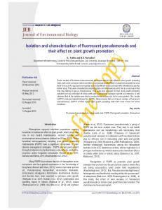
Construction and validation of MudY The MudK transposon and its delivery system have been described previously [15], and allow the straightforward construction of random lacZ translational fusions in Salmonella Typhimurium. For our application, we used recombineering [16] to exchange the lacZYA-npt part of MudK with a yfp-frt-cat-frt module, while making sure not to introduce stop codons upstream of the yfp open reading frame (Figure 1A). The resulting transposon was designated MudY, and can be used for random transposition in S. Typhimurium with the potential to create C-terminal translational fusions with the YFP fluorescent reporter protein. Furthermore, the frt-flanked chloramphenicol selectable cat marker can be readily flipped out by transiently expressing the site-specific Flp recombinase [23], in order to avoid polar effects or to reuse the cat marker in further strain engineering (Figure 1B). In order to validate random transposition and YFP fluorescence, a 6 kb plasmid containing a PBAD promoter was targeted with MudY. In total, 10 MudY insertions were mapped and all of them yielded different insertion sites in the plasmid, indicative for random transposition of the MudY transposon. Furthermore, one of the MudY insertions exhibited YFP fluorescence after addition of arabinose (data not shown). The MudY transposon is thus able to randomly transpose and can result in YFP fluorescence when the transposon hops in a gene in the correct orientation and reading frame.
Construction of iscR::yfp and iolR::yfp
Screening a random MudY insertion library for nucleoid reporters
Based on the MudY insertions in iscR and iolR, yfp translational fusions were de novo constructed at the 39 end of the same genes. For this, the yfp-frt-cat-frt cassette was PCR amplified (Phusion DNA polymerase; ThermoScientific) from plasmid pAC [18] with primers iscR_yfp_C-term_Fw (Table 2) and iscR_yfp_C-term_Rev_pAC, and iolR_yfp_C-term_Fw and iolR_yfp_C-term_Rev, respectively. The obtained amplicons were subsequently used to generate LT2 iscR::yfp and LT2 iolR::yfp via recombineering in LT2 and flipping out the cat cassette, using pKD46 [16] and pCP20 [23], respectively. In the latter strains, the YFP moiety is C-terminally fused to the IscR or IolR protein, respectively, with a SGGGG linker. The LT2 iscR::yfp recA1 strain was subsequently constructed by cotransducing the recA1 and srl-202::Tn10 alleles from TT521 (kindly provided by John Roth, University of PLOS ONE | www.plosone.org
After having validated the MudY transposon and delivery system, a random MudY transposition library was constructed in the S. Typhimurium LT2 chromosome in order to search for a useful nucleoid reporter. For this, the library of ca. 25.000 clones was first enriched in those clones displaying a clear YFP fluorescence through fluorescence activated cell sorting (FACS; selecting the 0.1% most fluorescent cells). Subsequently, after screening ca. 200 individual clones of this sub-library with fluorescence microscopy, two clones were retained in which YFP fluorescence clearly coincided with the nucleoid as stained with DAPI (Figure 2). Interestingly, the helix-like shape of the nucleoid, most recently described by Hadizadeh Yazdi et al. [24] and Fisher 4
April 2014 | Volume 9 | Issue 4 | e93785
Fluorescent Nucleoid Reporter in Salmonella
LT2 iscR::yfp was selected for further validation as a nucleoid reporter. Fluorescence microscopy experiments were conducted using several chemical components that are known to influence nucleoid structuring (Figure 3). In accordance with literature, it was observed that chloramphenicol condenses the nucleoid (Figure 3A) [26] while rifampicin decondenses the nucleoid (Figure 3B) [7]. Nalidixic acid inhibits DNA replication but not cell growth, leading to elongated cells with nucleoid-free regions toward the cell poles (Figure 3C) [27]. Mitomycin C is DNA crosslinking agent, inhibiting DNA replication and ultimately leading to double stranded breaks (Figure 3D) [28,29]. Furthermore, an LT2 iscR::yfp recA1 srl-202::Tn10 derivative was constructed, in which the defective RecA protein often leads to anucleate cells or cells with aberrant nucleoids (Figure 3E) [30]. In all cases, a perfect colocalization was observed when comparing the DAPI channel with the YFP channel.
Using the IscR::YFP nucleoid reporter to study nucleoid dynamics induced by high hydrostatic pressure stressed growth In a next step, we were interested to see how nucleoid dynamics were influenced by growth under HP (40–45 MPa, 37uC). The latter condition has been documented to cause excessive filamentation in mesophilic bacteria [10], although this phenotype remains poorly described. Using time-lapse fluorescence microscopy, it was observed that upon pressure release (after growth for 16–20 h at 40–45 MPa, 37uC) most of the filamentous cells initially elongated after which subsequent cell divisions took place leading to a viable microcolony (Figure 4A). Through monitoring the IscR::YFP nucleoid reporter, it could be demonstrated that immediately after HP release the nucleoids appeared as an unsegregated mass that nevertheless quickly started to segregate during cell elongation (Figure 4A). Subsequently, nucleoids were positioned and multiple cell divisions occurred, severing up the filamentous cell and resulting in mostly normal sized cells with the typical bilobed nucleoid appearance. Interestingly, it was observed that during this severing phase, cells with a ‘‘trilobed’’ nucleoid appearance occasionally occurred (Figure 5). Please note that in contrast to the IscR::YFP reporter, DAPI staining and corresponding UV excitation caused HP treated cells to quickly stop growing, leading to completely inactive cells after 60 min (Figure 4B). The latter observation clearly underscores the importance of non-invasive nucleoid reporters for studying proper nucleoid dynamics in DNA-stressed cells.
Figure 1. Construction and use of the MudY transposon. (A) During construction of the MudY transposon, the lacZYA-npt of MudK was replaced by an yfp-frt-cat-frt module through recombineering (blue and green crosses indicate the homologous regions involved in recombination). (B) The frt-cat-frt cassette can be readily flipped out using the Flp recombinase, thereby reducing possible polar effects of the cat marker. (C) Exact location and genomic context of two MudY insertions (iscR::MudY and iolR::MudY) in LT2 yielding endogeneous nucleoid reporters. doi:10.1371/journal.pone.0093785.g001
et al. [25], could be observed in a small fraction of cells without using any deconvolution analysis (inset Figure 2). The MudY insertion in the two clones could be mapped to the iscR (LT2 iscR::MudY, yielding the IscR144::YFP protein) and iolR (LT2 iolR::MudY, yielding the IolR159::YFP protein) gene, respectively (Figure 1C).
Impact of the IscR::YFP fusion on the fitness of LT2 While examining whether the presence of the IscR::YFP fusion affected the fitness of LT2 iscR::yfp, a small growth was defected for this strain (and LT2 iscR::MudY as well) in comparison with LT2 wild-type (Figure S2; in this setup the growth rate of the nucleoid reporter strains is ca. 33% lower than that of the wild type strain). Importantly, since IscR::YFP expression in LT2 iscR::yfp did not increase basal levels of the DNA damage response (as examined with a previously characterized SOS-reporter strain of LT2 [31]; data not shown), genotoxicity seemed not to be the cause of this growth defect. Upon further scrutinizing, however, we empirically observed that this growth defect could be mitigated upon construction of an LT2 iscR::yfp-frt-cat-frt mutant in which the cat gene and its promoter were oriented in the same direction as the iscR gene (Figure S3), without affecting this strain’s capacities as a nucleoid reporter. This latter observation indicates that IscR::YFP expres-
Validating LT2 iscR::yfp and LT2 iolR::yfp nucleoid reporter strains For further validation, both iscR::yfp and iolR::yfp translational fusions were reconstructed de novo by fusing the yfp moiety (this time with a SGGGG linker) to the penultimate codon of the iscR or iolR open reading frame on the chromosome, yielding LT2 iscR::yfp and LT2 iolR::yfp, respectively. Interestingly, the localization of this reconstructed IolR::YFP fusion protein differed from the original MudY mediated one, yielding small nucleoid associated foci instead of the typical ‘‘cloudy’’ appearance of the DAPI stained DNA (Figure S1). In contrast to IolR::YFP, the localization of the reconstructed IscR::YFP fusion did not differ from that of its IscR144::YFP counterpart. Moreover, since the IscR::YFP fusion protein was even more abundantly expressed,
PLOS ONE | www.plosone.org
5
April 2014 | Volume 9 | Issue 4 | e93785
Fluorescent Nucleoid Reporter in Salmonella
Figure 2. Representative images showing the colocalization of the IscR144::YFP and IolR159::YFP proteins with the DAPI stained nucleoid in LT2 iscR::MudY (A–D) and LT2 and iolR::MudY (E–H), respectively. Phase-contrast (A,E), DAPI (B,F), YFP (C,G), and merged (D,H) images are shown. Inset in panel C includes a larger image of an LT2 iscR::MudY cell showing the apparent helical shape of the nucleoid highlighted by IscR144::YFP. Scale bars correspond to 5 mm. doi:10.1371/journal.pone.0093785.g002
Figure 3. Representative images showing the colocalization of the IscR::YFP protein with the DAPI stained nucleoid in LT2 iscR::yfp cells stressed with (A) chloramphenicol, (B) rifampicin, (C) nalidixic acid, or (D) mitomycin C. (E) Representative images showing the colocalization of the IscR::YFP protein with the DAPI stained nucleoid in LT2 iscR::yfp recA1 srl-202::Tn10 cells, in which compromised RecA function leads to cells with an aberrant nucleoid positioning and morphology (indicated with arrows). For each condition, consecutive panels show phasecontrast, DAPI, YFP, and merged images. Scale bars correspond to 5 mm. doi:10.1371/journal.pone.0093785.g003
PLOS ONE | www.plosone.org
6
April 2014 | Volume 9 | Issue 4 | e93785
Fluorescent Nucleoid Reporter in Salmonella
Figure 4. Growth and nucleoid dynamics of HP stressed LT2. (A) Representative images showing cell growth and nucleoid dynamics of HP stressed LT2 iscR::yfp cells (grown overnight at 40–45 MPa and 37uC) at the indicated time points after pressure release. Merged phase contrast and YFP images are shown. (B) Representative images showing cell growth and nucleoid dynamics of a HP stressed LT2 iscR::yfp cell (grown overnight at 40–45 MPa and 37uC) at the indicated time points after pressure release in the presence of DAPI and intermittent UV excitation. Phase contrast (top panels), DAPI (middle panels) and YFP (lower panels) images are shown. Scale bars correspond to 5 mm. doi:10.1371/journal.pone.0093785.g004
studies [32–34], this is, to the best of our knowledge, the first time that this has been microscopically validated. The IolR repressor has been identified as the main regulator of the myo-inositol utilization island and binds at least four promoters located on this genomic island of 22.6 kb [32]. The IolR protein belongs to the RpiR family and has two predicted domains: an Nterminal helix-turn-helix-6 (HTH-6) motif and a C-terminal sugar isomerase domain predicted to bind phosphosugars. Comparison of the subcellular localization of IolR159::YFP and IolR::YFP, revealed an interesting difference: While IolR159::YFP colocalized with the typical cloudy appearance of the DAPI stained nucleoid (Figure 2 E–H), IolR::YFP showed distinctive chromosome associated foci (Figure S1). This suggests that lack of the Cterminus in the IolR159::YFP fusion protein, does not abolish the DNA binding ability completely but presumably causes a loss in specificity towards the cognate IolR promoter sites. It is tempting to assume that binding of a specific catabolite of myo-inositol to the phosphosugar binding domain of IolR, induces a conformational
sion itself is not toxic per se, and suggests that the downstream oriented cat promoter is able to counteract the potential polar effects of the iscR::yfp construct on the remainder of the isc operon.
Discussion In the search for a non-invasive fluorescent nucleoid reporter, we followed a straightforward rationale in which we first modified the well-characterized MudK transposon [15] of S. Typhimurium LT2 to encode the fluorescent YFP protein such that C-terminal YFP fusion proteins could be obtained after transposon mutagenesis (Figure 1). Random mutagenesis with this transposon (designated MudY) and a subsequent screen based on FACS and fluorescence microscopy yielded IolR159::YFP and IscR144::YFP fusion proteins to be specifically located on the nucleoid (Figure 2). While both proteins have previously been reported to be linked with the nucleoid through biochemical and crystallographic
Figure 5. Merged phase contrast and YFP images of growing LT2 iscR::yfp cells (previously grown overnight at 40–45 MPa and 376C) after pressure release, showing (A) typical cells with a bilobed nucleoid, and (B–F) occasionally occurring cells with a ‘‘trilobed’’ nucleoid (indicated by arrows). Scale bar corresponds to 5 mm. doi:10.1371/journal.pone.0093785.g005
PLOS ONE | www.plosone.org
7
April 2014 | Volume 9 | Issue 4 | e93785
Fluorescent Nucleoid Reporter in Salmonella
change in the protein leading to specific recognition of the promoter sites via the HTH-6 motif. However, further investigations are needed to confirm this hypothesis. The IscR protein has been studied extensively in Escherichia coli and was initially discovered as the negative autoregulator of the isc (iron-sulfur cluster) operon involved in Fe-S biogenesis [34]. However, it is now known that IscR is a global transcriptional regulator controlling at least 40 genes in 20 predicted operons dispersed throughout the genome [35]. The IscR protein of S. Typhimurium has a very high sequence identity (97%) with its E. coli homolog and is thus expected to have similar properties. Recently, crystallography data showed that IscR binds as a homodimer to DNA and comprises of two major domains: a DNA binding domain (winged HTH motif) and a dimerization helix [33]. Furthermore, the C-terminal domain of IscR seems not to be involved in the DNA binding properties of IscR, suggesting that a C-teminal YFP-tag will not influence its DNA binding capacity and thus supporting the fluorescence microscopy data obtained with IscR::YFP. While the IscR-YFP protein could be further validated as a nucleoid reporter through a number of colocalization experiments with the well-established DNA binding dye DAPI (Figure 3), it clearly outperformed this dye in time-lapse experiments that follow up nucleoid dynamics in conditions that are already stressful for the chromosome. In fact, UV/DAPI measurements further aggravated nucleoid stress imposed by HP growth, to the extent that it prevented the proper dynamics to be monitored (Figure 4). In contrast, readout of the IscR-YFP reporter did not impose this bias and allowed us to observe that after HP release the initially unsegregated nucleoid quickly segregated, ultimately leading to a viable microcolony with normal sized cells possessing the typical bilobed nucleoid appearance (Figure 5A). Interestingly, however, during the nucleoid segregation and cell septation process, cells with an unusual ‘‘trilobed’’ nucleoid occasionally emerged (Figure 5B–F), suggesting that during the defilamentation process, coordination of segregation and septation is somehow disturbed. Please note that our screen for nucleoid reporters was not exhaustive but could in principle be employed to map functional DNA binding domains (or other distinct cellular localization domains) throughout the Salmonella proteome, although insertions in some NAPs that are essential in nucleoid organization and regulation might compromise the viability of the corresponding clones and escape detection. Please note that during the preparation of this manuscript other nucleoid reporters such as
GFP-Fis And HupA-mCherry have been validated as well, and used to study chromosome organization and dynamics at high resolution [24,25]. In summary, we have adopted and validated a Mud-based random transposon delivery system to generate translational fusions to the yfp reporter gene throughout the S. Typhimurium chromosome, and have screened a library of such clones for a suitable nucleoid reporter. The resulting IscR-YFP nucleoid reporter was subsequently used to initiate studies on bacterial nucleoid dynamics resulting from HP stress. More generally, noninvasive fluorescent nucleoid reporters will allow proper nucleoid dynamics to be examined under DNA stressing physiology.
Supporting Information Figure S1 Representative images showing the localization of the IolR::YFP protein on the DAPI stained nucleoid in LT2 iolR::yfp cells. Phase-contrast (A), DAPI (B), YFP (C), and merged (D) images are shown. Scale bar corresponds to 5 mm. (TIF) Figure S2 Growth curves of LT2 wild-type, LT2 iscR::MudY and LT2 iscR::yfp., with growth monitored as an increase in optical density (OD630 nm) in time. Mean values of 3 independent experiments are shown, with standard deviations being,11%. (TIF) Figure S3 Growth curves of LT2 wild-type and LT2 iscR::yfp-frt-cat-frt, with growth monitored as an increase in optical density (OD630 nm) in time. Mean values of three independent experiments are shown, with standard deviations being,8%. (TIF)
Acknowledgments The authors would like to thank Sander K. Govers for his kind gift of the pGKBD plasmid.
Author Contributions Conceived and designed the experiments: IP AA. Performed the experiments: IP AG WC. Analyzed the data: IP AA. Contributed reagents/materials/analysis tools: AA CWM JL. Wrote the paper: IP AA.
References 9. Esnault E, Valens M, Espeli O, Boccard F (2007) Chromosome structuring limits genome plasticity in Escherichia coli. PLoS Genet 3: e226. 10. Zobell CE, Cobet AB (1964) Filament Formation by Escherichia Coli at Increased Hydrostatic Pressures. J Bacteriol 87: 710–719. 11. Sambrook J, Russell DW (2001) Molecular cloning : a laboratory manual. Cold Spring Harbor, N.Y.: Cold Spring Harbor Laboratory Press. 12. Davis R BD, Roth J (1980) Advanced bacterial genetics. New York: Cold Spring Harbor Laboratory Press. 254 p. 13. Schmieger H (1972) Phage P22-mutants with increased or decreased transduction abilities. Mol Gen Genet 119: 75–88. 14. Maloy SR, Stewart VJ, Taylor RK (1996) Genetic Analysis of Pathogenic Bacteria. Cold Spring Harbor Laboratory Press, Cold Spring Harbor, N. Y. 15. Hughes KT, Roth JR (1988) Transitory cis complementation: a method for providing transposition functions to defective transposons. Genetics 119: 9–12. 16. Datsenko KA, Wanner BL (2000) One-step inactivation of chromosomal genes in Escherichia coli K-12 using PCR products. Proc Natl Acad Sci U S A 97: 6640–6645. 17. Kleckner N, Bender J, Gottesman S (1991) Uses of transposons with emphasis on Tn10. Methods Enzymol 204: 139–180. 18. Lindner AB, Madden R, Demarez A, Stewart EJ, Taddei F (2008) Asymmetric segregation of protein aggregates is associated with cellular aging and rejuvenation. Proc Natl Acad Sci U S A 105: 3076–3081.
1. Wang X, Montero Llopis P, Rudner DZ (2013) Organization and segregation of bacterial chromosomes. Nat Rev Genet 14: 191–203. 2. Dorman CJ (2013) Genome architecture and global gene regulation in bacteria: making progress towards a unified model? Nat Rev Microbiol 11: 349–355. 3. Postow L, Hardy CD, Arsuaga J, Cozzarelli NR (2004) Topological domain structure of the Escherichia coli chromosome. Genes Dev 18: 1766–1779. 4. Wang W, Li GW, Chen C, Xie XS, Zhuang X (2011) Chromosome organization by a nucleoid-associated protein in live bacteria. Science 333: 1445–1449. 5. Petrushenko ZM, Cui Y, She W, Rybenkov VV (2010) Mechanics of DNA bridging by bacterial condensin MukBEF in vitro and in singulo. EMBO J 29: 1126–1135. 6. Rovinskiy N, Agbleke AA, Chesnokova O, Pang Z, Higgins NP (2012) Rates of gyrase supercoiling and transcription elongation control supercoil density in a bacterial chromosome. PLoS Genet 8: e1002845. 7. Cabrera JE, Cagliero C, Quan S, Squires CL, Jin DJ (2009) Active transcription of rRNA operons condenses the nucleoid in Escherichia coli: examining the effect of transcription on nucleoid structure in the absence of transertion. J Bacteriol 191: 4180–4185. 8. Cabrera JE, Jin DJ (2006) Active transcription of rRNA operons is a driving force for the distribution of RNA polymerase in bacteria: effect of extrachromosomal copies of rrnB on the in vivo localization of RNA polymerase. J Bacteriol 188: 4007–4014.
PLOS ONE | www.plosone.org
8
April 2014 | Volume 9 | Issue 4 | e93785
Fluorescent Nucleoid Reporter in Salmonella
19. Susskind MM (1980) A new gene of bacteriophage P22 which regulates synthesis of antirepressor. J Mol Biol 138: 685–713. 20. Cenens W, Mebrhatu MT, Makumi A, Ceyssens PJ, Lavigne R, et al. (2013) Expression of a novel P22 ORFan gene reveals the phage carrier state in Salmonella typhimurium. PLoS Genet 9: e1003269. 21. Kwon YM, Ricke SC (2000) Efficient amplification of multiple transposonflanking sequences. J Microbiol Methods 41: 195–199. 22. Wilson K (2001) Preparation of genomic DNA from bacteria. Curr Protoc Mol Biol Chapter 2: Unit 2 4. 23. Cherepanov PP, Wackernagel W (1995) Gene disruption in Escherichia coli: TcR and KmR cassettes with the option of Flp-catalyzed excision of the antibiotic-resistance determinant. Gene 158: 9–14. 24. Hadizadeh Yazdi N, Guet CC, Johnson RC, Marko JF (2012) Variation of the folding and dynamics of the Escherichia coli chromosome with growth conditions. Mol Microbiol 86: 1318–1333. 25. Fisher JK, Bourniquel A, Witz G, Weiner B, Prentiss M, et al. (2013) Fourdimensional imaging of E. coli nucleoid organization and dynamics in living cells. Cell 153: 882–895. 26. von Freiesleben U, Krekling MA, Hansen FG, Lobner-Olesen A (2000) The eclipse period of Escherichia coli. EMBO J 19: 6240–6248. 27. Marston AL, Errington J (1999) Dynamic movement of the ParA-like Soj protein of B. subtilis and its dual role in nucleoid organization and developmental regulation. Mol Cell 4: 673–682. 28. Nagashima K, Kubota Y, Shibata T, Sakaguchi C, Shinagawa H, et al. (2006) Degradation of Escherichia coli RecN aggregates by ClpXP protease and its implications for DNA damage tolerance. J Biol Chem 281: 30941–30946.
PLOS ONE | www.plosone.org
29. Keyamura K, Sakaguchi C, Kubota Y, Niki H, Hishida T (2013) RecA recruits SMC-like RecN to DNA double-strand breaks. J Biol Chem. 30. Zahradka K, Buljubasic M, Petranovic M, Zahradka D (2009) Roles of ExoI and SbcCD nucleases in ‘‘reckless’’ DNA degradation in recA mutants of Escherichia coli. J Bacteriol 191: 1677–1687. 31. Aertsen A, Tesfazgi Mebrhatu M, Michiels CW (2008) Activation of the Salmonella typhimurium Mrr protein. Biochem Biophys Res Commun 367: 435–439. 32. Kroger C, Fuchs TM (2009) Characterization of the myo-inositol utilization island of Salmonella enterica serovar Typhimurium. J Bacteriol 191: 545–554. 33. Rajagopalan S, Teter SJ, Zwart PH, Brennan RG, Phillips KJ, et al. (2013) Studies of IscR reveal a unique mechanism for metal-dependent regulation of DNA binding specificity. 34. Schwartz CJ, Giel JL, Patschkowski T, Luther C, Ruzicka FJ, et al. (2001) IscR, an Fe-S cluster-containing transcription factor, represses expression of Escherichia coli genes encoding Fe-S cluster assembly proteins. Proc Natl Acad Sci U S A 98: 14895–14900. 35. Giel JL, Rodionov D, Liu M, Blattner FR, Kiley PJ (2006) IscR-dependent gene expression links iron-sulphur cluster assembly to the control of O2-regulated genes in Escherichia coli. Mol Microbiol 60: 1058–1075. 36. Blattner FR, Plunkett G, 3rd, Bloch CA, Perna NT, Burland V, et al. (1997) The complete genome sequence of Escherichia coli K-12. Science 277: 1453–1462. 37. McClelland M, Sanderson KE, Spieth J, Clifton SW, Latreille P, et al. (2001) Complete genome sequence of Salmonella enterica serovar Typhimurium LT2. Nature 413: 852–856.
9
April 2014 | Volume 9 | Issue 4 | e93785