Two abundant fatty acid-binding proteins (MFB1 and MFB2) were isolated from the midgut cytosol of larval Manduca sexta. As isolated, MFBl and MFBS.
THEJOURNAL OF BIOLOGICAL CHEMISTRY 0 1992 by The American Society for Biochemistry and Molecular Biology, Inc.
Vol. 267, No. 1, Issue of January 5, pp. 380-384, 1992 Printed in U.S.A.
Isolation, Characterization, and cDNA Sequence of Two Fatty Acidbinding Proteins from the Midgut ofManduca sexta Larvae* (Received for publication, June 20, 1991)
Alan F. Smith, Kozo TsuchidaS,Eric HannemanQ,Teri C. Suzuki, and Michael A. Wells From the Biochemistry Department and Center for Insect Science, Biosciences West, University of Arizona, Tucson, Arizona 85721
Two abundant fatty acid-binding proteins (MFB1 and MFB2) were isolated from the midgut cytosol of larval Manduca sexta. As isolated, MFBl and MFBS were found to contain bound fatty acids in a 1:l molar stoichiometric ratio. Immunological screening demonstrated that MFBl and MFBB were restricted to the midgut in a gradient distribution, with MFBl more concentrated in the anterior two-thirds of the midgut and MFBS more concentrated in the posterior twothirds of the midgut. MFBl exchanged fatty acid more readily than did MFB2. MFBl was about 2%and MFBS about 12%of the cytosolic protein in the midgut. cDNA clones for MFBl and MFBB both encode proteins of 131 amino acids that are rich in lysine and acidic residues. Analysis of the amino acid sequence alignment of the MFBs with six mammalian fatty acidbinding proteins revealed a number of shared features: 9 conserved glycines, presumably important in turns of the &strands; a basic amino acid in a position corresponding to the residue reported to participate in binding the carboxyl group of the fatty acid (Arg in MFB 1 and Lys in MFB2); and conservation of many of the residues important in binding the aliphatic portion of the fatty acid.
acid uptake by cells, targeting fatty acids to organelles and specific pathways, altering the activity of enzymes involved in fatty acid metabolism, or protecting cellular proteins and membranes from the detergenteffects of fatty acids or their CoA derivatives (Ockner, 1990). In spiteof considerable effort, the physiological role(s) played by these proteins remains obscure. FABP has been recently identified in the flight muscle of the migratory locust, (Haunerland and Chisholm, 1990), and a cDNA clone from the blood fluke Schistosoma mansoni was found to encodea FABP(Moser et al., 1991).Because it processes large amounts of lipid, we investigated the possibility that the larval midgut of Manduca sexta might also contain FABP. In this paper we report the purification and characterization of two FABPs (MFB1, MFB2) and their cDNA sequences from the midgut of the larval tobacco hornworm, M. sexta. MATERIALS ANDMETHODS
M . sexta were raised on a high wheat germ diet as previously described (Prasad et al., 1986; Fernando-Warnakulasuriya et al., 1988). Benzamidine,and phenylmethylsulfonyl fluoride (PMSF) were from Aldrich. We obtained Sephadex G-75, Sephacryl S-300 HR, and high molecular weight standards for calibration of gel filtration columns from Pharmacia LKB Biotechnology, Inc.; DEAE-Trisacryl M from IBF Biotechnics(Villeneuve-la-Garenne, France);and CooFatty acid-binding proteins(FABP)’are low molecular massie Brilliant Blue R-250 from Pierce Chemical Co. [“CIOleic acid mass proteins(14-17 kDa), which are members of a superfam- was from Du Pont-New England Nuclear. Purification of Fatty Acid-binding Proteins-Midgut tissue from ily of cytoplasmic hydrophobic ligand-binding proteins day 2 fifth instar larvae was homogenized with 20 mM Tris-HCI, pH (Sweetser et al., 1987). Until recently, the only well charac- 7.5, containing 150 mM NaC1,0.5 mM PMSF, and 5 mM benzamidine terized proteins belonging to this family have been isolated (5 ml/g of tissue) in a polytronal homogenizer at setting 6 for 20 s from vertebrate tissues and consistof the following proteins: (Brinkmann Instruments, Inc., Westbury, NY), and the homogenate heart, liver, renal, and intestinal FABP;cellular retinol-bind- was centrifuged at 100,000 X g for 60 min in a Ti-60 fixed angle rotor. ing protein I and cellular retinol-binding protein 11; cellular In an ice bath, the 100,000 X g supernatant was adjusted to 75% saturation in ammonium sulfate and the suspension was centrifuged retinoic acid-binding protein; the P2 protein of peripheral (10,000 X g for 30 min). Thesupernatant was adjusted to 95% myelin; the p422 (aP2) adipocyte protein; bovine mammary- saturation in ammoniumsulfate; the precipitate was collected by derived growth inhibitor; and gastrotropin (Veerkamp et al., centrifugation, dialyzed against homogenization buffer, and the vol1991). The best studied of these proteins are the FABPs for ume was reduced to 5 ml/g of tissue, using a Diaflo Ultrafiltration which a number of roles have beenproposed facilitating fatty Membrane, YM-10 (Amicon Corp., Danvers, MA). During the development of the purification scheme, FABP was followed bylabeling it * This work was supported by National Institutes of Health Grant with radioactive fatty acid. This was accomplished by adding 5 pCi HL39116 (to M. W.) and National Institutes of Health Fellowship of [14C]potassium oleate to the solutioncontaining the dialyzed, GM13656 (to A. S.). The costs of publication of this article were resuspended 95% ammonium sulfate pelletand mixing for 4 h at 4 “C in a tube rotator. For large scale purifications, fractions were assayed defrayed in part by the payment of page charges. This article must therefore be hereby marked “aduertisement” in accordance with 18 for FABP using a fatty acid binding assay (see below). It should be noted that these procedures only detected MFB1. MFB2 was found U.S.C. Section 1734 solely to indicate this fact. The nucleotide sequence(s) reported in thispaper has been submitted when the low molecular weight proteins from midgut were analyzed totheGenBankTMjEMBLDataBankwith accession number(s) for fatty acid. The solution from the ammonium sulfate step was applied to a M77754 and M77755. Sephadex G-75 Superfine column (2.5 X 110 cm) equilibrated in 20 j: Present address: National Institute of Health, Tokyo, Japan. Present address: Department of Pathology, College of Medicine, mM Tris-HCI, pH 7.5, containing 150 mM NaCl, 0.5 mM PMSF, and 5 mM benzamidine and eluted with the same buffer at a flow rate of University of Arizona, Tucson, AZ 85724. ’ The abbreviations used are: FABP, fatty acid-binding protein; 11.5 ml/h and collected in 2.5-mlfractions. The fractions were assayed PMSF, phenylmethylsulfonyl fluoride; SDS-PAGE, sodium dodecyl for protein using the BCA method (Smith et al., 1985), and for fatty acid binding; selected fractions were subjected to SDS-PAGE. The sulfate-polyacrylamide gel electrophoresis.
380
Proteins Acid-binding Fatty
Midgut
381
low molecular weight fractions testing positive for fatty acids were pooled and applied to a DEAE-Trisacryl M column (2.5 X 8 cm) packed in the same buffer used for gel filtration, but without NaCI. Protein was eluted with a linear NaCl gradient (0-100 mM) in the same buffer a t a flow rate of 35 ml/h, and 1.6-ml fractions were collected. The fractions were assayed for protein, fatty acid binding, and selected fractions were subjected to SDS-PAGE. In addition to fractionscontainingimpure MFB1, the DEAE column gave pure preparations of two other low molecular mass proteins, which had molecular masses on SDS-PAGE of 15 and 17 kDa,respectively. As a fatty acid-binding protein shown below, the 15-kDa protein was also (MFB2). The fractions containing MFBlwere pooled and dialyzed against 25 mM Imidazole buffer, pH 7.4, and applied toa PBE 96 chromatofocusingcolumn (Pharmacia LKB Biotechnology Inc.) and eluted with pH 4.0 polybuffer/water 1%(v/v) according to the manufacturer's instructions. Fractions were assayed for protein, fatty acid binding, and pH. The fractions containing pure MFBl were concentrated by ammonium sulfate precipitationas described above. Fatty Acid Binding Assay-Fatty acid binding was measured bya modification of the methodof Morrow and Martin(1983). The sample containing FABP andvarying amounts of ['4C]potassium oleate(11.6 pCi/pmol) in 200 pl of 10 mM Tris, pH7.5, containing 150 mM NaCI, was incubated for 1 h at 25 "C. In order to remove unbound fatty acid, 50 pl of a 2% suspension of charcoal (Norit A) in 0.2% Dextran T 70 was added and the samplewas centrifuged ina microfuge for 5 min;thesupernatant wasassayedfor proteinand radioactivity. Control experimentsshowed that 97% of added FABPwas recovered in the supernatant andonly 1%of labeled fatty acid remained in the supernatant. In some experiments the timeof incubation or pHwere varied as detailed in the figure legends. Immunology-Antibodies for immunoblotting were raised by injecting the protein-adjuvant mixture (Ribi Immunochem Research, Hamilton, MT) into the breast muscle of laying hens.IgY was purified from egg yolks using the methodof Polson et al. (1985). SDS-PAGEseparated proteins were electrophoretically transferred to nitrocellulose and immunoblotted according to the methodof Burnette (1981) using IgY and rabbit anti-chicken IgY coupled to horseradish peroxidase (Jackson Immuno Research Laboratories, West Grove, PA). Antisera for cDNA library screening were produced in New Zealand Whiterabbits by intramuscular injection of the protein-adjuvant mixture (Ribi Immunochem Research, Hamilton, MT). The serum was stored a t -70 'C. Tissue Distribution-The following samples were used whole midgut, midgut divided into three sections (anterior to posterior), fat body and muscle from day 2 fifth instar larvae, and eggs from day 2 adults. For each sample a 100,000 X g supernatant was prepared as described ab05 e, and 100 pg of protein was separated by SDS-PAGE, transferred tonitrocellulose, and immunoblotted. Electrophoresis-SDS-polyacrylamide gel electrophoresis was car-
-12 a a
- 8 b
0 ELUTION VOLUME,
-0
ml
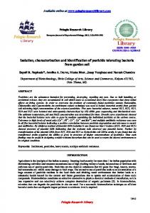
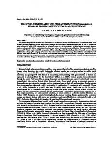
FIG. 2. Chromatography on DEAE-Trisacryl M. The fractions containing MFBl and MFBZ from the Sephadex separation were chromatographed on a 2.5 X 8-cm column of DEAE-Trisacryl M packed in 20 mM Tris-HCI, pH 7.5, containing 0.5 mM PMSF and 5 mM benzamidine. After washing the column with the same buffer, the column was eluted with a linear gradient of NaCl (0-100 mM). The flow rate was 35 ml/h and 1.6-ml fractions were collected. The fractions were assayed for protein (0)and radioactivity (A). Selected fractions were analyzed by SDS-PAGE using the same molecular weight standards as inFig. 1. Note thepresence of the 17-kDa protein (fractions 140-160) and MFBZ (fractions 200-240).
W
gY
.4
!i! .2
0
ELUTION VOLUME.
rnl
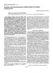
FIG. 3. Chromatofocusing of MFB1. TheMFBlcontaining fractions from the DEAE-Trisacryl M column were separated on a PBE 96 chromatofocusing column. Fractions were assayed for protein (O), radioactivity (A), and pH (0).Selected fractions were analyzed by SDS-PAGE using the same molecular weight standards as in Fig. 1.
0.8
P 0.4
4E
-
n
.. 100
200
400
ELUTIONVOLUME. rnl
FIG. 1. Chromatography of midgut supernatant on Sephadex G-75 superfine. The supernatant, concentrated by ammonium sulfate precipitation, was labeled with ['4C]oleic acid and applied to a 2.5 X 110-cm column of Sephadex packed in 20 mM Tris-HCI, pH 7.5, containing 150 mM NaCI, 0.5 mM PMSF, and 5 mM benzamidine and eluted with the same buffer a t a flow rate of 11.5 ml/h and 2.5ml fractions were collected. The fractions were assayed for protein (0)and radioactivity (A). Selected fractions were subjected to SDSPAGE using the following molecular markers: bovine serum albumin (a); egg albumin (b); glyceraldehyde-3-phosphate dehydrogenase (c); carbonicanhydrase (d); trypsinogen (e);trypsininhibitor ( f ) ; alactalbumin (g).
ried out using the methodof Laemmli (1970) in slab gels containing either 15% or a 4-15% linear gradient of polyacrylamide. Gels were stained with Coomassie Brilliant Blue R-250. Lipid Analysis-The fatty acid content of proteins was determined as previouslydescribed (Fernando-Warnakulasuriya et al., 1981), using 5 mg of protein. RNA Isolation-Total RNA was prepared from the midgut of a day 2 fifth instar male larva using theprocedure of Savakis et a/. (1986). PolyadenylatedRNA wasselected by twopassages throughan oligo(dT)-cellulose column accordingto the method of Aviv and Leder (1972). cDNA Library Construction and Screening-Five pg of midgut polyadenylated RNA was used to prepare cDNA and a directional library from a commercialkit (ZAP-cDNA Synthesis Kit; Stratagene, La Jolla, CA). The cDNA library was screened with rabbit antisera using goat antirabbit IgG and an alkaline phosphatase color development system (Bio-Rad). DNA Sequencing-Both MFB clones were subcloned into pBluescript SK- plasmid using an in vivo excision protocol (Stratagene, LaJolla, CA). Both single anddouble-stranded DNA were sequenced by the dideoxy chain termination method (Sanger et al., 1977). Sequence Alignments-Protein sequences were aligned using the multiple sequence alignment procedure of Feng and Doolittle (1987).
Midgut Fatty Acid-binding Proteins
382
TABLE I Summary of the purificationof MFBl from M. sexta larval midgut Step
Total protein
Total activity'
Specific activityb
w 1.1 100,000 X g supernatant' 5320 4669 (100%) 3.6 4.0 75-9596 ammonium sulfate fraction 952 3840 (72.2%) 10.3 Sephadex G-75 2600 253 (48.9%) 22.8 25.1 DEAE-trisacryl M (42.5%) 2260 89.9 52.6 1900 (35.7%) 57.9 Chromatofocusine 32.8 Total nanomoles of fatty acid bound in fatty acid binding assay. Percent recovery in parentheses. Nanomoles of fatty acid bound/mg protein. From 267 midguts.
Fold purification
1.o
9.4
TABLE I1 Fatty acid content and compositionof midgut fatty acid-binding proteins Five mg of MFBl (0.34 pmol) and MFB2 (0.35 pmol) were quantitatively analyzed for fatty acid content and composition. The data are means f S.D. ( n = 3). mol fatty acid/mol protein Fatty acid
MFB2
16:O 16:l 180
MFBl
0.39 18:2 18:3
0.17 f 0.16 0.05 0.04 f 0.01 0.04 f 0.02 0.03 0.10 f 0.09 0.02 f 0.47 0.06 0.09 k 0.03
f 0.04 0.07 f 0.01 f 0.01 f 0.01 f 0.07 0.08 f 0.03
Total
0.83 f 0.05
0.90 f 0.04
181
oleic acid concentration
(pH)
FIG.4. Binding of ['4C]oleic acid to M F B l and MFBP. The proteins were incubated with the indicated amount of ["C]oleic acid a t pH 7.5 for 1 h at 25 "C. The protein was separated from the free of fatty acid bound fatty acid as described in the text and the amount to the protein determined by scintillation counting. The points represent the mean and the bars the standarddeviation for four measurements. Open circles, MFBl; closed circles, MFB2. cDNA sequences were aligned using the GAP program of the GCG sequence Analysis Software Package (Genetics Computer Group, Inc., Madison WI)
RESULTS AND DISCUSSION
Isolation and Characterization FABPs-Figs. 1-3 illustrate the purification of MFBl in which the purification was followed using [I4C]oleic acid to label the protein. Table I presents a summary of the large scale purificationin which MFBl was followed using a fatty acid binding assay. These quantitative data, with a recovery of 35% and a purification of 50fold, show that MFBl comprises approximately 2% of the soluble protein of the midgut tissue. The elutionprofile presentedin Fig. 2shows that,in addition to MFBl there were two other low molecular weight proteins in themidgut supernatant with molecular masses of 17 and 15 kDa. The latter protein (MFB2) isclearly a major protein in themidgut and appears tobind a small amount of [I4C]oleic acid(see Fig. 2). In the large scale preparation detailed in TableI, we isolated 250 mg of MFB2. Byassuming
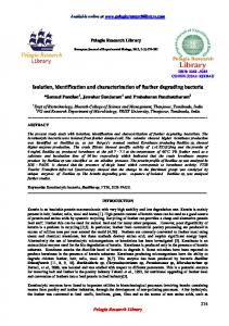
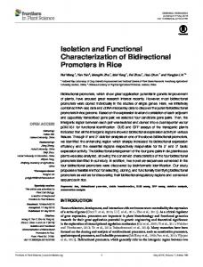
FIG. 5. Tissue distribution of MFB1, MFBP, and the 17kDa protein. A 100,000 X g supernatant was prepared from whole midgut (I); the anterior one-thirdof the midgut (2);the middle onethird of the midgut ( 3 ) ;the posterior one-third of the midgut ( 4 ) ;fat body ( 5 ) ;muscle (6);and eggs (7). Except for eggs, all samples were from day 2 fifth instar larvae. Samples (100 pg) were separated by SDS-PAGE on a 15% acrylamide gel. Gel A was stained with Coomassie Brilliant B. For gels B-D, the samples were separated by SDSPAGE and transferred to nitrocellulose. Immunodetection was carried using chicken anti-MFBI ( B ) , chicken anti-MFB2 ( C ) , or chicken anti-I7 kDa and anti-chicken IgY conjugated to horseradish peroxidase and then developed with 4-chloro-1-naphthol.
a recovery similar to MFBl from the DEAE column, MFB2 comprises about 12%of the soluble protein in themidgut. In the same preparation we recovered 14.5 mg of the 17-kDa protein. The fact that the MFBsmajor are components of the cytosolic proteins is consistent with that reportedfor mammal FABPs (Veerkampet al., 1991). In order to further characterize these three proteins, we measured fatty acid and otherlipid content. MFBl andMFB2 both contained 1 mol of free fatty acid/mol of protein, but the 17-kDa protein containednegligible fatty acid (Table 11). A 1:1molar ratioof fatty acid to protein is in accordance with that reported for the vertebrate and locust FABPs (Veerkamp et dl., 1991; Haunerland and Chisholm, 1990). The two MFBs have essentially the same fatty acid composition, which corresponds closely to the fattyacid composition of the artificial diet (Fernando-Warnakulasuriyaet al., 1988). Neither MFBl nor MFB2 contained any lipids other than fatty acids when the lipid extracts were examined by thin layer chromatography (data not shown). In general fatty acids predominate in vertebrate FABPs, with theliver and kidney forms being the exceptions (Veerkamp etal., 1991). Fig. 4 compares the bindingof [I4C]oleicacid to MFBl and MFB2. These datashow that MFBl hasa single high affinity binding site with an apparentdissociation constant of about 14 PM. It should be kept in mind that we are measuring exchange of fatty acid, not truedissociation constants in these experiments, since the protein already has a bound fattyacid when isolated. This is quite apparent in analyzing the data for MFB2. We were never able to saturate the protein with labeled fatty acid and the estimated dissociation constant was greater than 100 PM. These data initially lead us t o believe
Proteins Acid-binding Fatty
Midgut
383 c
1
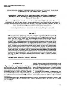
FIG.6. Partial restricti.on map, sequencing strategy, and nucleotide and deduced amino acid sequence of MFBl (top) and MFBB (bottom). Capitalletters on the full line indicate R = endonuclease restriction sites: EcoRI; S = SalI; K = KpnI; C = ScaI. The lines below the full line show the length of the subclones sequenced full lines represent clonessequenced from left to right, and dotted lines are clones
K I
HFB2 I
I
sequenced from right to left.
t .
thirds of the midgut. The absenceof MFB2 from the anterior one-third of the midgut is evident even in the Coomassiestained gel. Hence MFBl andMFB2 are similar to the mammalian liver andintestineFABPs which exhibit atissue .- -.-..........-. . . ... . . .... .- .... SLGVGFATROVASWlKPTT~lEKNW~LTlKl distribution gradient (Glatz and Vusse, 1990). The 17-kDa protein was found in the midgut, where it had a distribution TDDWOGGLPIKTTYTMG WFBl NTVTOWN SAOGSAlFKREYNGDELKVTlTSSEWGVAYRYYKA similar to thatfor MFBZ, and in small amountsin the egg. FODVICACESVKSMYTMG N W T H W K GDAGVATFKKEYNGDDLVVTITSSNWGVARRYYKA WFB2 cDNA Cloning-From 30,000 recombinant plaques, 23 CELETWTCEKVKAWDIEGDNKWVTTFK GIKSVTEFNGDTITNTWTLG DIVYKRVSGRI RFBL DlVFKRlSKRl HFBL CELETMTGEKVKTWOLEGDNKLVTTFK YIKSVTELNGDIITWTMTLG MFBl and 150 MFBB positive cDNA clones were identified RFBl fAVSLADGTELTGTLTWEG YKLVGKFKRWNGYELIAVREISGNELIOTYTYE GVEAKRIFKK CVEAKRIFKKD fNYNLMGTELRGTUSLEC HFBl NKL1GKFKRTDNGNELNTVREllCOELVOTYVYE by immunological screening. One positive clone from each of HFBH fOETTADDRKVKSlVTLDG GKLVHLOKU DGOETTKVRELIDGKLILTLTHG TAVSTRTYEKEA RFBH FDEVTADDRKVKSWTLDG GKLVHVOKU DGOETTLTRELSDGKLILTLTHG NWSTRTYEKEA the two screenings was purified, subcloned into pBluescript 1 1 f T SK- plasmid, restriction mapped, and sequenced (Fig. 6).The Percent i d e n t i t y f o r mino acid a l i g m m t : cDNAs were of similar size (491 and465 bases for MFBl and MFB1 WFB2 RFBL HFBL RFBl HFBI HFBH RF0H 25.0 25.0 30.7 29.0 55.7 MF0l MFB2, respectively) and encoded proteins of 131 amino acid 3 4 . 7 3 3 . 9 2 0 . 3 2 0 . 3 3 2WFB2 .3 29.0 61.4 2 5 . 6 2 6 . 4 2 4 . 0 2 0 . 2 0 RFBL 1.8 43.7 40.7 residues(Fig. 6). The deduced amino acidcompositionre2 0 .200 . 0 2 4 . 0 2 7 . 4 0 5 . 2HFBL 44.2 49.7 32.0 32.0 RFBI 39.9 36.9 4 2 . 64 3 . 1 00.8 vealed that both proteins are rich in acidic amino acids and 3 2 .363 . 3 0 2HFBL .421 .432 .399 .357 . 9 _ _ _ _ .... ".. .". .". HF0H 90.1 lysine with molecular masses of 14.7 and 14.1 kDa (MFB1 4 7 . 50 0 .RF0H 4 5 . 43 4 . 40 0 . 49 2 . 0 ---Percent i d e n t i t y for cDNA a l i g m t : and MFB2, respectively), not unlike the vertebrate FABPs FIG. 7. Alignment of MFBl and MFBS protein sequence (Maatman, 1991). The cDNAfor MFBl contains an open with other members of the FABP family. The amino acid se- reading frame beginning with an ATG codon at position 12 quences were alignedby the progressive methodof Feng and Doolittle and extending to position 407, followed by an &-base pair 3'(1987). Percent identities for the amino acid sequences are derived untranslated sequence (Fig. 6). The open reading frame for from the generated alignments and are shown in the upper triangle of the table. Thelower triangleof the table gives the percent identities the MFB2 cDNA begins with an ATG codon at position 6 for the cDNA sequences derivedfrom painvise alignments(the cDNA with the translatedregion extending toposition 401 and a 64sequence for human heart FABP has not been reported). Identical base pair 3'-untranslated region (Fig. 6). All FABPs isolated residues are marked by an asterisk; residues involved in fatty acid to date have an acetyl group at the NH,-terminal aminoacid binding RFBI are indicated by t. Sequences are: Manduca FABP 1 (Bernier and Jollk, 1987). Several attempts to sequence the (MFB1); Manduca FABP 2 ( M F B Z ) ;rat FABP-liver (RFBL);human FABP-liver (HFBL); rat FABP-intestine (RFBZ); human FABP- MFBs by Edman degradation were unsuccessful, suggesting intestine ( H F B I ) ; human FABP-heart ( H F B H ) ; rat FABP-heart that the NH2 terminus of these proteins may also be acetylated. (RFBH). Analysis of the progressive amino acid alignment of the that MFBS was not a fatty acid-binding protein. Since these MFBs with six mammalian FABPs by the method of Feng experiments measure exchange only under one set of condi- and Doolittle (1987) revealed a number of shared features (Fig. 7). The two MFBs are 55.7% identical in amino acid tions, one must be cautious about interpreting the apparent acid identities between dissociation constants. In fact, the apparent dissociation con- sequence. Whereas the percent amino stants are too high to be consistent with the fact that both mammalian FABPs from the same tissuesof different species are high, ranging from 80 to 90%, mammalian FABPs from FABPs are isolated with 1 mol of bound fatty acid. to other (24-33.3%) Fig. 5 presents data on the tissue-specific distribution of different tissues are about as similareach MFBl andMFB2. The FABPswere found only in themidgut as they are to the MFBs (20.3-33.3%). Similar conclusions and not in the fat body, muscle, or eggs. Interestingly, MFBl were reached fromanalysis of the painvise alignment of cDNA was found predominately in the anterior two-thirds of the sequences (see table in Fig. 7). midgut, whereas MFB2 was found only in the posterior twoThe occurrence of two FABPs within a single organ has WFBl WFB2 RFBL HFBL
.1 20.4
AYLCKVYKFDREENFDtFLKSIGLSEEOVOKYLOYKPSSOLVKEGDKYKYlSVS~GTKETVFESGVE SYLCKVYSLVKOEYFDGFLKSAGLSDDKlOALVSDKPTOKWEANGDSYSlTSTGLGGERTVSFKSGVE NFSGK YOVOSOEUFEPF~KAWGLPEDLlOKGKDlKGVSElVHEGKKVKLTlTYGSKVlHNEFTLGEE SFSGK YOLOSOEYFEAFWKAlGLPEELlOKGKDlKGVSElVONGKHFKFTlTAGSKVlONEFTVGEE
""
384
Midgut Fatty Proteins Acid-binding
been observed in vertebrate tissues: liver and intestine FABP elucidate the structureof MFBZ. of rats and humans are both found in the intestine (for review, Acknowledgments-We thank Drs. D. Frohlich, J. Soulages, and seeVeerkamp et al., 1991) and two types are reported in Kanost for critical review of the manuscript. We thank Mary mammalian kidneys (Maatman etal., 1991; Lam et al., 1988). M. Hernandez for animal care. However, the percent amino acid identity between the liver and intestine forms is considerably lower (28.2-24.8%) than REFERENCES the similarity between the two MFBs (55.7%). Aviv H., and Leder P. (1972) Proc. Natl. Acad. Sci.U. S. A . 69,1408Elucidation of the three-dimensional structureof rat intes1412 tinal FABP through x-ray crystallography (Sacchettinial., et Bernier I., and Jo1li.s P. (1987) Biochemie 6 9 , 1127-1152 Burnette W. N. (1981) Anal. Biochem. 1 8 , 5294-5299 1990) permits the identification of several important conD. P., Sacchettini J . C., Banaszak L. J., Walsh M. T., and served residues among the FABPs. Rat intestinal FABP con-Cistola Gordon J. I. (1989) J. Biol. Chem. 264,2700-2710 sists of two orthogonally oriented 0-sheets with glycines fa- Feng D. F., and Doolittle R. F. (1987) J . Mol. Euol. 2 5 , 351-360 cilitating turns between P-strands. These 7 glycines are con- Fernando-Warnakulasuriya G. J. P., Tsuchida K., and Wells M. A. (1988) Insect Biochem. 18,211-214 served in the MFBs (Fig. 7). Also highly conserved are the residues involved in binding of the fatty acid. An arginine Glatz J . F. C., and van der Vusse G . J. (1990) Mol. Cell. Biol. 9 8 , 237-251 participates in the electrostatic interaction with thecarboxyl Haunerland N. H., and Chisholm J. M. (1990) Biochim. Biophys. Acta group of the fatty acid (Sacchettini etal., 1990). This residue 1047,233-238 is conserved in MFBl and is substituted with another basic Laemmli U. K. (1970) Nature 227,680-685 amino acid, lysine, in MFB2. This change may account for Lam K. T., Borkan S., Claffey K. P., Schwartz J. H., Chobanian A. V., and Brecher P. (1988) J. Biol. Chem. 2 6 3 , 15762-15768 the high affinity of MFB2 for fatty acids. We are currently R. G. H. J., Van Kuppevelt T. H. M. S. M., Veerkamp J. investigating this phenomenon. It is interesting to note that Maatman H. (1991) Biochem. J. 2 7 3 , 759-766 the basic amino acid ( i e . arginine) purported to act in the Morrow F. D., and Martin R. J. (1983) J. Lipid Res. 24,324-331 fatty acid carboxyl groupis not conserved in theliver FABPs Moser D., Tendler M., Griffiths G., and Klinkert M.-Q. (1991) J . Biol. Chem. 266,8447- 8454 where the carboxylgroup is reported to interact near the aqueous surfaceof the protein (Cistola al., et 1989). Moreover, Ockner R. K. (1990) Mol. Cell. Biochem. 98,3-9 Prasad S. V., Ryan R. O., Law J. H., and Wells M. A. (1986) J. Biol. of the 18 residues associatedwith the binding of the fatty acid Chem. 26 1,558-562 hydrocarbon chain, 10 and 12 residues of MFBl and MFB2, Sacchettini J. C., Banazak L. J., and Gordon J. I. (1990) Mol. Cell. Biochem. 98, 81-93 respectively, are either retained or conservatively substituted. Based on the physiochemical and cDNA data, we conclude Sanger F., Nicklen S., and Coulson A. R. (1977) Proc. Natl. Acad. Sci. U. 5'. A . 74,5463-5467 that MFBl and MFBZ are fatty acid-binding proteins. The Savakis C., Ashburner M., and Willis J. H. (1986) Deu. Biol. 114, MFBs have molecular weights, amino acid residue numbers 194-207 and composition, and lipid composition in agreement with Smith P. K., Krohn R. I., Hermanson G. T., Mallia A. K., Gartner F. H., Provenzano M. D., Fujimoto E. K., Goeke N. M., Olson B. J., those described for the vertebrate and locust FABPs (Ockner, and Klenk D. C. (1985) Anal. Biochern. 150,76-85 1990; Haunerland and Chisholm,1990). Aminoacid sequence Sweetser D. A., Heuckeroth R. O., and Gordon J. I. (1987) Annu. alignments (Fig. 7) suggest that the three-dimensional struc- Reu. Nutr. 7, 337-359 ture of the MFBs and the FABP may be similar. Presently x- Veerkamp J. H., Peeters R. A,, and Maatman R. G. H. J. (1991) ray crystallographic studies are being conducted which will Biochim. Biophys. Acta 1081,1-24