with a buffered inhibitor solution containing 1 M KH2P04, 0.2 M Nas. EDTA, and 0.2 M ..... Isolation of C3"The data for recovery of protein and C3 functional ...
THCJ O U R N A L
OF
BIOLOGICAL CHEMISTRY
Vol. 256, No. 8. Issue of April 25, p p . 3995-4006, 1981 Prlnted m U.S.A.
Large Scale Isolationof Functionally Active Components of the Human Complement System* (Received for publication, August 18, 1980,and in revised form, November 24, 1980)
Carl H. Hammer$& George H. Wirtzsq, Lois RenferS, Hattie D. Gresham$, and BrianF. Tack\\** From the +Laboratory of Clinical Investigation, National Institute of Allergy and Infectious Diseases and the I/Laboratory of Chemical Biology, National Institute of Arthritis, Metabolism andDigestive Diseases, National Institutes of Health, Bethesda, Maryland 20205
In pursuingwork on thebiological functions of complement, In the present work a scheme is presented for the isolation of multiple componentsof human complement it became apparent that simpler and moreefficient methods in a functionallyand biochemically pure state and with were required for preparationof milligram quantities of funcfull hemolytic activity. These preparative procedures tionally active, biochemically pure, and well characterized allow high recovery of milligram and gram quantities components of the human complement system. Early experiofparticular complement componentsfrom a large pool ments proved the feasibility of greatly simplified methods. (2-11 liters) of fresh EDTA plasma in no more than four The purpose of this paper is to present an approach for large chromatographicsteps. Many components (C3bINA, scale purification of multiple complement components from a C5,C3, ClEI, C4, and C9) are recovered functionally single pool of human plasma that meets these criteria. pure or highly purified following the first chromatoIt wasshownthat individual pools of inhibitor-treated graphic step employing DEAE-Sephacel and may be human plasma (2-11 liters) can be processed to yield at least utilized as reagents withno further purification. Prior to anion exchange, individual units of plasma 14 complement components early in the procedure that are generally stable and amenable to further purification. The are treated with inhibitors of complement activation and serum proteases, the pooled plasma is fractionated excellentresolution obtained by an initial DEAE-Sephacel with polyethylene glycol, depleted of plasminogen on ion exchange chromatographic step allowed the isolation of Sepharose-lysine, and rapidly ultrafilteredto low ionic many complement components in a state of high functional strength and high proteinconcentration. The high de- purity that canbe used directly as complement reagents. The gree of resolution of the components on DEAE-Sepha- utility of this procedure was established by the subsequent cel subsequently obtainedis demonstrated by the func- isolation of homogeneous and functionally pure components tional recovery and purification in a representative C3, C5, C7, and C8 in good yield with no more than three experiment as indicated (in their orderof elution) for additional chromatographic stepsfor each protein. the following proteins: C3bINA (24%,l&fold), C2 (74%, In developing efficientprocedures to maintain recovery and 12-fold), C7 (87%, 14-fold), factor B (55%, 8.7-fold), C8 purity while limiting inactivation of individual complement (50%, 16-f0ld), C6 (82%, 25-f0ld),j31H (39%, 12-fold), C5 components, we have taken measures first to eliminate acti(62%, Ill-fold), C3 (99%, 64-fOld), ClEI (42%, 135-f0ld), vation of both theclassical and alternative pathways and also C9 (80%, 297-fold), and C4 (78%, 164-fold). Other com- to block intrinsic or potential proteolytic activities of human ponents separatedby these procedures include Clq and plasma during the purification. In our approach to the resoC4 binding protein. lution andisolation of pure complement components,we have Additional steps described, which demonstrate the used the usual physicochemical and immunological parameutility and effectiveness of this preparative scheme, ters to assess purity but, following the approach of Nelson et have allowed isolation of C3, C5, and C7 as pure components with fullhemolytic activity as judged by func- al. (1) and Vroon et al. (2) we have used, as well, sensitive allow the detectionof contaminants far tional, immunochemical, and physicochemical criteria. functional assays that below the usual level of resolution. C8, also isolated a s a homogeneous protein, was recovered with partial hemolytic activity. All these comMATERIALS AND METHODS ponents were recovered in high yield and in the purification as indicated: C3 (61%, 103-fold),C5 (24% 1350Complement Components and AssayProcedures fold), C7 (19%, 2260-fold),and C8 (328, 547-fold). ComIn this purification the chromatographic behavior of the various plement components C6, PlH, factor B, and C2 in ad- components was followed by functional hemolytic assays. For five dition to C3blNA, ClEI, C4, and C9 are recovered par- components (CBbINA,’ factor B, C5. C3, and C4), antigenic assays tially purified with good activity and are amenable to further purification. ”
’
* T h e costs of publication of this article were defrayed in part by the payment of page charges. This article must therefore be hereby marked “advertisement” in accordance with 18 U.S.C. Section 1734 solely to indicate this fact. 5 To whom correspondence should be addressed. 7 American Cancer Society Scholar, Grant No. SG-102. * * Established investigator of the American Heart Association. Present address, Children’s Hospital Medical Center, Harvard Medical School, Boston,MA 02115.
The abbreviations used are: C3bINA, C3b inactivator; ClEI, C1 esteraseinhibitor;symbols E, A, and C represent, respectively,a sheep erythrocyte, one molecule of IgM, or two molecules of IgG antibody, and complement componentsbeing designated by number, i.e.C1, C2, etc. The letters “a” and “b” are used to designate fragments of the complement molecules. EAC-7 is a shorthand designation for an intermediatelysible by C8 and C9; PMSF, phenylmethanesulfonyl fluoride; SDS-PAGE, sodiumdodecyl sulfate-polyacrylamide gel electrophoresis; PEG, polyethylene glycol; mS, milliSiemen-conductance measurementwhere a Siemen is equivalent to areciprocal ohm; EACA, e-amino-n-caproic acid.
3995
3996
Isolation Complement o Components f Human
were performed as well. In most cases, standard functional assays were utilized. For these assays guinea pig C1 and C9 were isolated as previously described (3, 4). Functionally pure guinea pig C5, C6, C7, and C8 were prepared as in Ref. 5. Guinea pig C2 and C3 were purchased from CordisLaboratories, Miami,FL. The human components used in these assayswere obtained or prepared as follows: C4, C2, C3, and C6 were purchased fromCordis Laboratories. Biochemically pure human C3 was prepared by the method of Tack and Prahl (6) with an added immunoadsorbent step to remove contaminating C5, IgG, and IgA. C5 was prepared following hydroxylapatite chromatography also, as in Ref. 6. Contaminating IgG and IgA were removed by immunochemical depletion. The human C5b6 complex was used in preparation of EAC-7 from EAC43b. This complex was prepared as in Ref. 7.
Methods of Functional Assay
the Sepharose 4B-coupled IgG was treated with 1 mM PMSF for 30 min at 37 "C prior to use.
Polyacrylamide Gel Electrophoresis Crude and purified protein preparations were examined by polyacrylamide gel electrophoresis in the presence of 0.1%sodium dodecyl sulfate. T h e monomer acrylamide concentration of the gel was 7.58, with an acrylamide/bisacrylamide ratio of 37.5:l. Samples and standard molecularweight markers (Bio-Rad) were prepared and run concurrently under both reducing and nonreducing conditions.Details of the method are asdescribed by Maize1 (17).
Concentration and Buffer Equilibration of Protein Small volumes of protein solutions (up to 1 liter) were concentrated and theirbuffer composition adjusted by using Amicon ultrafiltration cells with P M 30 membranes or by precipitation with PEG 4000 (J. T . Baker Chemical Co.) at levels determined tomaximize recovery of the desired component(s) with subsequent solubilization in the appropriate buffer. Volumes of several hundred milliliters or less were concentrated in collodion bags (25,000- or 75,000-dalton exclusion). The processing of large volumes of protein solution (2-11 liters), in particular the preparation of plasma for chromatography on DEAESephacel, was accomplished with a Pellicon Ultrafiltration System using 20 square feet of 30,000-dalton exclusion membrane, high volume heads, and pump (Millipore).
Buffers were prepared as described by Hammer et al. (7) except that buffer D" contained 0.20 M glucose. Human C2 was assayed by a modification of the assay of Borsos et al. (8) using EAC14 (9) prepared with guinea pig C1 and human C4. Human C4 was assayed by a minor modification of the assay of Gaither et al. (IO). Human C3, C5, C6, and C7 were titrated by modifications of the method of Hammer et al. (11).Human C8 and C9 were titrated using EAC-7 cellsalso as described in Ref. 11. Titers for C2 through C9 are expressed as theaverage of theproduct 2 X reciprocaldilution obtained from three or more experimental measurements in the linear Purification Procedure portion of the dose-response curve, i.e. generally below 2 = 1 and where 2 = -In (1 - y), y = % lysis. One unit/ml corresponds to 1.5 General Approach and Methodology X 10' effective molecules in a system containing 0.1 ml of indicator cells (1.5 X 10⁸/ml), 0.2 ml of sample dilution, and0.2 ml of converting The purification scheme is presented in two sections. The first reagent. ClEI was assayed by the method of Gigli et al. (12). Human describes the preparation of human plasma for chromatography on C3bINA and PlH were assayed as in Ref.13. DEAE-Sephacel and the chromatographic separationof the components. At this step a high degree of resolution of components was Antisera to Components achieved with a very high yield of functionally active protein. For convenience, the column eluent was divided into eight distinct comFor analytical purposes, small volumes of the antisera to human proteins listed below were obtained from the designatedcommercial plement-containing pools. The second section presents the further laboratories: goat anti-human serum and rabbit anti-fibrinogen (Cap- purification of complement components C3, C5, C7, and C8 obtained pel Laboratories, Downingtown, PA); goat anti-albumin, C4, and C5 from select DEAE-Sephacelpools. All procedures were performed a t 4 "C unless otherwise indicated. (Meloy Laboratories,Springfield, VA); rabbit anti-IgG, a,-antitrypsin, Centrifugation of PEG-precipitated solutionswas carried outat 14,000 aa-macroglobulin,a,-acid glycoprotein,plasminogen, C3, andClq IgM X g for 25 min in a Sorvall RC2B centrifuge. The conductivity of (Behring Diagnostics,Sommerville, NJ); rabbitanti-IgAand (Calbiochem-Behring Corp., La Jolla, CA); goat anti-transferrin, a2- buffers and solutions was measured a t 0 "C. All stock buffers and human serum-glycoprotein ceruloplasmin, P2-glycoprotein,C-reactive solutions were Millipore-filtered prior to dilution and use. The data protein, ClEI, PIH, factor B, and properdin (Atlantic Antibodies, presented, except where otherwise indicated,refer to the preparation Scarborough, ME); goat anti-C3bINA (Kent Laboratories,No. Van- of components from a 2-liter pool of plasma. In some experiments volumes of upto 11 liters of plasmawereprocessed andthese couver, B.C.); goat anti-Cls (Kallsted Laboratories, Chaska, MN). Monospecific antLC8 and anti-C4binding protein were gifts of Drs. preparations wereused where indicated as the source of several components which were purified further. Hans J. Muller-Eberhard (Department of Molecular Immunology, Scripps Clinic and Research Foundation, La Jolla, CA) and Victor Nussensweig (New York University School of Medicine, New York, NY), respectively. When large volumes of monospecific or polyvalent antisera were burros, or required (i.e. for immunoadsorbentpreparation)sheep, wereimmunizedwith the goats housed at theNIHanimalfarm appropriate immunogens. Fifty to 500 pg of protein were emulsified in 50% Freund's complete adjuvant (2 ml) and injected intradermally at sites along the animals' back.
Part A:Preparation of Human Plasma forChromatography on DEAE-Sephacel Treatment of Plasma withInhibitor Solutions-A volume ofabout 500 ml of platelet-free EDTA human plasmawas obtained from each of four donors whowasmedication-free andhadfasted prior to plasmaphoresis. Each unit of fresh plasma was diluted while stirring with a buffered inhibitor solution containing 1 M KH2P04, 0.2 M Nas EDTA, and 0.2 M benzamidine-HC1 that had been adjusted to pH 7.4 with NaOH (20 parts plasma to 1 partinhibitor). Following the addition of inhibitors the plasma was pooled and 0.1 M PMSF in Immunological Methods anhydrous isopropyl alcohol was added to a final concentration of 1 Antigenic levels of selected proteins in plasma and purified frac- mM. The pH was adjusted to 7.4, if required. tions were quantified by radial immunodiffusion according to Mancini Fractionation of Plasma with PEG 4000-The inhibitor-treated et al. (14).Immunoelectrophoresis wasperformed in 1% agarose plasma was made 5% (w/v)inPEG 4000by slow addition,with (Sigma),Veronal-acetate buffer, pH 8.2, containing 0.03% sodium stirring, of the solid powder and allowed to equilibrate for 1 h. The azide and 10 mM EDTA. Ouchterlony analyses of column fractions precipitate which formed was removed by centrifugation, suspended were carried outon diffusion plates preparedwith phosphate-buffered in a 2-fold concentrated Sepharose-lysine buffer to be described, and saline containing 0.03% Na azide, 10 mM EDTA, and 1% agarose. stirred overnight a t 4 "C. The 5% PEG supernatant containing the bulk of the plasma proteins and complement components was adAntisera Fractionation and Immunoadsorbent Preparation justed toa specific conductivity of 12 mS/cm by the addition of solid NaCl prior to plasminogen depletion. A two-step procedure for using octanoic acid and DEAE-cellulose The solubilized precipitate was cleared by centrifugation at 14,000 for theisolation of IgG from mammalian seradeveloped by Steinbuch x g for 1 h. This solution containing Clq and C4 binding protein was and Audran (15)wasemployed to fractionate antisera and obtain pure IgG antibody. Antibody at 10 mg/ml was coupled to Sepharose filteredthrough glass wool, broughtto 1 mM PMSF, and rapidly 4B (Pharmacia) by the BrCN method of March et al. (16). Following frozen into small 10O-pl volume pellets by pumping into liquid nitrocoupling, the unreacted groups were masked with a solution of 0.1 M gen, and stored at -65 "C. ethanolamine, pH 9.0. As a precaution against proteolytic degradation Plasminogen Depletion of the 5% PEG Supernatant-Sepharose was prepared by coupling 200 g of L-lysine/liter of the adsorbed material due to exposure to the immunoadsorbent, 4B-~-lysine adsorbent
Isolation of Human Complement Components of BrCN-activated Sepharose 4B. About 1.1 liters of this biospecific adsorbent were packed into a glass column 13 cm in diameter. The plasminogen capacity of the Sepharose-lysine adsorbent prepared as described was sufficient to deplete up to 3.5 liters of plasma/liter of adsorbent. A 50 mM K/Na phosphate buffer (pH 7.4, 11.8 mS/cm) containing 10 mM EDTA and 150 mM NaCl was used to equilibrate the column. The 5% PEG supernatant was applied and collected a t a flow rate of 1300 ml/h. Residual nonspecifically bound protein was removed from the column by washing with a 2-fold concentrated buffer and included in the adsorbed plasmapool. Following severalcolumnwashes with the 2-fold concentrated buffer, thebound plasminogenwas eluted with theconcentrated buffer containing 200 mM EACA and stored frozen at -65 "C. The adsorbent was regenerated by sequentialtreatment with10 mM NaOH and10 mM phosphoric acid. After washing withstarting buffer, the gel was stored in the cold in the presence of 0.02% azide. Concentration and Ionic Strength Adjustment of the Plasminogen-Depleted P E G Supernatant-The post Sepharose-lysine pool (13.2 mS/cm) was made 33 mM in EACA by the addition of solid inhibitor and allowed to warm to about 15 "C. A diluent solution of pH 7.4, 1.1 mS/cm, and containing 6.7 mM EDTA, 6.7 mM benzamidine-HCI, and 33.3 mM EACA was prepared and prior to use was to "C. The plasminogen-depleted made 1 mM in PMSF and warmed 15 PEG supernatant was rapidly reduced in ionic strength by repeated sequential additions (4 times) of the 1.1 mS/cm diluent to a total volume of 4 liters and subsequent concentrationof the adjustedpool to 2 liters using a Pellicon Ultrafiltration System a t a n average rate of 5 liters/h. When the adjusted pool approached a specific conductivity of 1.35 mS/cm, the volume of the pool was reduced to 500 ml and transferred to a 1-liter graduate. T h e concentrator was rinsed with two 500-1111 portions of ice-cold DEAE-Sephacel buffer to be described, each concentrated to 200 ml, and these were added to the adjusted pool now a t 4 "C. The specific conductivity and pH were adjusted with solid NaCl and 1 N NaOH to 1.35 mS/cm and 7.4, respectively. DEAE-Sephacel Chromatography-The concentrated plasminogen-depleted 5% PEG supernatant was applied a t 220 ml/h to a (4850ml bed) glass column (6.5 X 146 cm) of DEAE-Sephacel (Pharmacia), equilibrated with a 3.2 mM K/Na phosphate buffer (pH 7.4, 1.37 mS/ cm) containing6.4 mM EDTA, 6.4 mM benzamidine-HCI, and 31.8 mM EACA. Following application of the sample, the columnwas washed with the same buffer at the same flow rate until 3 liters of effluent had been collected, the first2400 ml of which were discarded, and the subsequent 600 ml, enriched in IgG, were pooled and stored frozen a t -65 "C. Fraction collection (22 ml/tube) was initiated and thecolumn was developed with a 12-liter linear salt concentration gradient. The limit buffer was identical in composition with the wash buffer except that in addition it contained300 mM NaCl(16 mS/cm). Theflow rate was maintained at 200 ml/h. Following application of theentire gradient, several liters of limit buffer were applied to complete the elution profile. The absorbance at280 nm was monitored for protein after adjusting for interference by benzamidlne. Complement component activities were identified by functional assays as described earlier on appropriate dilutionsof column fractions. Wheremonospecific antisera wereavailable, double diffusion assayswere run to identify regions of complement component antigen prior to performing functional assays. Ceruloplasmin was detected by its blue color and monitored optically a t 600 nm. Pools of individual complement components or multiple component pools were prepared as described under "Results" (Table I, Step A5). P a r t B: Isolation Procedures for Purificationof Complement Components C3, C5, C7, a n d C8 C3 Purification: Gel Filtration on Sepharose CL-GB-The C3containing fractions from the DEAE-Sephacel column were pooled and the C3 was precipitated by addition of solid PEG to 16% (w/v). After 1 h of equilibration the C3 precipitate was collected by centrifugation and dissolved in a 100 mM K phosphatebuffer (pH 7.4) containing 150 mM NaCl, 5 mM EDTA,and 50 mM EACA. This procedure was repeated one time to further concentrate and purify the c 3 preparation. This C3 pool was applied to a column (10 x 110 cm) containing SepharoseCL-GB (Pharmacia), equilibratedwith the buffer, and eluted at a flow rate of 160 ml/h, 20-1111 fractions being collected. The C3-containing fractions were pooled. C3: Immunoadsorption of C5, ZgG, a n d ZgA-Detectable amounts of C5 activity, IgG, and IgA antigen in the C3 preparation were removed by selective adsorption on a Sepharose 4B anti-C5, IgG, and IgA immunoadsorbent. Ten per cent of the post-Sepharose CL-GB C3
3997
pool was applied at 4 "C to acolumn containing about 30 ml of immunoadsorbent equilibratedwith a 100 mM K/Na phosphate buffer (pH 7.4) containing 150 m M NaCl and 5 mM EDTA. The column was developed and washed at 60 ml/h. Protein was monitored by absorbance at 280 nm and the C3 collected as a pool. C5 Purification: Gel Filtration on Sepharose CL-GB-The C5containing fractions from the DEAE-Sephacel column were pooled and the C5 was precipitated at pH 7.4 by the addition of solid PEG to 16% (w/v). Following equilibration for 1 hr at 4 "C, the C5 precipitate was collected by centrifugation and reso1ubi:ized inDEAESephacel(pH 7.4) wash buffer containing 500 mM NaCl. For gel filtration, a small volume (I5 ml)of the concentrated C5 preparation was adjusted with PMSF to a final concentration of 1 tnM and applied CL-GB. The gel column to a column (5 X 85 cm) containing Sepharose was equilibrated and developed at a flow rate of 125 mi/h using a pH 7.4, 5 mM Na phosphate buffer (8.5 mS/cm) containing 150 mM NaCl and 5 mM EDTA. The 10-ml fractions were collected. C5-containing fractions were pooled and concentrated. C5: Hydroxylapatite Chromatography-The C5 pool prepared as above was equilibrated with a 5 mM Na phosphate buffer (pH 7.4) containing 100 mM NaCl and 5 mM EDTA (6.0 mS/cm). The sample was applied to a hydroxylapatite (Gallard-Schlesinger Chem. Mfg. Corp., Carle Place, NY) column (1.5 X 26 cm) equilibrated with the same buffer and washed with one column volume at 20 ml/h. The C5 was eluted by application of a linear salt concentration gradient of 200 ml collecting 2-ml fractions. The limit buffer was of the same composition as the wash buffer except that it contained 1 M NaCI. Elution of residual protein from the column was completed by appliM NaCl and a terminal cation of an additional linear salt gradient2 to kick buffer (pH 7.4) containing 125 mM Na phosphate, 100 mM NaCI, and 5 mM EDTA. C5-containing fractions were pooled and concentrated to about4 ml on a collodion bag of 75,000-dalton exclusion. C5: Immunoadsorption of I g G a n d IgA-Contamination of the post-hyroxylapatite C5 with IgG and IgA was removed by selective adsorption of the C5 preparation on a Sepharose 4B anti-DlH, IgG, and IgA containingimmunoadsorbent. The procedure was asdescribed for immunoadsorption of C3 except that the C5 preparation wasapplied to acolumn (1.5 X 8 cm)containing 14ml of the adsorbent. The C5 drop-through was collected as a pool. C7 Purification: DEAE-Sephacel Chromatography-The C7-containing fractions, devoid of most C2 activity, but containing factor B antigen from the DEAE-Sephacel column (Part A) were pooled and concentrated. T o 49 ml of the C7 pool was added an equalvolume of pH 7.5, 5 mM Na phosphate buffer (5.6 mS/cm) containing 90 mM NaCl, 2 mM EDTA, and0.005%gelatin, and enough solid PEG togive a final concentration of 20%' (w/v). After stirring for 1 h in the cold, the precipitated C7 was collected by centrifugation and dissolved in diluent buffer and applied to acolumn (2.5 X 90 cm) containing DEAE-Sephacel equilibrated with the same buffer. The column was developed by washing with three column volumes of starting buffer at 24 ml/h and terminated following elution with one column volume of the buffer with 230 mM NaCl added. The C7-containing fractions were pooled, concentrated, and dialyzed against a pH 6.0, 5 mM Na phosphate buffer containing 15nlM NaCI, 2 mM EDTA, and 0.0059 gelatin (1.0 mS/cm). C7: CM-cellulose Chromatography-The C7 pool was applied to a column (1.6 X 98 cm) of CM-52 cellulose (Whatman) equilibrated with the dialysis buffer. The column was washed with 200 ml of this buffer and C7 was eluted with a linear salt concentration gradient of 600 ml at 20 ml/h collecting 8-ml fractions. The limit buffer contained 150 mM NaCl (8.7 mS/cm). C7: Gel Filtration on Sepharose CL-6B"To removemacromolecular protein, identified by SDS-PAGE from the C7 preparation, the post-CM-cellulose C7 pool was applied to a column (2.5 X 98 cm) of Sepharose CL-GB equilibrated with a pH 7.5, 3.4mM Na phosphate buffer containing 77mM NaCl, 5 mM EDTA, and 0.00541gelatin (5.0 mS/cm). The C7was eluted a t a flow rate of 8 ml/h, collecting 7 ml/ tube. C8 Purification: Gel Filtration on SephadexG-200-The C8-containing fractions from the DEAE-Sephacel column overlapped with both ,B1H and C6 functional activities. Thus, fractions containing all of these activities were pooled, adjusted to pH6.0 with 1 N HC1, and solid PEG was added to a final concentration of 16%(w/v). Following equilibration for 1h at 4 "C, the precipitate containing the C8, BlH, and C6 activities was recovered by centrifugation and dissolved in a minimum volume of 5 mM Na phosphate buffer (pH 6.0) containing 150 mM NaCI. T o resolve /3IH from C6 and C8,40 ml of this pool was made 5T in dextrose and applied (60 ml/h) to a column (7.2 X 97 em)
Isolation of Human Complement Components
3998
of Sephadex G-200 equilibrated with the same buffer. Fractionsof 20 ml were screened for P1H antigen and also for C6 and C8 functional activities. Two pools were made, one containing B1H and the other C6 and C8 activities,and were adjustedto 16% PEG (wt/v) by addition of the solid and stirred for 1 h at 0 "C. The precipitateswere collected by centrifugation. The P1H precipitate was solubilized in the above phosphate buffer at pH 7.0. The precipitate containingthe C6 and C8 protein was suspended in a small volume of 4 M NaCl solution and subsequently dissolved by dilution with a pH 6.0, 5 mM Na phosphate buffer containing 100 mM NaCl and 0.005%gelatin. The conductance of this solution was then adjusted to 2.6 mS/cm by the additionof distilled water. C8: CM-cellulose Chromatography-Theconcentrated C6- and C8-containing post-SephadexG-200 pool was applied toa column (1.6 X 95 cm) of CM-cellulose equilibrated with a pH6.0, 5 m M Na phosphate buffer containing 40 mM NaCl and 0.005% gelatin(2.6 mS/ cm). The column was washed with200 ml of starting buffer at 30 ml/ h, collecting 12-ml fractions. The C6 and C8 proteins were eluted by application of a 580-ml linear salt gradient, thelimit buffer containing 270 mM NaCl(14.7 mS/cm). The C6- and C8-containing fractions
were pooled separately. RESULTS
P a r t A: Preparation of the Plasma for DEAE-Sephacel Chromatography Pooled plasma adjusted with PEG to a final concentration of 5% (w/v) lost all C4 functional activity andmore than 90% of the C2 activity. It was critical to mix individual units of plasma with EDTA and benzamidine at final concentrations of 10 mM each before they were pooled, adjusted to 1 mM PMSF, and fractionated with PEG yield to a stabilized supernatant solutionwhich contained a t least 12 complement components of interest. As shown in Table I, Step A2, this latter procedure allowed recovery of 10 complement componentsin the 5% PEG supernatant with a functional yield of 95% or more. The only exception was C5, in which 83% of the functional activity was retained. Factor B antigen was present at 97% of the level in the original 2-liter plasma pool. Plasminogen depletion of plasma reported (6, 18, 19) to be necessary for the subsequent good recovery of C3 and C5 could be achieved using 1 liter of Sepharose-lysine biospecific adsorbent/3.5 liters of plasma. One liter of adsorbent/2 liters of plasma was used here to insure completeremoval of plasminogen. No plasminogen could be detected by double diffusion analysis in the concentrated post-Sepharose-lysine pool (88.8 A2,,,/ml). In order to obtaina concentrated soluble pool of low ionic strength for application to the DEAE chromatographic column, it was critical to use the Pellicon Ultrafiltration System as described earlier. If direct rapid decrease in volume of the complement pool was attempted, the concentration of residual PEG led to precipitation of protein and loss of components, especially C5 and PlH. The post-concentration pool contained 69% ofthe initial plasma protein, while only three complement components, P l H , C5, and C7, were reduced to this level of functional activity (Table I, Step A4). All other components lost no or little activity when maintained under theconditions of low ionic strength (1.5 mS/cm) and high protein concentration (89 A28,,/ml) required for subsequent DEAE-Sephacel chromatography. DEAE-Sephacel Chromatography-Although it is known thatDEAE-52(Whatman) cellulose chromatography of guinea pig serum will resolve functional C3 and C5,2 this is not so when human serum is substituted (6). Initial experiments utilizing DEAE-Sephacel, however, demonstrated that human C5 could be readily separated from human C3 and other complement components. In addition, other complement factors (CSbINA, C4, and C9) werealsoindividually 'l
C. H. Hammer, unpublished observation.
resolved. When the adjusted post-Sepharose-lysine pool was chromatographed on DEAE-Sephacel and elution the position of all the complement components identified by antigenic and/orfunctional assay, theresults shown in Fig. 1 were obtained. In addition to C3bINA eluting a t 1.35 mS/cm, C5 (5.5 mS/cm), C9 (10.0 mS/cm), and C4 (11.6 mS/cm) were individually resolved and pooled as such. While C2 (1.7 mS/ cm) was almost completely separated from C7 and factor B (co-migrating a t 2.2 mS/cm), these three components were pooled together to obtainmaximal recovery. C8 (2.6 mS/cm), C6 (2.8 mS/cm), andP l H (3.4 mS/cm) overlapped sufficiently with each other so that one pool of these three components was made. In some experiments the elution position ofC6 varied such that itwould co-chromatograph with PIH rather than C8. C3 (6.6 mS/cm) was effectively separated from C5 as expected, but overlapped with about 30% of the ClEI (7.4 mS/cm), and theywere pooled together ((23, ClEI-1). A pool (ClEI-2) was made of the remainder of the ClEI (57%) that co-chromatographed with ceruloplasmin (7.8 mS/cm). A summary of the purification of Part A, including stepwise recovery of protein and functional activity for each component, is presentedin Table I. The C3bINA, the ClEIfrom C3 (ClEI-l), ClEI-2, C9, and C4 pools were adjusted with the solid PEG as indicated in Table I, Step A5, and therespective components were recovered and solubilized. A comparison of the amount of functional activity thatwas present in each of the dilute pools (not shown) with that obtained following concentration with PEG demonstrated, with the exception of factor B, the effectiveness inusing appropriate concentrations of PEG for rapid and quantitative recovery of the components. It was found by radial immunodiffusion analysis that approximately half of the factor B antigen was not precipitable by found optimal for recovPEG at 20% (w/v), the concentration ery of C7. The functional contamination of components isolated following DEAE-Sephacel chromatography, but purified nofurther, is shown in Table 11. The lack of significant functional cross-contamination with other complement components for thesingle component pools of C3bINA, ClEI, C9, and C4 clearly demonstrated the high degree of resolution obtained following this first chromatographic step.
Part B: Isolation of Pure Components Data for complete quantitationof each component at each step of the purification in Part A are shown for asingle 2-liter plasma pool. Because complete quantitation of each component in Part B was not available for this 2-liter purification, thequantitation of components intypical pools of other preparations isshown. Isolation of C3"The data for recovery of protein and C3 functional activityfollowing each step aregiven in Table 111." The second 16% PEG precipitation shown in Step A5 was effective in removing 5% of theprotein including trace amounts of ceruloplasmin and was not required if care was taken to exclude this protein whenmaking the C3-containing pool following DEAE-Sephacel chromatography. Gel filtration of the C3 on Sepharose CL-GB removed a small amount of high molecular weight contamination as shown in Fig. 2. Functional and antigenic analysis of this C3 preparation revealed contamination with 4,200 units/ml of C5, 0.24 mg/ml of IgG, and a trace of IgA. These contaminations were com,'Portions of this paper (including Figs.2 to 9 and Tables 111 to VI) are presented in miniprint at the end of this paper. Miniprint is easily read with the aid of a standard magnifying glass. Fullsize photocopies are available fromthe Journal of Biological Chemistry,9650 Rockville Pike, Bethesda, MD20014.RequestDocument No. 80M-1758, cite author(s), and include a check or money order for $5.60 per set of photocopies. Full size photocopies are also included in the microfilm edition of the Journal that is available from Waverly Press.
Isolation of Human Complement Components
3999
9
L
I
C
I
T C 0 0 - u
mmmm
Isolation Complement o fComponents Human
4000
ClEl
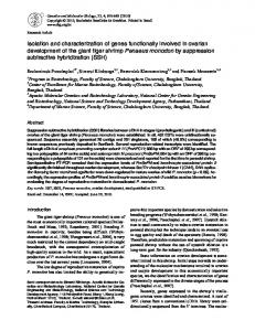
FIG. 1. DEAE-Sephacel chromatography of the concentrated, plasminogendepleted, 5% PEG supernatant of fresh humanEDTA plasma on a columnbed (6.5 X 146 cm). Equilibration and wash buffer:3.2 mM K/Na phosphate (pH7.4) containing 6.4 mM EDTA, 6.4 mM benzamidine-HCl, and 31.8 mM EACA,1.37rnS/crn.Elution buffer: 12-liter linearsalt gradient to 300 mM NaCl, 16 mS/cm in wash buffer. Flow rate: 220 ml/h; 22 ml/tube. All curves denoting complement elution positions weredetermined by functional assayexceptforfactor B (FB), which was detected by immunologic test. Ceruloplasmin (Cerulop.)was monitored by its absorbance at 600 nm.
,
TABLEI1 Functional purity of select DEAE-Sephacel pools Individual componentpools that were purifiedno further followiing preparation fromthe fist DEAE-Sephacel chromatographic step.Titrated Titer in complepooled ment corn- plasma human ponent
Titer in uoofs
C3bINA CIEI-I
ClEI-2
C9
c4
acttuity (units/ml)
c2
2,820
122
0
3
389
c4
22,800 129,000
0 0
0 66
0 c7 101,ooo 14 C8 121,000 6 c9 95,000 9 C3bINA 30,800 ClEl 18,700 163 PIH 3,400 13
2
c3 49,000
5,290
c5 C6
101,Ooo
3
31 129
0 122 0
7
24
19 32 1,940 2,130,000
15
128
3 21 1566
0
33 89
39 0 26
252 998,000
180
0
6,500
0 2,800
0
5
156,000 41,200
pletely removed by selective immunoadsorption (Table 111, Step B2). The specific activity of the final product was 30,000 and represented a 103-fold purification. The recovery of C3 functional activityfor this preparationwas 61% of that which was present in the startingpool of plasma. Isolation of C5-A summary of the C5purification scheme giving protein and functional activityrecoveries for each step is presented in Table IV. Concentration of the DEAE-Sephacel C5 pool by precipitation with 16% PEG (w/v) resulted in a 2.5-fold purification. The next step, gel filtration with Sepharose CL-GB shown in Fig. 3, was effective in resolving macromolecular protein as was evident from the elution profile. Approximately 73% of the protein not containing C5 activity was removed with an apparent gain inpurity of 4.3-fold (Table IV, Step B3). Immunochemical analyses demonstrated that the C5 preparation still contained C3, PlH, human serum albumin, IgA, and significant amounts of IgG antigen. The following step, fractionationon hydroxylapatite, removed not only the contaminating PlH,C3, and human serum albumin, but 15%of the C5 functional activity aswell (Fig. 4). Adsorption of the hydroxylapat.ite C5 poolwitha Sepharose 4B immunoadsorbent of the appropriate specificity gave a pure product freeof IgG and IgA when tested at 0.43 mg of C5/ml
and resulted in a further purification of 2.3-fold. Recovery of C5 was 24% with an overall purification of 1350-fold (Table IV, Step B5). Isolation of C7-When C7 wasisolatedfroma pool of fractions obtainedfollowing DEAE-Sephacel chromatography devoid of most C2 activity, purification to homogeneity was V, achieved in three sequential chromatographic steps (Table Steps B6, 7, and 8). With respect to C7, the C2-deficient C7 pool was 18-fold purified over plasma; treatment with PEG prior to further chromatographyremoved 64% of the Alxobut little C7 functional activity (TableV, Step A5b). Chromatography of the C7 preparation a second time on DEAE-Sephacel shown in Fig. 5 and subsequently on CM-cellulose shown in Fig. 6 gave a product almost pure as determined by SDSPAGE (not shown). The only significant contamination detectable by Coomassie blue protein stain was a pair of bands just entering the gel. Both these high molecular weight components reduced to a pair of diffuse bands when electrophoresed in the presence of mercaptoethanol, one of which comigrated with albumin. Chromotography of C7 on DEAESephacel was very efficient with respect torecovery of functional activity; chromatographyusing CM-cellulose, however, resulted in a substantial loss of activity (Table V, Step B7). The high molecular weight contaminants in the C7 preparation were effectively removed by gel filtration on Sepharose CL-GB as shown in Fig. 7. The final C7 product was 2260-fold purified over thepooled plasma witha recovery of 19%.Trace contamination of this preparation with IgG and IgA could be removed by selective immunoadsorption on Sepharose-coupled anti-IgG and anti-IgA. Isolation of CB-The DEAE-Sephacelchromatography pool containing C8, C6, and P1Hwas used as the source of C8 for purification. The quantitative data (Table VI) of protein and functional recovery are presented for both C8 and C6; while C6 requires additional purification to obtain homogeneous protein, theC6 product was recovered in good yield and in a functionally clean state. In order to separate P1H and high molecular weightprotein fromC6 and C8, advantage was taken of the propertyof P1H to elutefrom Sephadex G-200 as a 300,000 dalton component (20). As shown in Fig. 8, P1H antigen was detected earlyin the elutionprofile with the bulk of the protein and well resolved from functional C6 and C8
Isolation of Complement Components Human
4001
TABLE VI1 activities. Separation of p1H from C6 and C8 was not attainable if the gel filtration was performed in low ionic strength Functional purity of complement componentsisolated from DEAESephacelpools buffer (2.6 mS/cm) conditions tobe used inthe following step, CM-cellulose chromatography. Under these circumstances C8 Titrated Titer,in Titer purified in component comole~, was found to aggregate and consequently was filtered early ' c3 c5 C6 c7 C8 resulting in a broad elution profile that overlapped bothB1H nent ~'."...d and C6. This C8 aggregation was found to be reversible if the activity (unitdml) aggregated C8 wassubsequently refiltered under conditions of 0 0 0 3 0 c 2 2,820 normal ionic strength. The recovery of C6 and C8 functional 0 5 35 0 18,400 99,000 activity from thegel filtration step was complete and resultedcc 43 0 0 0 2 0 129,000 in a 10-fold increase in purity for both components (Table VI, c 5 0 1,300,000 0 0 0 135,000 Step B9). The following step, chromatographic separation on C6 4 155,200 32 16 100 49,000 1 177 7,330,000 29 CM-cellulose, resulted in an additional 2-fold purification for c 7 1 68,200 221 0 354 74 151,000 121,000 C6 and 3.3-fold purification for C8 (Fig. 9). Based in part on C8 36 0 0 9 0 95,000 SDS-PAGE patterns of these purified proteins, it was appar- c 9 0 0 15 0 10 5,290 ent that the C6 preparation still contained significant contam- C3MNA 1 2 1 1 9 ClEI 18,700 inating noncomplement protein. This C6 preparation was502- RlH 0 0 0 0 0 3,400 fold purified over plasma at this stage (TableVI, Step B10). However, the C8 preparation purified 547-fold over plasma when analyzed by SDS-PAGE was effectively pure as judged a three-chain pattern upon analysis compatible with the reby Coomassie bluestaining (Fig. 10). The final yield of C8 and ported structure of this component (24). Functional Purity of the Complement Components-The C6 functional activity was 32% and 39%, respectively. Homogeneity a n d PhysicochemicalCharacterization of C3 (9.9 X lo4 units/ml), C5 (1.3 X 10" units/ml), C6 (1.5 X 10; the Purified Proteins-The purified preparations of C3, C5, units/ml), C7 (7.3 X 10' units/ml), and C8 (1.5 X lo5units/ C7, and C8revealed the polypeptide chain structures as shown ml) preparations wereassayedfor contamination by other The results of hemolytic tests in Fig. 10 a, 6, c, and d,respectively, when subjected to SDS- complementcomponents. shown in Table VI1 demonstrate thehigh degree of functional PAGE analysis. Under reducing conditions C3 and C5 revealed subunit structures consisting of two chains each, a purity obtained for these proteins when analyzed at concenchains, estimated M , = 127,000 and 123,000, and p chains, trations up to 100 times that found in plasma. Immunochemical Analysis-Ouchterlony analysis of the estimated M , = 75,000 and 80,000, respectively, in agreement with previously reported values (6, 21, 22). C7 demonstrated purified components was performed at the following concena single polypeptide chain of 103,000 daltons similar to that trations: C3,3.3; C5, 1.5; C7, 1.7, and C8, 2.2 mg/ml. Of the 25 reported by Podack et al. (23).Under nonreducingconditions, monospecific antisera indicated under "Materials and MethC8 showed two subunitsof 99,000 ( a - y ) and 70,000 ( p ) daltons. ods" used to test for contamination, none reacted with the Reduction of C8 with mercaptoethanol (not shown) produced homogeneous preparations of C3 or C5. The C7 preparation reacted onlyweakly withanti-factor B and gavea trace response to anti-IgG and IgA. The C8 preparation produced a trace response only to anti-plH of all antisera tested. ~
~
~
DISCUSSION
In this report we describe a preparative schema for the isolation of multiple components of complement from one large pool of human plasma in which the purified products arecharacterized functionally,physicochemically, andimmunochemically. The fine resolution and degree of purity of complementcomponentsobtained in thefirstchromatographic steputilizing DEAE-Sephacel ion exchange cellulose are shown in Fig. 1. The distribution of complement compoantigenic analysis nents as identified by functional activity and has been greatly improved in comparison to the resolution obtained in multicomponent preparations reported by others (1, 2). It has been shown by several investigators that human C3 and C5 do not resolve well by chromatography on certain types of weak anion exchangecelluloses (2, 6, 19, 25, 26), presumably a result of their similar molecular structures (22). It hasbeen reported that humanC6 and C7 which have very similar physicochemical properties are consequentlyalso difficult to resolve usinganion exchange chromatography (23,27, 28). The resolution of these four components, however, is c3 c5 c7 possible when DEAE-Sephacel is utilized as described here FIG. 10. The subunit structure and purity of the purified (Fig. 1).Furthermore, the C5 and C3 post-DEAE preparations complement components C3, C5, C7, and C8 as revealed by were determined to be 20% and 86% pure, respectively. The SDS-PAGE. Twenty pg each of the final stage C3, C5,and C7 and 40 biochemical purity of the post-DEAE C4 preparation was pg of C8 are shown, respectively, on gels: ( a ) reduced C3, a chain 127,000 and p chain 75,000 daltons; ( b ) reduced C5, a chain 123.000 estimated tobe >90%, as judged by SDS-PAGE (not shown). and fl chain 80.000 daltons; (e) reduced C7, 103,000 daltons; and (d) A three-chain polypeptide structurewas identified as 97,000, nonreduced C8, a-y chain 99,OOO, and p chain 70,000 daltons. 72,000, and 36,000 daltons, respectively, for the a, p, and y
-
4002
Isolation of Human Complement Components
chains in agreement with others (29,30). SDS-PAGEanalysis discrete componentfollowing C7 activity. The factorB in this of the post-DEAE C9 preparation (not shown) indicated a pool retained biological activity as assessed by its ability to single, major polypeptide chainof 64,000 and better than50% reconstitute factor B-depleted serum and lyse rabbit erythpurity as judgedby Coomassie blue staining. rocytes. C2 activity was showntoprecedeelution of C7 To establish the feasibility of further purifying the compo- following the repeat DEAE-Sephacel step (Fig. 5). Following nent pools obtained from the DEAE-Sephacel step, we pro- gel filtration on Sepharose CL-GB, the overall recovery of C7 ceededwith complementcomponents C3,C5,C7, and C8. was 19%of the startingplasma. The purification observed for There aretwo previous reports that describe methods for the C7 was 2270 andrepresents a gain in specific functional isolation of multiple componentsof complement (1,2). Nelson activity of almost 2-fold. The purification of C8 from the post-DEAE-Sephacelpool et al. (1) in 1966 were the first to outline methods for the preparation of individual complement component pools from (Table VI) also containing C6 and P1Hwasaccomplished guinea pig serum. Later,using similar techniques, Vroon et al. with only two additional steps: filtration on SephadexG-200, (a),with somewhat largervolumes of human serum (200 ml), followed by chromatography on CM-cellulose. The C8 prep547-fold ascalculated from the also reported methods for isolating functionally pure compo- aration waspurifiedonly nents from a single pool of human serum. The purity of the specific activity increase shown in Table VI, Step B10. When individual component preparationswas assessed onlyby func- analyzed by SDS-PAGE, the C8 preparation appeared pure tional hemolyticassays; no attempt was made to establish the as judged by Coomassie blue staining of the gel (Fig. 104. Thus, we suspect that as much35% as ofthe specific functional biochemical purity of their preparations. Further, these reports did not provide quantitative information regarding each activity was lost.The overall recoveryof C8 activity, however, component that would allow for characterization of the puri- was 32% of the activity presentin plasma. Methods used here fied material aswell as a comparison of the component quality also allowed the recovery of both P1H and C6. The overall recovery of C6 activity was 39%. It is shown in Fig. 8 t h a t P l H for repeat preparations. Although methods are availablefor the preparation of one can be effectively resolved from C6 and C8 activities by gel G-200. In preliminary experiments (not or at most two pure complement components in fair yield fitration on Sephadex from a single pool of serum, many of these original protocols shown) the ,BlH recovered was further purified to homogepresent no data on functional purity and only partial func- neity by isolation from an anti-plH Sepharose immunoadtional activity is retained by the purified protein. There are sorbent. The product is biologically active as determined by no protocols for the preparationof multiple components with its ability to participate in C3b inactivation. The results of functional assays (Tables I1 and VII), imhigh resolution from a single pool of serum or plasma. of the purified Thus, C3 isolated from pooled human plasma as described munologic tests,andSDS-PAGEanalysis here is obtained witha 103-fold purification as a homogeneous complement component preparations (Fig. lo), establish the protein (Fig. loa). The product was recovered with a 1.6-fold high degree of purity obtained by the proceduresdescribed in we have developedmethodologywhich gain in specific-hemolytic activity, accounting for 61% of the thisreport.Thus, provides pure complement components in high yield from a activity in plasma (Table 111, Step B2). single plasma pool. The availability of this protocol should The procedures used to purify human C5 (Table IV) are of theinteractionand biological similar tothosepresented inRef. 21. We found that to facilitatefurtherstudies maintain C5 as well as /31H solubility and obtaingood protein function of the various complement components. recovery, the adjustmentof the plasminogen-depleted plasma Acknowledgments-We are grateful to Dr. Michael M. Frank for pool to low ionic strength for chromatography on DEAEhis critical comments and advice in preparation of the manuscript. Sephacel required theuse of the Pellicon Ultrafiltration Sys- We thank Dr. Maria Santaella for her assistance in preparation and tem as described earlier. The low ionic strength (1.35 mS/cm) titration of C5; Thelma A. Gaither and Kathy Katusha for their for the pre-DEAE plasmapool was required for resolution of technical assistance in performing functional titrations of /31H and components eluting earlier than C5, as shown in Fig. 1. The CSbINA, and ClEI, respectively; and Mardell Wilson for her assistof this manuscript. immunochemically contaminant-depletedC5product was ance in the typing shown tobe a homogeneous protein by SDS-PAGE (Fig. lob) REFERENCES with 24% of the functional activity present in plasma (Table 1. Nelson, R. A., Jr., Jensen, J., Gigli, I., and Tamura, N. (1966) IV, Step B5). This C5 prepraration was 1350-fold purified as Immunochemistry 3, 111-135 2. Vroon, D. H., Schultz, D. R.. and Zarco, R. M. (1970) Immunecalculated by the increase in specific activity and represents chemistry 7,43-61 a 1.5-fold gain in specific functional activity over that present in plasma. HumanC5 was first isolated by Nilssonand Muller- 3. Borsos, T., and Rapp, H. J . (1963) J. Immunol. 91,851-858 4. Tamura. N.. and Shimada, A. (1971) Immunology 20,415-425 Eberhard in 1965 (25) and later by Nilsson et al. (19) in an 5. Hammer, C. H., Nicholson, A., and Mayer, M. M. (1975) Proc. improved three-step procedure. Natl. Acad. Sei. U . S. A. 72, 5076-5080 A method for theisolation of highly purified human C7 was 6. Tack, B. F., and Prahl, J. W. (1976) Biochemistry 15, 4513-4521 first developed in 1973 (31). The product was reported, how7. Hammer, C. H., Abramovitz, A. S., and Mayer, M. M. (1976) J. Immunol. 117,830-834 ever, to retain little of its original hemolytic activity (23). 8. Borsos, T., Rapp, H. J., and Mayer, M. M. (1961) J. Immunol. 87, Nilsson (28) reported a procedure that allowed separation of 310-325 human C6, C7, and C8, but these products were not pure as 9. Borsos, T., and Rapp, H. J. (1967) J . Immunol. 99,263-268 assessed by physicochemical and immunological criteria. Re- 10. Gaither. T.A.. Alling. D. W., and Frank,M. M. (1974) J. Immunol. cently,Podacket al. (23, 27) have described methodsfor 113, 574-583 purification of C6 and C7 from outdated human serum. Our 11. Hammer. C. H.. Shin, M. L.. Abramovitz, A. S., and M a w , M. M. (1977) J . Immunol. 119, 1-8 procedure for purification of C7 requires four chromatographic steps if care is taken to exclude most of the C2 functional 12. Gigli, I., Ruddy, S., and Austen, K. F. (1968) J . Immunol. 100, 1154-1164 activity when preparing theC7 pool following DEAE-SephaM. M. (1979) J . 13. Gaither, T. A,, Hammer,C.H.,andFrank, cel chromatography (Table V). Factor B antigen was shown Immunol. 123, 1195-1204 to co-chromatograph withC7 through the2nd DEAE-Sepha- 14. Mancini, G., Carbonwa, A. 0.. and Heremans, J . F. (1965) Imcel step but was effectively resolved by chromatography on munochemistry 2, 235-254 CM-52 cellulose (Table V, Step B7; Fig. 6), eluting as a 15. Steinbuch, M., and Audran, R. (1969) Arch. Biochem. Biophys. ~
Isolation of Human Complement Components 134,279-284 16. March, S. C., Parikh, I., and Cuatrecasas, P. (1974) Anal. Biochem. 60, 149-152 17. Maizel, J . V., Jr. (1971) Methods Virol. 5, 179-246 18. Bokisch, V. A., Muller-Eberhard, H. J., and Cochrane, C. G . (1969) J. Exp. Med. 129, 1109-1130 19. Nilsson, U. R., Tomar, R. H., andTaylor, F. B., Jr. (1972) Immunochemistry 9, 709-723 20. Whaley, K., and Ruddy, S . (1976) J. Exp. Med. 144, 1147-1163 21. Tack, B. F., Morris, S. C., and Prahl, J. W. (1979) Biochemistry 18, 1490-1497 22. Nilsson, U. R., Mandle, R. J., Jr., and McConnell-Mapes, J. A. (1975) J . Zmmunol. 114,815-822 23. Podack, E. R., Kolb, W. P., and Muller-Eberhard, H. J. (1976) J. Immunol. 116, 263-269
4003
24. Kolb, W. P., and Muller-Eberhard,H. J. (1976) J . Exp. Med. 143, 1131-1139 25. Nilsson, U. R., and Muller-Eberhard, H. J. (1965) J . Exp. Med. 122, 277-298 26. Harrison, R. A., and Lachmann, P. J. (1979) Mol. Immunol. 16, 767-776 27. Podack, E. R., Kolb, W. P., Esser, A. F., and Muller-Eberhard, H. J. (1979) J. Immunol. 123, 1071-1077 28. Nilsson, U. (1967) Acta Pathol. Microbial. Scend. 70,469-480 29. Schreiber, R. D., and Muller-Eberhard, H. J. (1974) J . Exp. Med. 140, 1324-1334 30. Bolotin, C., Morris, S., Tack, B., and Prahl,J . (1977)Biochemistry 16,2008-2015 31. Arroyave, C. M., and Muller-Eberhard, H. J. (1973) J. Immunol. 111,302
4004
Isolation of Human Complement Components
r1
t L
I
i
Isolation of Human Complement Components
4005
b
Isolation of Human Complement Components
4006
Y
I
_m
"0 "0
"" a m