The Journal of Immunology
Loss of Tolerance and Autoimmunity Affecting Multiple Organs in STAT5A/5B-Deficient Mice1 Jonathan W. Snow,*† Ninan Abraham,* Melissa C. Ma,* Brian G. Herndier,‡ Alexander W. Pastuszak,*§ and Mark A. Goldsmith2*§ STAT5 has previously been reported to be dispensable for the maintenance of tolerance in vivo. However, in examining hemopoiesis in mice lacking both isoforms of STAT5, STAT5A, and STAT5B, we noted that a subset of these mice demonstrated dramatic alterations in several bone marrow progenitor populations concomitant with lymphocytic infiltration of the bone marrow. In addition, cellular infiltration affecting the colon, liver, and kidney was observed in these mice. Survival analysis revealed that STAT5A/5Bⴚ/ⴚ mice exhibited early death. The increased mortality and the pathology affecting multiple organs observed in these mice were abrogated on the recombination-activating gene 1ⴚ/ⴚ background. In light of the similarities between STAT5A/ 5B-deficient mice and mice unable to signal through the IL-2R, we hypothesized that the tolerizing role of STAT5A/5B was triggered via activation of the IL-2R. In agreement with this, we found that IL-2R chain-deficient mice exhibited similar hemopoietic abnormalities. Because IL-2 signaling is thought to contribute to tolerance through maintenance of a CD4ⴙCD25ⴙ regulatory T cell population, we examined these cells and observed a numerical reduction in STAT5A/5Bⴚ/ⴚ mice along with a higher rate of apoptosis. These data provide strong evidence for a requirement for STAT5 in the maintenance of tolerance in vivo. The Journal of Immunology, 2003, 171: 5042–5050.
A
utoreactive T cells normally are eliminated by central mechanisms of tolerance in the thymus, or are subsequently destroyed or rendered anergic by the complex system of peripheral tolerance. Tolerance in the periphery involves multiple cell types and signaling networks to avoid the generation of cell-mediated or humoral immune responses against self-Ags by lymphocytes that have escaped central tolerance mechanisms (1– 3). Cytokines that use type I cytokine receptors, such as IFN␥, IFN␣, IL-2, IL-4, IL-10, and IL-12, play a critical role in this system by regulating the type, strength, and duration of normal immune responses, and in arresting the initiation and progression of pathogenic immune responses (2, 4 – 6). In particular, the role of IL-2 and its receptor in maintaining tolerance in lymphocytes is well established, because mice deficient for IL-2 or any one of its receptor components spontaneously develop autoimmunity (7–10). The downstream signaling components used by the IL-2R in regulating peripheral tolerance in vivo have not been elucidated (2, 11–13). One signal transduction pathway used by the IL-2R to transmit signals into biological actions of target cells is the Janus
*Gladstone Institute of Virology and Immunology, and Departments of †Microbiology and Immunology, ‡Pathology, and §Medicine, University of California, San Francisco, CA 94114 Received for publication October 15, 2002. Accepted for publication September 10, 2003. The costs of publication of this article were defrayed in part by the payment of page charges. This article must therefore be hereby marked advertisement in accordance with 18 U.S.C. Section 1734 solely to indicate this fact. 1 J.W.S. is supported by the Dean’s Health Sciences Fellowship at the University of California, San Francisco. N.A. is supported by Damon-Runyon Fellowship 1548. B.G.H. is supported by P30 MH59037. This work was supported in part by National Institutes of Health Grant GM54351 (to M.A.G.) and the J. David Gladstone Institutes (to M.A.G.). 2 Address correspondence and reprint requests to Dr. Mark A. Goldsmith, Gladstone Institute of Virology and Immunology, P.O. Box 419100, San Francisco, CA 941419100. E-mail address:
[email protected]
Copyright © 2003 by The American Association of Immunologists, Inc.
kinase (JAK)3-STAT pathway. STATs are cytoplasmically located, latent transcription factors that dimerize upon tyrosine phosphorylation by an activated receptor complex, translocate into the nucleus, and modulate the transcription of target genes by binding to specific DNA sequence motifs (14, 15). Disruption of a number of STAT factors such as STAT3 in myeloid cells (16) and STAT6 (17), normally activated by IL-10 and IL-4, respectively, has revealed that the activation of these transcription factors can be important for the maintenance of tolerance by these cytokines. However, the main STAT family member activated by the IL-2R, STAT5, has not been shown to be important in the maintenance of tolerance in vivo (11, 12). Initial characterization of mice deficient for STAT5A/5B detected no evidence of autoimmunity (18), despite the presence of activated lymphocytes in these mice. STAT5 is activated by diverse cytokine receptors involved at multiple levels within the hemopoietic system. The widespread engagement of its two isoforms, STAT5A and STAT5B, has implicated STAT5 as a potentially important component of cytokine receptor signaling in this context. Surprisingly, therefore, mice deficient for either STAT5A (19) or STAT5B (20) were found to exhibit only subtle alterations in the regulation of hemopoietic cells (21–23). In contrast, mice deficient for both the STAT5A and STAT5B isoforms, were shown to have multiple defects in hemopoietic development at all stages of differentiation from the hemopoietic stem cell (HSC) (24 –26) to lineage-committed progenitors (27–29). In addition, some abnormalities in the function of mature hemopoietic cells have been described in mice both singly and doubly deficient for the STAT5 isoforms (18, 29 –35). These findings provided a basis for considering the possibility that abnormalities in T cell maturation and/or homeostasis in the setting of impaired activation could lead to dysregulation and aberrant cell function.
3 Abbreviations used in this paper: JAK, Janus kinase; HSC, hemopoietic stem cell; RAG, recombination-activating gene; BrdU, 5-bromo-2⬘-deoxyuridine; GITR, glucocorticoid-induced TNFR; PFA, paraformaldehyde.
0022-1767/03/$02.00
The Journal of Immunology
5043
In our studies of the role of STAT5 in hemopoietic development, we noticed significant variability in the expressivity of multiple hemopoietic phenotypes that prompted us to investigate the possibility that defects in cells other than the hemopoietic progenitors contribute to the spectrum of hemopoietic abnormalities in STAT5A/5B-deficient mice. We found that a subset of STAT5A/ 5B-deficient mice exhibited autoimmune pathology very similar to mice lacking IL-2 or its receptor components, characterized by lymphocytic infiltration of multiple organs, including the bone marrow. This disease correlated with decreased numbers of CD4⫹CD25⫹ regulatory T cells, which undergo apoptosis at increased rates in the absence of STAT5. Our findings provide definitive evidence that STAT5 is critical for the maintenance of tolerance in vivo. In addition, our findings indicate that regulatory T cells require the activation of STAT5, most likely by the IL-2R, to maintain their own homeostasis in vivo, and that reduction of this population may contribute to the loss of tolerance evident in these mice.
Francisco) (36)) along with other Abs to surface markers in 1.5 mM CaCl2. For intracellular staining of BrdU, cells were stained for expression of surface markers, and then fixed and permeabilized for 1 h at 25°C in 1% paraformaldehyde (PFA) with 0.1% Tween 20, washed, incubated with 10 mg/ml DNase 1 for 30 min at 37°C, washed, stained for 30 min at 25°C with anti-BrdU Ab at 20 l per sample with 7 l of FCS, and then washed. For intracellular staining of Bcl-2, cells were stained for expression of surface markers, and then fixed and permeabilized for 1 h at 25°C in 1% PFA with 0.1% Tween 20, washed, stained for 30 min at 25°C with anti Bcl-2 Ab at a 1/5 dilution, and then washed. All FACS analyses were performed using a FACSCalibur, and all sorting was performed using a FACSVantage (BD Biosciences, Mountain View, CA).
Materials and Methods
Histology
Handling and characterization of mice STAT5A/5B⫺/⫺ mice (18) were obtained from Dr. J. Ihle (St. Jude Children’s Research Hospital, Memphis, TN) and backcrossed onto a C57BL/6 background for at least six generations. IL-2R⫺/⫺ mice (7) were generously provided by Dr. T. Mak (Ontario Cancer Institute, Toronto, Ontario, Canada). Recombination-activating gene (RAG)1⫺/⫺ mice and C57BL/6 mice were purchased from The Jackson Laboratory (Bar Harbor, ME). Mice were housed in a pathogen-free rodent barrier facility and received mouse chow and acidified water ad libitum. All studies were performed on 6- to 8-wk-old mice unless otherwise specified, and littermates were always used as wild-type controls. Bone marrow was harvested by flushing femurs and tibias into 6 ml of PBS containing 2% FBS and 2 mM EDTA, and cell counts were determined after ACK (NH4Cl) lysis by trypan blue exclusion. Single-cell suspensions of spleens were generated by passing them through a 70-m nylon mesh strainer into 5 ml of PBS containing 2% FBS and 2 mM EDTA, and cell counts were determined after ACK (NH4Cl) lysis by trypan blue exclusion.
Flow cytometry All Abs were obtained from BD PharMingen (San Diego, CA), and the following clones were used: Sca-1 (E13-161.7), Ly-76 (Ter-119), Gr-1 (RB6-8C5), CD3 (145-2C11), B220 (RA3-6B2), CD11b (M1/70), c-kit (2B8), CD4 (RM4-5), CD8 (53-6.7), CD25 (7D4), CD62L (MEL-14), CD44 (IM7), Bcl-2 (3F11), 5-bromo-2⬘-deoxyuridine (BrdU) (3D4), CD45RB (16A), and CD103 (M290), except goat polyclonal antiglucocorticoid-induced TNFR (GITR), which was purchased from R&D Systems (Minneapolis, MN). For blocking nonspecific binding to FcRs, purified Ab to CD16/CD32 (2.4G2) was used at 1:100 for 3 min. Subsequently, Abs to surface markers of interest were used at 1/60 dilution. Apoptosis staining was performed using annexin V-green fluorescent protein (generously provided by Dr. J. Ernst (University of California, San
CD4⫹ cell transfer Lymph node and spleen cells were harvested from C57BL/6 mice as described above. CD4 cells were purified using CD4⫹ isolation kit (Miltenyi Biotec, Auburn, CA) per the manufacturer’s instructions, and sometimes further purified using Ab to CD4 (RM4-5) by FACS. Purified CD4⫹ cells were always ⬎90%. A total of 1 ⫻ 106 CD4⫹ cells or no cells were injected i.p. into neonates (⬍96 h postbirth), and survival was assessed on STAT5A/5B-deficient mice.
Tissues were prepared by fixation in 1% PFA. Leg bones were decalcified in 19% EDTA for 10 days with changing of solutions every other day. Tissues were then dehydrated and embedded in paraffin. H&E staining was performed on 5-m-thick sections.
Statistical analysis Data are presented as mean ⫾ SEM when appropriate. Statistical significance was assessed by two-sided Student’s t test.
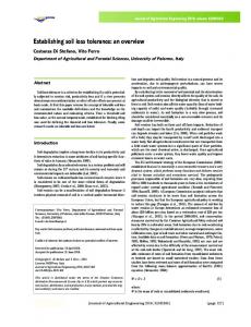
Results Severe alterations in bone marrow progenitor populations in a subset of STAT5A/5B-deficient mice, coincident with infiltration of memory CD4⫹ and CD8⫹ T cells FACS analysis of the bone marrow compartment of STAT5A/5Bdeficient mice at 6 –7 wk of age revealed a wide range in the severity of alterations in the representation of two progenitor subsets found among cells that have low expression of lineage-commitment markers: 1) the Sca-1⫹c-kit⫹ population, which contains the HSC; and 2) the Sca-1⫺c-kit⫹ post-HSC myeloid progenitors, which include the common myeloid progenitor, megakaryocyteerythroid progenitor, and granulocyte-macrophage progenitor (37, 38). Contour plots of c-kit and Sca-1 expression portray the normal distribution of the progenitor subsets from a typical STAT5A/ 5B⫹/⫹ animal, and abnormal distribution in a moderately affected STAT5A/5B⫺/⫺ animal and a severely affected STAT5A/5B⫺/⫺ animal (as based on the criteria below) (Fig. 1). These plots reveal a
FIGURE 1. The HSC and post-HSC abnormalities in STAT5A/5B⫺/⫺ mice exhibit variable expressivity. Subsetting was performed according to surface expression of lineage markers (Lin), Sca-1, and c-kit for the LindimSca-1⫹c-kit⫹ population, which contains the HSC, and the LindimSca-1⫺c-kit⫹ population, containing post-HSC myeloid progenitors. Representative contour plots showing Sca-1 and c-kit surface expression by lineage marker-dim cells for a typical STAT5A/5B⫹/⫹ animal, a moderately affected STAT5A/5B⫺/⫺ animal, and a severely affected STAT5A/5B⫺/⫺ animal.
5044
AUTOIMMUNITY IN STAT5A/5B-DEFICIENT MICE
dramatic increase in the relative representation of the HSC-containing population as well as a substantial decrease of the postHSC myeloid progenitor population in the severely affected animal; similar, although less severe, changes were evident in the moderately affected STAT5A/5B-deficient animal. When we examined these subsets in absolute terms, we found that the STAT5A/ 5B⫺/⫺ mice could be separated into two groups based on the absolute number of cells in the Sca-1⫹c-kit⫹ HSC-containing population. We classified 4 of 10 of STAT5A/5B⫺/⫺ mice as severely affected and the remaining 6 of 10 STAT5A/5B⫺/⫺ mice as moderately affected. There was a ⬎2-fold increase in the Sca-1⫹ckit⫹ population in the severely affected STAT5A/5B⫺/⫺ mice compared with either the moderately affected STAT5A/5B⫺/⫺ mice or STAT5A/5B⫹/⫹ mice (Table I). We further found that all STAT5A/ 5B⫺/⫺ mice possessed numbers of Sca-1⫺c-kit⫹ cells that were well below those of wild-type littermates. Because the Sca-1⫺ckit⫹ post-HSC myeloid progenitor population represents a direct product of the Sca-1⫹c-kit⫹ HSC population, we calculated the number of the post-HSC myeloid progenitors per HSC in each of these groups as another means to represent the severity of the block at this stage of hemopoiesis. Severely affected STAT5A/ 5B⫺/⫺ animals had a greatly decreased number of post-HSC myeloid progenitors per HSC (0.684 ⫾ 0.28) compared with moderately affected STAT5A/5B⫺/⫺ animals (2.02 ⫾ 0.15) or STAT5A/ 5B⫹/⫹ animals (6.83 ⫾ 0.687). These data reveal that, based on the criterion of absolute number of cells in the Sca-1⫹c-kit⫹ population, the STAT5A/5B⫺/⫺ genotype segregates into two fundamental groups. To determine whether abnormalities in mature lymphocytes in a subset of animals might contribute to the existence of a bimodal distribution in STAT5A/5B-deficent mice, we examined lymphocyte subsets within the bone marrow of these mice. FACS analysis revealed that the mice with the most severe alterations in bone marrow progenitor subsets also had a substantial increase in the absolute numbers of memory CD4⫹ (Fig. 2A) and CD8⫹ (B) T cells in the bone marrow compartment (severely affected animals; E) compared with the moderately affected STAT5A/5B⫺/⫺ mice (F) and wild-type littermates. There was a 4-fold increase in the number of memory CD3⫹CD4⫹ (CD62LlowCD44high) T cells in the severely affected animals (36.5 ⫾ 9.71 ⫻ 104 cells) compared with the moderately affected animals (7.94 ⫾ 2.93 ⫻ 104 cells) and STAT5A/5B⫹/⫹ animals (6.43 ⫾ 0.678 ⫻ 104 cells). Similarly, there was a 4-fold increase in the number of memory CD3⫹CD8⫹ (CD62LlowCD44high) T cells in the severely affected animals (16.3 ⫾ 5.66 ⫻ 104 cells) compared with the moderately affected animals (3.66 ⫾ 0.823 ⫻ 104 cells) and the STAT5A/5B⫹/⫹ animals (3.63 ⫾ 0.375 ⫻ 104 cells). The presence of increased numbers of memory T cells in bone marrow implied that these cells had been activated and homed to this tissue in response to some foreign Ag or self-Ag localized to the bone marrow. The association of this infiltration with striking abnormalities in progenitor subsets im-
FIGURE 2. STAT5A/5B⫺/⫺ mice with severe hemopoietic abnormalities exhibit bone marrow infiltration by memory CD4⫹ and CD8⫹ memory cells. Subsetting was performed according to surface expression of CD4 or CD8 in combination with CD44 and CD62L. Absolute values were obtained by multiplying gated percentages by total nucleated cell numbers. F, Animals from the STAT5A/5B⫹/⫹ group and from the STAT5A/5B⫺/⫺ moderate group; E, animals from the STAT5A/5B⫺/⫺ severe group in Table I. A, Memory CD4⫹ cells in bone marrow. B, Memory CD8⫹ cells in bone marrow.
plied that the presence of memory T cells might contribute to the phenotypic alterations observed in the bone marrow. Cellular infiltration is observed in other organs of severely affected STAT5A/5B-deficient mice To determine whether the T cell infiltration we observed in the bone marrow represented systemic pathology, we performed histological analyses of other organs, including liver, colon, and kidney. We observed significant cellular infiltration in all organs examined from the STAT5A/5B-deficient mice that exhibited increased numbers of T cells in the bone marrow, as described earlier. The colon was the most severely affected organ, with massive mononuclear and polymorphonuclear cell infiltration of the lamina propria (Fig. 3A). The inflammation was associated with disruption of the architecture of the tissue including elongation of the crypts and widening of the gap between crypts. In the kidney,
Table I. Absolute number of cells in Lindim progenitor subsets (⫻104)a
STAT5A/5B⫹/⫹ STAT5A/5B⫺/⫺ Moderate Severe
Sca-1⫹/c-kit⫹
Sca-1⫺/c-kit⫹
4.1 ⫾ 0.232
27.7 ⫾ 2.54
5.09 ⫾ 1.24 13.4 ⫾ 1.87b,c
9.75 ⫾ 1.84b 8.45 ⫾ 2.75b
a Total nucleated cells obtained from hind legs. Absolute values were obtained by multiplying gated percentages and total nucleated cell numbers. b p ⬍ 0.05 compared to STAT5A/5B⫹/⫹. c p ⬍ 0.05 compared to STAT5A/5B⫺/⫺ moderate.
FIGURE 3. Mononuclear cell infiltration in multiple organs of severely affected STAT5A/5B-deficient mice. Histological analysis of H&E sections from colon (A), kidney (B), and liver (C) of both STAT5A/5B⫹/⫹ and STAT5A/5B⫺/⫺ mice that were previously defined as severely affected reveal infiltration of these tissues by mononuclear cells in the STAT5A/5B⫺/⫺ mice. All images are at ⫻20 magnification.
The Journal of Immunology
5045
there was mononuclear cell infiltration of the tubular interstitium (large arrow) and sporadic alterations of glomerular architecture; primarily cell loss and apparent shrinkage (small arrow) were also noted (Fig. 3B). The liver exhibited mononuclear cell infiltration surrounding the vasculature of the portal triad with few cells seen invading the parenchyma (Fig. 3C). We did not observe infiltration of either the colon or the kidney in moderately affected STAT5A/ 5B-deficient mice or in STAT5A/5B wild-type littermates. However, we did observe a cellular infiltration of the liver parenchyma in one of the moderately affected STAT5A/5B-deficient animals, which had the characteristics of extramedullary hemopoietic foci (data not shown). Mononuclear and polymorphonuclear cell infiltration of multiple organs observed coincidentally with histologic destruction of the tissues in affected organs suggested the possibility that a more global breakdown of the maintenance of tolerance might be occurring in STAT5A/5B-deficient mice. Reduced survival in STAT5A/5B-deficient mice is rescued on an immunodeficient RAG1-deficient background We found that STAT5A/5B-deficient mice died spontaneously as early as 6 wk of age and that ⬎70% (14 of 19) of these mice were dead by 20 wk of age (Fig. 4A). Based on the mononuclear cell infiltration we detected in the livers, colons, and kidneys as well as the CD4⫹ and CD8⫹ T cell infiltration we observed in the bone marrow, we hypothesized that the premature deaths were linked to infiltration by lymphocytes. To test the hypothesis of a cause-andeffect relationship, we crossed the STAT5A/5B-deficient mice onto an immunodeficient RAG1⫺/⫺ background. Preliminary evidence indicated that both STAT5A/5B⫹/⫺ and STAT5A/5B⫺/⫺ mice on the RAG1⫺/⫺ background were more susceptible to infection compared with mice on a RAG1⫹/⫹ background. This phenomenon was likely due to the neutropenia (Ref. 24 and data not shown) and NK cell deficiencies observed in these mice. To reduce the confounding effects of infection-related deaths on our assessment of the role of lymphocytes in STAT5A/5B-deficient mice, we measured survival curves for the various STAT5A/5B genotypes on both RAG1⫹/⫹ and RAG1⫺/⫺ backgrounds in the presence of antibiotics administered through the drinking water. We observed that, although ⬎70% (5 of 7) of STAT5A/5B⫺/⫺ mice on the RAG1⫹/⫹ background died by 20 wk, only 9% (1 of 11) of the STAT5A/5B⫺/⫺ on the RAG1⫺/⫺ background had succumbed by the same time period (Fig. 4B). Furthermore, examination of euthanized STAT5A/5B-deficient mice on the RAG1⫺/⫺ background revealed no evidence of mononuclear cell infiltration in any organs (data not shown). In addition, we found no alteration in the fractional representation (Fig. 4C) of the HSC or post-HSC myeloid progenitor subsets in the bone marrow of these mice compared with that of STAT5A/5B⫹/⫹ animals on the RAG1⫺/⫺ background. To determine whether lymphocytes might contribute to other hematologic abnormalities observed in these mice, we performed RBC counts on STAT5A/5B-deficient mice on RAG1⫹/⫹ and RAG1⫺/⫺ backgrounds. We found that the presence of severe anemia in STAT5A/5B-deficient mice (Fig. 4D) was rescued on a RAG1⫺/⫺ background (E), indicating that lymphocytes are indispensable for this pathology. These data demonstrate that the pathologies observed in multiple tissues, including alterations of bone marrow progenitor subsets and severe anemia, are dependent on the presence of lymphocytes. In addition, because the removal of lymphocytes rescued the early death exhibited by STAT5A/5Bdeficient mice, these data suggest that the early death is linked to infiltration of these organs by lymphocytes. Finally, these results indicate that more animals will likely develop the pathology than initially thought based on the 6 wk time point. However, because ⬃25% of animals do not develop disease, although our initial es-
FIGURE 4. An immunodeficient background fully abrogates the lethality, bone marrow progenitor effects, and anemia of STAT5A/5B deficiency. Subsetting was performed as above. A, Survival analysis of STAT5A/5B⫹/⫹ mice (n ⫽ 18) and STAT5A/5B⫺/⫺ mice (n ⫽ 19). B, Survival analysis of STAT5A/5B⫹/⫹ mice (n ⫽ 9) and STAT5A/5B⫺/⫺ mice (n ⫽ 11) on the RAG1⫹/⫹ background and STAT5A/5B⫹/⫹ mice (n ⫽ 14) and STAT5A/ 5B⫺/⫺ mice (n ⫽ 11) on the RAG1⫺/⫺ background in the presence of antibiotics. C, Representative contour plots showing Sca-1 and c-kit surface expression by lineage marker-dim cells for STAT5A/5B⫹/⫹ mice and STAT5A/5B⫺/⫺ mice on the RAG1⫺/⫺ background are shown. D and E, RBC per microliter for STAT5A/5B⫹/⫹ mice and STAT5A/5B⫺/⫺ mice on the RAG1⫹/⫹ background (D) and on the RAG1⫺/⫺ background (E) are shown. F, Survival analysis of STAT5A/5B-deficient neonates receiving CD4⫹ cells (n ⫽ 5) or no cells (n ⫽ 5) is shown.
timates of the penetrance are underestimates, the disease still appears to follow a bimodal distribution. Transfer of wild-type CD4 T cells is sufficient to abrogate disease To assist in assessing the role of T cells in the pathology we observed, we transplanted either 1 ⫻ 106 CD4⫹ cells from wild-type
5046
AUTOIMMUNITY IN STAT5A/5B-DEFICIENT MICE
mice or no cells into STAT5A/5B-deficient neonates and followed survival over 16 wk. We observed that, whereas 67% of STAT5A/ 5B⫺/⫺ mice receiving no cells succumbed within 16 wk, no STAT5A/5B⫺/⫺ mice that received CD4⫹ cells died during the same period (Fig. 4F). These results indicate that wild-type CD4 cells are sufficient to provide protection from early death to STAT5A/5B⫺/⫺ mice.
In addition, as in the STAT5A/5B⫺/⫺ mice, these abnormalities in the bone marrow were associated with a substantial increase in the absolute numbers of memory CD4⫹ and CD8⫹ T cells in the bone marrow compartment (Fig. 5). These data, in conjunction with other similarities noted in these animals, suggest that loss of tolerance in these two mouse models results from defects in the same mechanism of maintaining tolerance.
Mice deficient for IL-2R chain exhibit similar abnormalities in the bone marrow
STAT5A/5B-deficient mice have reduced numbers of CD4⫹CD25⫹ regulatory T cells in secondary lymphoid organs and in peripheral tissue
Previous reports indicate some hemopoietic progenitor abnormalities in mice lacking IL-2 (39, 40), with mononuclear cell infiltration in the colon and liver and anemia in mice deficient for IL-2 (9), IL-2R␣ (8), or IL-2R (7) similar to that observed by us in STAT5A/5B-deficient mice. We speculated that, if IL-2 signaling was responsible for the tolerizing effect of STAT5, mice deficient in IL-2 signaling would manifest the same specific bone marrow alterations that we had observed in the STAT5A/5B-deficient mice. Indeed, we found that, at 6 wk of age, mice deficient for IL-2R exhibited bone marrow abnormalities including T cell infiltration and alterations in the representation of lineage marker-dim progenitor populations. The phenotypes appeared more severe in these mice and displayed a higher penetrance, because all IL-2R-deficient mice examined were markedly affected (Fig. 5).
FIGURE 5. IL-2R-deficient mice manifest severe hemopoietic abnormalities like those in STAT5A/5B-deficient mice. Subsetting and generation of absolute values was performed as above. A, Representative contour plots showing Sca-1 and c-kit surface expression by lineage marker-dim cells for an IL-2R⫹/⫹ and an IL-2R⫺/⫺ animal are shown. B–E, Scatter plots showing absolute values for HSC (B), post-HSC myeloid progenitors (C), memory CD4⫹ cells in bone marrow (D), and memory CD8⫹ cells in bone marrow of both IL-2R⫹/⫹ and an IL-2R⫺/⫺ animal (E) are shown.
Because the multiorgan autoimmunity seen in the STAT5A/5B⫺/⫺ mice was strikingly similar to that seen in IL-2R⫺/⫺ mice, we hypothesized that the same mechanism for disruption of tolerance might be involved in both types of gene-targeted mice. One model to account for the loss of tolerance in these mice, is that a reduction in an IL-2-dependent subset of CD4⫹CD25⫹ regulatory T cells causes a disruption in the maintenance of peripheral tolerance to multiple self-Ags (41– 45). Therefore, we examined STAT5-deficient mice for the number of T cells expressing these markers both in a secondary lymphoid organ, the spleen, and in an affected endorgan, the bone marrow. We observed that there was a ⬃4-fold decrease in both CD25⫹ and CD25⫺ CD4⫹ cells in the spleen,
FIGURE 6. STAT5A/5B-deficient mice have reduced numbers of CD4⫹CD25⫹ regulatory T cells. Total nucleated cells obtained from either the spleen or the bone marrow were analyzed. Subsetting was performed according to surface expression of CD4 and CD25. A and B, Plots showing CD4⫹CD25⫺ and CD4⫹CD25⫹ subsets in both the spleen (A) and the bone marrow (B) are shown. C, Surface expression of CD62L on CD4⫹CD25⫹ cells in the spleen is shown.
The Journal of Immunology such that the relationship of CD4⫹CD25⫹ to CD4⫹CD25⫺ cells was preserved (Fig. 6A). In contrast, the absolute number of CD4⫹CD25⫺ cells in the bone marrow was unaffected in STAT5A/ 5B-deficient mice, whereas the absolute number of CD4⫹CD25⫹ in this tissue was reduced 2-fold in STAT5A/5B⫺/⫺ mice compared with wild-type mice (Fig. 6B). To exclude the possibility that CD4⫹CD25⫹ cells had down-regulated CD62L and thus represented activated naive cells that confounded quantitation of regulatory T cells (46), we also examined the expression of CD62L. We found that 78.1% of CD4⫹CD25⫹ cells in the spleens of STAT5A/5B⫹/⫹ animals were CD62Lhigh, whereas only 33.8% of these cells in the spleens of STAT5A/5B⫺/⫺ mice were CD62Lhigh (Fig. 6C). Therefore, by this analysis, there was in fact a relative reduction in the actual number of regulatory T cells vs nonregulatory T cells in the spleens of STAT5A/5B-deficient animals. Similar results for CD62L expression were observed in the bone marrow. Because CD25 is regulated by STAT5, we wished to
5047 demonstrate that the reduction in regulatory T cells we observed was not in fact due simply to down-regulation of surface markers in the absence of STAT5. We found that there was a representational decrease in CD4⫹CD62Lhigh cells with a CD45RBlow (Fig. 7, C and D) or GITRhigh phenotype (G and H), as well as in CD4⫹ cells with a CD103⫹ phenotype (E and F), all characteristic of regulatory T cells (47) in STAT5A/5B-deficient mice compared with wild-type mice. In accordance with our model and the conclusion of others (41, 43) that IL-2 was important for the regulation of this population, we found that the number of CD4⫹CD25⫹ CD62Lhigh cells in the spleen of IL-2R⫺/⫺ mice was reduced compared with wild-type littermates (data not shown). Thus, the overall reduction of regulatory T cells in STAT5A/5B⫺/⫺ mice may contribute to a loss of peripheral tolerance in these mice, thereby causing the multiorgan autoimmunity observed in a manner similar to mice that lack all IL-2R signaling. CD4⫹CD25⫹ regulatory T cells have an increased rate of apoptosis and decreased Bcl-2 expression in STAT5A/5Bdeficient mice To determine how STAT5 may contribute to maintenance of CD4⫹CD25⫹ cells, we examined both proliferation and apoptosis of this subset in vivo. We observed a ⬃2-fold increase in the proportion of CD4⫹CD25⫹CD62Lhigh cells in the spleen staining positive for annexin V in STAT5A/5B⫺/⫺ mice compared with wild-type mice (Fig. 8A). In addition, in vivo labeling with BrdU revealed that the proportion of CD4⫹CD25⫹CD62Lhigh cells in the spleen that were BrdUhigh, and thus had incorporated BrdU during DNA synthesis in the periphery, was increased in STAT5A/ 5B⫺/⫺ mice compared with STAT5A/5B⫹/⫹ mice (data not shown). Therefore, despite an apparent ability to undergo homeostatic proliferation in secondary lymph organs, the regulatory T cell population in STAT5A/5B⫺/⫺ mice has a higher rate of cell death, which correlates with the reduction in their numbers in these animals. In addition, we also observed a ⬃2-fold increase in the proportion of CD4⫹CD25⫹CD62Lhigh cells in the spleen staining positive for annexin V in IL-2R⫺/⫺ mice compared with wildtype mice (data not shown). To determine whether Bcl-2 might
FIGURE 7. Representational decrease in regulatory T cells by multiple surface criteria. Lymph node cells from STAT5A/5B⫹/⫹ (A, C, E, and G) and STAT5A/5B⫺/⫺ (B, D, F, and H) mice were stained for CD4, CD62L, and CD25 (A and B), CD45RB (C and D), CD103 (E and F), and GITR (G and H). Representational plots are shown.
FIGURE 8. Increased apoptosis and decreased Bcl-2 expression in regulatory T cells from STAT5A/5B-deficent mice. CD4⫹CD25⫹CD62Lhigh splenocytes were stained for either the surface expression of annexin V (A) or intracellular Bcl-2 (B) with dotted lines representing isotype control and solid lines representing anti-Bcl-2 Ab staining.
5048 play a role in the regulation of apoptosis in regulatory T cells, we then examined its expression in these cells by intracellular FACS. We found that nearly all of these CD4⫹CD25⫹CD62Lhigh cells in the spleen expressed Bcl-2 protein in wild-type mice (Fig. 8B, left panel), whereas fewer than half of CD4⫹CD25⫹CD62Lhigh cells in the spleen of STAT5A/5B-deficient mice expressed Bcl-2 protein above background (B, right panel). An even more striking reduction in the proportion of CD4⫹CD25⫹CD62Lhigh cells expressing the Bcl-2 protein above background was observed in IL-2R⫺/⫺ mice compared with wild-type mice (data not shown). These data indicate that STAT5 contributes to the homeostasis of CD4⫹CD25⫹ regulatory T cells in part through survival effects that may be mediated through Bcl-2. Similar results observed in IL-2R⫺/⫺ mice suggests that IL-2 may be the cytokine that provides the STAT5-dependent survival signals to the regulatory T cells that are critical for their homeostasis. In addition, we found that, when whole spleen and lymph node cells were cultured for 24 – 48 h in the presence of IL-2 or IL-2 and PMA, CD4⫹CD25⫹CD62Lhigh cells from STAT5-deficient mice displayed increased rates of apoptosis, as assessed by annexin V staining, compared with these cells from wild-type mice (data not shown), further supporting a model in which IL-2 mediates survival of these cells through activation of STAT5.
Discussion Because STAT5 is activated by a diverse array of cytokine receptors, we hypothesized that defects that are non-cell autonomous to early progenitors might contribute to the complex, and variably expressed, hematologic defects in STAT5A/5B-deficient mice. Specifically, we posited that a loss of tolerance in the adaptive immune system might lead to autoimmune-mediated effects on hemopoiesis. Three lines of evidence led us to this hypothesis: 1) an autoimmune etiology has been postulated to be responsible for a number of human diseases that affect the function of hemopoietic progenitor cells and interfere with their ability to produce mature cells of the hemopoietic lineages, such as aplastic anemia, pure red cell aplasia, and paroxysmal nocturnal hemoglobinuria (48 –51); 2) the role of IL-2 and its receptor system in the maintenance of tolerance has been well established (7–10, 13), and mice lacking IL-2 manifest hemopoietic abnormalities affecting bone marrow (39, 40); and 3) the importance of STAT5 activation by the IL-2R system has been well documented in vitro and in vivo (11, 12). In a higher resolution examination of the early progenitor compartment of the bone marrow of STAT5A/5B-deficient mice, we established two phenotypic categories, which we termed moderately affected and severely affected. The two most striking characteristics of the severely affected mice were dramatic increases in the HSC-containing population and low production of post-HSC myeloid progenitors despite the increased HSC population. We found that the severely affected mice exhibited a dramatic increase in the number of memory CD4⫹ and CD8⫹ T cells infiltrating the bone marrow. Because STAT5A/5B⫺/⫺ mice are lymphopenic, this infiltration of T cells represents a heightened recruitment of lymphocytes into this tissue rather than a new equilibrium reached with an increased total T cell pool. T cells localized aberrantly in the bone marrow could cause the progenitor effects observed either by inhibiting the productive capacity of HSC or by reducing the number of post-HSC. Further study is required to determine whether these effects are caused by Ag-specific autoimmunity or by bystander effects and whether the effector cells that are responsible for pathology are CD4⫹ or CD8⫹ T cells (4, 52–54). We found that the immune cell infiltration was a more general phenomenon, because we observed it in all organs examined, including colon, kidney, and liver. In both the colon and the kidney,
AUTOIMMUNITY IN STAT5A/5B-DEFICIENT MICE this infiltration resulted in architectural abnormalities, which could have resulted in compromised function of these tissues. These severe abnormalities in multiple organs most likely contribute to the 70% incidence of premature death observed in STAT5A/5B-deficient mice by 20 wk of age. The longitudinal analysis indicates that the cross-sectional analysis performed at 6 –7 wk of age detected the presence of disease in only a fraction of those animals that would ultimately develop pathology. Crossing the STAT5A/5B-deficient mice onto the RAG1⫺/⫺ background abrogated severe anemia, the incidence of early death, and any visible alterations in bone marrow, colon, kidney, and liver in STAT5A/5B⫺/⫺ animals. In addition, addition of wild-type CD4⫹ cells into STAT5A/5B⫺/⫺ mice abrogated early death in these mice, demonstrating that CD4 cells are sufficient to prevent disease. These observations suggest that defects in STAT5A/5B⫺/⫺ lymphocytes, probably within CD4⫹ cells in particular, are responsible for the observed pathology. The complex effects of STAT5 deficiency on other cell types such as the bone marrow progenitors (18, 24, 25, 27–31) may also play a role in the pathology observed in these organs. The lymphocytic infiltration of the colon and liver as well as the recognized role of STAT5 in IL-2 signaling implicated impaired signaling by this cytokine as the cause of loss of tolerance in STAT5A/5B-deficient mice. In further support of this, all IL2R⫺/⫺ mice possessed bone marrow characteristics similar to the severely affected STAT5A/5B⫺/⫺ mice. In addition, regulatory T cells from STAT5A/5B-deficient mice did demonstrate reduced survival in response to IL-2 in vitro. A previous study found that IL-2R⫺/⫺ mice expressing a mutant IL-2R transgene that is unable to activate STAT5 did not develop autoimmune disease. However, this model used a transgene on a knockout background that may not faithfully recapitulate endogenous IL-2R expression in certain subpopulations. In addition, the measurement of T cell activation and autoimmune disease was done at 8 wk. It is possible that the occurrence of pathology is delayed in these mice compared with IL-2R⫺/⫺ mice. It is interesting to note that the JAK-binding S region appears to be able to function with either the H or A region to prevent autoimmune disease in these mice. It might be interesting to know whether the JAK-binding S region alone would suffice as well. Additional experiments must be undertaken to reconcile these discordant pieces of data (55). In addition, the severity of the abnormalities in the IL-2R-deficient mice was markedly higher compared with the defects in STAT5A/5B-deficient mice. The increased penetrance and severity of pathology in the IL2R⫺/⫺ mice might be explained by one or more differences between STAT5A/5B⫺/⫺ and IL-2R⫺/⫺ mice. First, loss of STAT5 signaling may not fully disable IL-2 signaling competence, whereas loss of the  subunit of the IL-2R would have more profound effects on the complex downstream signaling pathways. This distinction might allow persistence of a portion of the tolerogenic effect of IL-2R signaling in the STAT5A/5B-deficient mice. Second, loss of STAT5 has more wide-ranging effects on lymphocytes than loss of IL-2R (18, 24, 30, 31), which may result in reduced incidence of autoimmunity or a reduced ability of effector populations to cause damage. In any event, it would appear that STAT5 is an important mediator of IL-2-mediated tolerance maintenance. Currently, loss of tolerance in the absence of IL-2 or any of its receptor components is thought to be due to loss of two tolerizing mechanisms. First, activation of the IL-2R is important for upregulation of Fas and activation-induced cell death (6, 34, 56, 57). Second, there is a requirement for this cytokine in the maintenance of a population of CD4⫹CD25⫹ regulatory T cells (41– 43, 58, 59). Reduced number and/or function of these regulatory cells have been observed in multiple mouse models of autoimmunity
The Journal of Immunology (44, 45, 60). Furthermore, such cells possess the capacity to inhibit responses of CD4⫹CD25⫺ T cells in vitro (61– 63). The dependence of such regulatory T cells on the presence of IL-2 and intact receptor components has been shown both in vivo and in vitro (41, 43– 45). However, the signaling molecules downstream of IL-2 and its cognate receptor that are important for the regulation of these cells and the mechanisms by which these signals affect cell number are not well understood (13). A representational loss of CD4⫹CD25⫹ cells has been reported in the spleens of DO11.10 STAT5A-deficient mice (33), but the relevance of this observation to the role of STAT5 in the maintenance of tolerance has not been explored. In the present study, we found that there were reductions in the number of regulatory T cells in both the spleen and lymph nodes by multiple cell surface criteria, and in an end-organ tissue, the bone marrow, by CD4⫹CD25⫹ phenotype, in STAT5A/5B⫺/⫺ animals. These data demonstrate that STAT5 is indispensable for the maintenance of this population in vivo, which may contribute significantly to the loss of tolerance in these mice. In addition, the results of the CD4⫹ add-back experiment provide evidence that a population of wild-type CD4⫹ cells can rescue the defects in these mice, which supports the hypothesis that loss of regulatory T cells could be involved in the loss of tolerance observed. Interestingly, although all STAT5A/5B-deficient animals exhibited reduced numbers of these cells, not all of the mice developed autoimmune pathology or succumbed to premature death. Because many of the phenotypes seen in STAT5A/5B-deficient mice exhibit varying degrees of expression in individual mice, development of autoimmunity may require a convergence of multiple threshold effects. The mechanism by which IL-2 contributes to the maintenance of these cells in vivo is under intense investigation. Defects in thymic development, or in peripheral proliferation or survival, could all be involved. Our finding that a greater proportion of CD4⫹ CD25⫹CD62Lhigh regulatory T cells were apoptotic in STAT5A/ 5B-deficent animals compared with littermates implies that STAT5 may be important for the survival of the cells. These data are consistent with earlier work by ourselves and others (24, 27–29) that showed STAT5 to be an important mediator of survival signals in multiple cell types. In addition, this hypothesis was further supported by the observation that a smaller proportion of the CD4⫹CD25⫹CD62Lhigh cells were positive for Bcl-2 protein, which may be an important target of STAT5 in providing survival signals. IL-2 has been shown to provide antiapoptotic signals to cell lines in vitro (64) and to activate Bcl-2 transcription through STAT5 (65). These findings are consistent with the known role for STAT5 as a mediator of survival signals in other cell types (27– 29). However, other STAT5-dependent mechanisms could be important for providing increased survival in these cells, such as upregulation of other Bcl-2 family members or the IL-2R␣ chain (66). The finding that these cells in IL-2R⫺/⫺ mice also have increased apoptosis and decreased expression of Bcl-2 suggest that the IL-2R mediates effects on regulatory T cells in part though modulation of their survival, perhaps through STAT5-dependent effects. Additionally, these data suggest a novel role of apoptosis in the cytokine-mediated homeostasis of this cellular population (44, 45). Our data support a model in which IL-2 contributes to the survival of CD4⫹CD25⫹CD62Lhigh regulatory T cells in vivo, through receptor-mediated activation of STAT5. The reduction in the number of these cells contributes to loss of tolerance in STAT5A/5B-deficient animals, leading to a lymphocyte-dependent autoimmune disease affecting multiple organs. One sequela of this disease is the lymphocyte-dependent perturbation of the hemopoi-
5049 etic system, contributing to the complex hematologic phenotypes seen in STAT5A/5B-deficient mice. These data provide the first evidence that STAT5 is important for the maintenance of tolerance in vivo, which adds a new function to the growing list that this transcription factor plays in the context of the organism. Whether STAT5 plays its tolerogenic role downstream of the IL-2R exclusively, or whether other receptors are involved, must be further explored. In addition, the extent of the cell types involved in STAT5-dependent maintenance of tolerance and the downstream genes through which STAT5 mediates these effects need further study.
Acknowledgments We thank Dr. James Ihle for kindly providing STAT5A/5B⫺/⫺ mice and Dr. Tak Mak for providing IL-2R⫺/⫺ mice. We gratefully acknowledge the technical assistance of the Gladstone Flow Cytometry Core and the Gladstone Histology Core in the conduct of these experiments and the University of California, San Francisco, Laboratory Animal Resource Center animal care staff. Also, we thank Mauricio Montano for assistance in experiments; Dr. Oliver Keppler, Dr. Andreas Jeckle, and Jason Kreisberg for critical review of the manuscript; Dr. Kevin Shannon, Dr. Warner Greene, and Dr. Stephen Chan for scientific advice; and Heather Gravois, Sue Cammack, Robin Givens, Jack Hull, Chris Goodfellow, and John Carroll for their assistance in the preparation of this manuscript.
References 1. Marrack, P., J. Kappler, and B. L. Kotzin. 2001. Autoimmune disease: why and where it occurs. Nat. Med. 7:899. 2. O’Shea, J. J., A. Ma, and P. Lipsky. 2002. Cytokines and autoimmunity. Nat. Rev. Immunol. 2:37. 3. Durkin, H. G., and B. H. Waksman. 2001. Thymus and tolerance: is regulation the major function of the thymus? Immunol. Rev. 182:33. 4. O’Garra, A., L. Steinman, and K. Gijbels. 1997. CD4⫹ T-cell subsets in autoimmunity. Curr. Opin. Immunol. 9:872. 5. Falcone, M., and N. Sarvetnick. 1999. Cytokines that regulate autoimmune responses. Curr. Opin. Immunol. 11:670. 6. Van Parijs, L., and A. K. Abbas. 1998. Homeostasis and self-tolerance in the immune system: turning lymphocytes off. Science 280:243. 7. Suzuki, H., T. M. Ku¨ ndig, C. Furlonger, A. Wakeham, E. Timms, T. Matsuyama, R. Schmits, J. J. Simard, P. S. Ohashi, H. Griesser, et al. 1995. Deregulated T cell activation and autoimmunity in mice lacking interleukin-2 receptor . Science 268:1472. 8. Willerford, D. M., J. Chen, J. A. Ferry, L. Davidson, A. Ma, and F. W. Alt. 1995. Interleukin-2 receptor ␣ chain regulates the size and content of the peripheral lymphoid compartment. Immunity 3:521. 9. Sadlack, B., H. Merz, H. Schorle, A. Schimpl, A. C. Feller, and I. Horak. 1993. Ulcerative colitis-like disease in mice with a disrupted interleukin-2 gene. Cell 75:253. 10. Nakajima, H., E. W. Shores, M. Noguchi, and W. J. Leonard. 1997. The common cytokine receptor ␥ chain plays an essential role in regulating lymphoid homeostasis. J. Exp. Med. 185:189. 11. Gaffen, S. L. 2001. Signaling domains of the interleukin 2 receptor. Cytokine 14:63. 12. Lin, J. X., and W. J. Leonard. 2000. The role of Stat5a and Stat5b in signaling by IL-2 family cytokines. Oncogene 19:2566. 13. Nelson, B. H. 2002. Interleukin-2 signaling and the maintenance of self-tolerance. Curr. Dir. Autoimmun. 5:92. 14. Bromberg, J., and J. E. Darnell, Jr. 2000. The role of STATs in transcriptional control and their impact on cellular function. Oncogene 19:2468. 15. Ihle, J. N. 1996. STATs: signal transducers and activators of transcription. Cell 84:331. 16. Takeda, K., B. E. Clausen, T. Kaisho, T. Tsujimura, N. Terada, I. Forster, and S. Akira. 1999. Enhanced Th1 activity and development of chronic enterocolitis in mice devoid of Stat3 in macrophages and neutrophils. Immunity 10:39. 17. Homann, D., A. Holz, A. Bot, B. Coon, T. Wolfe, J. Petersen, T. P. Dyrberg, M. J. Grusby, and M. G. von Herrath. 1999. Autoreactive CD4⫹ T cells protect from autoimmune diabetes via bystander suppression using the IL-4/Stat6 pathway. Immunity 11:463. 18. Teglund, S., C. McKay, E. Schuetz, J. M. van Deursen, D. Stravopodis, D. Wang, M. Brown, S. Bodner, G. Grosveld, and J. N. Ihle. 1998. Stat5a and Stat5b proteins have essential and nonessential, or redundant, roles in cytokine responses. Cell 93:841. 19. Liu, X., G. W. Robinson, K. U. Wagner, L. Garrett, A. Wynshaw-Boris, and L. Hennighausen. 1997. Stat5a is mandatory for adult mammary gland development and lactogenesis. Genes Dev. 11:179. 20. Udy, G. B., R. P. Towers, R. G. Snell, R. J. Wilkins, S. H. Park, P. A. Ram, D. J. Waxman, and H. W. Davey. 1997. Requirement of STAT5b for sexual dimorphism of body growth rates and liver gene expression. Proc. Natl. Acad. Sci. USA 94:7239.
5050 21. Feldman, G. M., L. A. Rosenthal, X. Liu, M. P. Hayes, A. Wynshaw-Boris, W. J. Leonard, L. Hennighausen, and D. S. Finbloom. 1997. STAT5A-deficient mice demonstrate a defect in granulocyte-macrophage colony-stimulating factorinduced proliferation and gene expression. Blood 90:1768. 22. Nakajima, H., X. W. Liu, A. Wynshaw-Boris, L. A. Rosenthal, K. Imada, D. S. Finbloom, L. Hennighausen, and W. J. Leonard. 1997. An indirect effect of Stat5a in IL-2-induced proliferation: a critical role for Stat5a in IL-2-mediated IL-2 receptor ␣ chain induction. Immunity 7:691. 23. Imada, K., E. T. Bloom, H. Nakajima, J. A. Horvath-Arcidiacono, G. B. Udy, H. W. Davey, and W. J. Leonard. 1998. Stat5b is essential for natural killer cell-mediated proliferation and cytolytic activity. J. Exp. Med. 188:2067. 24. Snow, J. W., N. Abraham, M. C. Ma, N. W. Abbey, B. Herndier, and M. A. Goldsmith. 2002. STAT5 promotes multilineage hematolymphoid development in vivo through effects on early hematopoietic progenitor cells. Blood 99:95. 25. Bunting, K. D., H. L. Bradley, T. S. Hawley, R. Moriggl, B. P. Sorrentino, and J. N. Ihle. 2002. Reduced lymphomyeloid repopulating activity from adult bone marrow and fetal liver of mice lacking expression of STAT5. Blood 99:479. 26. Zhang, S., S. Fukuda, Y. Lee, G. Hangoc, S. Cooper, R. Spolski, W. J. Leonard, and H. E. Broxmeyer. 2000. Essential role of signal transducer and activator of transcription (Stat)5a but not Stat5b for Flt3-dependent signaling. J. Exp. Med. 192:719. 27. Socolovsky, M., H. Nam, M. D. Fleming, V. H. Haase, C. Brugnara, and H. F. Lodish. 2001. Ineffective erythropoiesis in Stat5a⫺/⫺5b⫺/⫺ mice due to decreased survival of early erythroblasts. Blood 98:3261. 28. Socolovsky, M., A. E. Fallon, S. Wang, C. Brugnara, and H. F. Lodish. 1999. Fetal anemia and apoptosis of red cell progenitors in Stat5a⫺/⫺5b⫺/⫺ mice: a direct role for Stat5 in Bcl-xL induction. Cell 98:181. 29. Kieslinger, M., I. Woldman, R. Moriggl, J. Hofmann, J. C. Marine, J. N. Ihle, H. Beug, and T. Decker. 2000. Antiapoptotic activity of Stat5 required during terminal stages of myeloid differentiation. Genes Dev. 14:232. 30. Moriggl, R., V. Sexl, R. Piekorz, D. Topham, and J. N. Ihle. 1999. Stat5 activation is uniquely associated with cytokine signaling in peripheral T cells. Immunity 11:225. 31. Moriggl, R., D. J. Topham, S. Teglund, V. Sexl, C. McKay, D. Wang, A. Hoffmeyer, J. van Deursen, M. Y. Sangster, K. D. Bunting, et al. 1999. Stat5 is required for IL-2-induced cell cycle progression of peripheral T cells. Immunity 10:249. 32. Kagami, S., H. Nakajima, K. Kumano, K. Suzuki, A. Suto, K. Imada, H. W. Davey, Y. Saito, K. Takatsu, W. J. Leonard, and I. Iwamoto. 2000. Both Stat5a and Stat5b are required for antigen-induced eosinophil and T-cell recruitment into the tissue. Blood 95:1370. 33. Kagami, S., H. Nakajima, A. Suto, K. Hirose, K. Suzuki, S. Morita, I. Kato, Y. Saito, T. Kitamura, and I. Iwamoto. 2001. Stat5a regulates T helper cell differentiation by several distinct mechanisms. Blood 97:2358. 34. Van Parijs, L., Y. Refaeli, J. D. Lord, B. H. Nelson, A. K. Abbas, and D. Baltimore. 1999. Uncoupling IL-2 signals that regulate T cell proliferation, survival, and Fas-mediated activation-induced cell death. Immunity 11:281. 35. Sexl, V., R. Piekorz, R. Moriggl, J. Rohrer, M. P. Brown, K. D. Bunting, K. Rothammer, M. F. Roussel, and J. N. Ihle. 2000. Stat5a/b contribute to interleukin 7-induced B-cell precursor expansion, but abl- and bcr/abl-induced transformation are independent of Stat5. Blood 96:2277. 36. Ernst, J. D., L. Yang, J. L. Rosales, and V. C. Broaddus. 1998. Preparation and characterization of an endogenously fluorescent annexin for detection of apoptotic cells. Anal. Biochem. 260:18. 37. Akashi, K., D. Traver, T. Miyamoto, and I. L. Weissman. 2000. A clonogenic common myeloid progenitor that gives rise to all myeloid lineages. Nature 404:193. 38. Kondo, M., I. L. Weissman, and K. Akashi. 1997. Identification of clonogenic common lymphoid progenitors in mouse bone marrow. Cell 91:661. 39. Contractor, N. V., H. Bassiri, T. Reya, A. Y. Park, D. C. Baumgart, M. A. Wasik, S. G. Emerson, and S. R. Carding. 1998. Lymphoid hyperplasia, autoimmunity, and compromised intestinal intraepithelial lymphocyte development in colitisfree gnotobiotic IL-2-deficient mice. J. Immunol. 160:385. 40. Reya, T., N. V. Contractor, M. S. Couzens, M. A. Wasik, S. G. Emerson, and S. R. Carding. 1998. Abnormal myelocytic cell development in interleukin-2 (IL-2)-deficient mice: evidence for the involvement of IL-2 in myelopoiesis. Blood 91:2935. 41. Papiernik, M., M. L. de Moraes, C. Pontoux, F. Vasseur, and C. Penit. 1998. Regulatory CD4 T cells: expression of IL-2R ␣ chain, resistance to clonal deletion and IL-2 dependency. Int. Immunol. 10:371.
AUTOIMMUNITY IN STAT5A/5B-DEFICIENT MICE 42. Furtado, G. C., M. A. De Lafaille, N. Kutchukhidze, and J. J. Lafaille. 2002. Interleukin 2 signaling is required for CD4⫹ regulatory T cell function. J. Exp. Med. 196:851. 43. Malek, T., A. Yu, V. Vincek, P. Scibelli, and L. Kong. 2002. CD4 regulatory T cells prevent lethal autoimmunity in IL-2R-deficient mice: implications for the nonredundant function of IL-2. Immunity 17:167. 44. Maloy, K. J., and F. Powrie. 2001. Regulatory T cells in the control of immune pathology. Nat. Immunol. 2:816. 45. Shevach, E. M. 2002. CD4⫹CD25⫹ suppressor T cells: more questions than answers. Nat. Rev. Immunol. 2:389. 46. Chatenoud, L., B. Salomon, and J. A. Bluestone. 2001. Suppressor T cells— they’re back and critical for regulation of autoimmunity! Immunol. Rev. 182:149. 47. McHugh, R. S., M. J. Whitters, C. A. Piccirillo, D. A. Young, E. M. Shevach, M. Collins, and M. C. Byrne. 2002. CD4⫹CD25⫹ immunoregulatory T cells: gene expression analysis reveals a functional role for the glucocorticoid-induced TNF receptor. Immunity 16:311. 48. Young, N. S., and J. Maciejewski. 1997. The pathophysiology of acquired aplastic anemia. N. Engl. J. Med. 336:1365. 49. Young, N. S., J. L. Abkowitz, and L. Luzzatto. 2000. New insights into the pathophysiology of acquired cytopenias. In American Society of Hematology, Vol. Hematology 2000. American Society of Hematology, San Francisco, p. 18. 50. Croisille, L., G. Tchernia, and N. Casadevall. 2001. Autoimmune disorders of erythropoiesis. Curr. Opin. Hematol. 8:68. 51. Rosti, V. 2000. The molecular basis of paroxysmal nocturnal hemoglobinuria. Haematologica 85:82. 52. Santamaria, P. 2001. Effector lymphocytes in autoimmunity. Curr. Opin. Immunol. 13:663. 53. Vizler, C., N. Bercovici, A. Cornat, C. Cambouris, and R. S. Liblau. 1999. Role of autoreactive CD8⫹ T cells in organ specific autoimmune diseases: insight from transgenic mouse models. Immunol. Rev. 169:81. 54. Suri, A., and J. D. Katz. 1999. Dissecting the role of CD4⫹ T cells in autoimmune diabetes through the use of TCR transgenic mice. Immunol. Rev. 169:55. 55. Fujii, H., K. Ogasawara, H. Otsuka, M. Suzuki, K. Yamamura, T. Yokochi, T. Miyazaki, H. Suzuki, T. W. Mak, S. Taki, and T. Taniguchi. 1998. Functional dissection of the cytoplasmic subregions of the IL-2 receptor c chain in primary lymphocyte populations. EMBO J. 17:6551. 56. Van Parijs, L., A. Ibraghimov, and A. K. Abbas. 1996. The roles of costimulation and Fas in T cell apoptosis and peripheral tolerance. Immunity 4:321. 57. Van Parijs, L., A. Biuckians, A. Ibragimov, F. W. Alt, D. M. Willerford, and A. K. Abbas. 1997. Functional responses and apoptosis of CD25 (IL-2R␣)-deficient T cells expressing a transgenic antigen receptor. J. Immunol. 158:3738. 58. Suzuki, H., Y. W. Zhou, M. Kato, T. W. Mak, and I. Nakashima. 1999. Normal regulatory ␣/ T cells effectively eliminate abnormally activated T cells lacking the interleukin 2 receptor  in vivo. J. Exp. Med. 190:1561. 59. Wolf, M., A. Schimpl, and T. Hunig. 2001. Control of T cell hyperactivation in IL-2-deficient mice by CD4⫹CD25⫺ and CD4⫹CD25⫹ T cells: evidence for two distinct regulatory mechanisms. Eur. J. Immunol. 31:1637. 60. Asano, M., M. Toda, N. Sakaguchi, and S. Sakaguchi. 1996. Autoimmune disease as a consequence of developmental abnormality of a T cell subpopulation. J. Exp. Med. 184:387. 61. Takahashi, T., Y. Kuniyasu, M. Toda, N. Sakaguchi, M. Itoh, M. Iwata, J. Shimizu, and S. Sakaguchi. 1998. Immunologic self-tolerance maintained by CD25⫹CD4⫹ naturally anergic and suppressive T cells: induction of autoimmune disease by breaking their anergic/suppressive state. Int. Immunol. 10:1969. 62. Itoh, M., T. Takahashi, N. Sakaguchi, Y. Kuniyasu, J. Shimizu, F. Otsuka, and S. Sakaguchi. 1999. Thymus and autoimmunity: production of CD25⫹CD4⫹ naturally anergic and suppressive T cells as a key function of the thymus in maintaining immunologic self-tolerance. J. Immunol. 162:5317. 63. Thornton, A. M., and E. M. Shevach. 2000. Suppressor effector function of CD4⫹CD25⫹ immunoregulatory T cells is antigen nonspecific. J. Immunol. 164:183. 64. Zamorano, J., H. Y. Wang, R. Wang, Y. Shi, G. D. Longmore, and A. D. Keegan. 1998. Regulation of cell growth by IL-2: role of STAT5 in protection from apoptosis but not in cell cycle progression. J. Immunol. 160:3502. 65. Lord, J. D., B. C. McIntosh, P. D. Greenberg, and B. H. Nelson. 2000. The IL-2 receptor promotes lymphocyte proliferation and induction of the c-myc, bcl-2, and bcl-x genes through the trans-activation domain of Stat5. J. Immunol. 164:2533. 66. Kim, H. P., J. Kelly, and W. J. Leonard. 2001. The basis for IL-2-induced IL-2 receptor ␣ chain gene regulation: importance of two widely separated IL-2 response elements. Immunity 15:159.