Depth-of-Interaction-Encoding High-Resolution PET Detector with Sub-Nanosecond Timing Kent C. Burr, Adrian Ivan, Member, IEEE, Donald E. Castleberry, Richard A. Thompson, James W. LeBlanc, Member, IEEE, Kanai S. Shah, and Richard Farrell
Abstract- We present an evaluation of a depth-of-interaction (DOI) encoding PET detector based on dual-end read-out of a scintillator array. One end of the scintillator array is read out using a position sensitive avalanche photodiode (PSAPD), and the other end is read out using a single-channel photomultiplier tube (PMT). The PSAPD signals are used for crystal identification, the PMT signal is used for timing measurement, and the PSAPD and PMT signals are combined to determine the energy and DOI. This design was investigated because it provides the possibility of significant improvements in timing resolution over designs which use PSAPDs to read out both ends of the scintillator array. This detector provided identification of all 64 crystals in the scintillator array, energy resolution of 16.9% FWHM at 511 keV, and timing resolution of 430 ps FWHM vs. a fast reference detector. I.
INTRODUCTION
high sensitivity PET applications, such as small animal Inimaging, the use of small gantry diameters and long narrow scintillator crystals leads to a radial blurring effect if the depth of interaction (DOI) in the crystals is not recorded. We have previously reported on the evaluation of a prototype DOI-encoding PET detector using dual-end read-out of the scintillator crystals with position sensitive avalanche photodiodes (PSAPDs) [1], [2]. Using this approach, the DOI resolution was better than 3 mm FWHM, but the timing resolution of approximately 4 ns FWHM [vs. a plastic scintillator coupled to a photomultiplier tube (PMT)] was not optimal for some applications. Significant progress in modifying the PSAPD design and processing to improve the timing resolution has been made recently by Radiation Monitoring Devices, Inc. (RMD). As a potentially faster alternative to the dual-PSAPD approach we are also investigating a mixed-photodetector approach in which one end of the scintillator block is read out by a PSAPD and the other end is read out by a single-channel PMT [3]. The PSAPD signals are used for crystal identification, the PMT signal is used for timing measurement, and the PSAPD and PMT Manuscript received November 1, 2004. Kent C. Burr, Adrian Ivan, Donald E. Castleberry, Richard A. Thompson, and James W. LeBlanc are with General Electric Research, Niskayuna, NY 12309, USA (telephone: 518-387-4286, e-mail:
[email protected],
[email protected],
[email protected],
[email protected],
[email protected]). Kanai S. Shah and Richard Farrell are with Radiation Monitoring Devices, Inc., Watertown, MA 02472, USA (telephone: 617-926-1167, e-mail:
[email protected],
[email protected]).
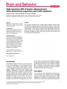
signals are combined to determine the energy and DOI of each event. II. DETECTOR MODULE DESCRIPTION The components of the DOI-encoding high-resolution fast PET module are summarized in Fig. 1. The detector consists of a fast single-channel PMT (Hamamatsu H6533), a PSAPD (RMD) with a sensitive area of 14 mm × 14 mm, and an 8 × 8 scintillator array of mixed lutetium silicate (MLS) crystals. The crystals had dimensions of 1.65 mm × 1.65 mm × 22.0 mm and were packed in an array of reflective polymer foil (VM2000, by 3M) as in [4]. One end of the scintillator array was optically coupled to the PMT using National Diagnostics Histomount [5]. The other end of the scintillator array was coupled to the PSAPD using Cargille Labs Meltmount. We used Meltmount on the PSAPD side because the PSAPD can
PSAPD • Radiation Monitoring Devices, Inc. • 14 mm x 14 mm active area • provides crystal identification • combined with signal from PMT for Energy and DOI measurement
PSAPD Scintillator array
PMT
Scintillator Array • MLS scintillator • 8 x 8 array • each crystal measures 1.65 mm x 1.65 mm x 22.0 mm • 1.75 mm pitch • VM2000 (3M) reflector separating each element
PMT • Hamamatsu H6533 • provides timing signal • combined with signal from PSAPD for Energy and DOI measurement
Detector Module Fig. 1. Overview of detector components.
0-7803-8701-5/04/$20.00 (C) 2004 IEEE
z
Detector Module MLS scintillator slab 22 Na source
PSAPD Scintillator array
Reference PMT
PMT
III. DEPTH OF INTERACTION MEASUREMENTS For the DOI calibration and DOI resolution measurements we used a setup similar to the one presented in [2] and shown schematically in Fig. 2. Using this setup, a DOI calibration is performed for each crystal [2]. A set of DOI profiles (averaged over all 64 crystals) for six z-positions is shown in Fig. 3. For these measurements an energy window of 250 keV to 650 keV was used. The FWHM of these profiles ranges from about 3 mm to 4 mm, but the width of the profiles is largely determined by the width of the collimated beam that is used in the measurements. Using the same technique as in [2], the beam width was found to be approximately 2.7 mm FWHM. IV. ENERGY AND TIMING RESOLUTION
coincidence Fig. 2. Electronic collimation geometry used for DOI measurements. The detector module is translated in the z-direction to measure the response at different DOIs.
In order to measure the timing and energy resolution performance, we have used the top-down coincidence setup shown in Fig. 4. Another fast PMT (Hamamatsu H6533) was coupled to a thin MLS scintillator slab and positioned above the DOI detector. With this measurement and the previously calibrated DOI function, we were able to determine the energy and position (x,y,z) for the recorded events, as well as the coincidence timing distribution with respect to the top PMT. Fig. 5 shows a histogram map of the (x,y) positions for events in a 250 keV to 650 keV energy window, with all 64 crystals readily identifiable. The overall energy resolution was
Normalized Count Rate
1
0.8
Fast PMT (Hamamatsu H6533)
0.6
0.4
22Na
source
PSAPD
0.2
MLS scintillator slab
coincidence
Scintillator 0
0
4
8
12
16
20 22
array
Detector Module
Depth (mm)
Fig. 3. Depth profiles (summed over all 64 crystals) measured at six positions. The FWHM of the profiles varies from about 3 to 4 mm, and it is dominated by the width of the collimated beam. An energy window of 250 to 650 keV was applied.
be repositioned repeatedly simply by heating the detector module to approximately 60ºC. For future detectors, however, we plan to use Histomount on both ends of the scintillator array because it appears to have superior mechanical properties. The detector was operated in an enclosure with controlled air temperature at 10ºC.
PMT
Fig. 4. Setup used for timing and energy resolution measurements and position histograms.
0-7803-8701-5/04/$20.00 (C) 2004 IEEE
220 200
Average Delay (ps)
180 160 140 120 100 80 60 40 20 0 Fig. 5. Position histogram generated using the geometry shown in Fig. 4. An energy window of 250 to 650 keV was applied, and events at all depths within the crystals were used. 14000
5
10
15
20
Depth (mm) Fig. 7. Average delay as a function of depth for all 64 crystals. Zero delay is defined as the delay at z = 22 mm (near the PMT surface).
The uniformity of the timing response across the detector is demonstrated in Fig. 9, which shows the timing distributions for each crystal in the 8 × 8 array. The good uniformity of the time response is also evidenced in Fig. 10 by a histogram of the FWHM of the 64 crystals.
12000
10000
Counts
0
V. DISCUSSION 8000
FWHM = 16.9%
6000
4000
2000
0
0
200
400
600
800 1000 1200 1400 1600 1800
Energy (channel #) Fig. 6. Energy spectrum for all 64 crystals recorded using the geometry shown in Fig. 4. The FWHM of the energy spectrum is 16.9%.
16.9% FWHM at 511 keV as indicated in Fig. 6. The average time delay (defined in [6]) between events recorded at different depths z is shown in Fig. 7 for all crystals. The coincidence timing distribution is presented in Fig. 8(a) without any crystal-specific or DOI corrections. Its FWHM was 457 ps. The crystal-dependent offset and DOI-corrected time distribution is shown in Fig. 8(b) with the FWHM reduced to 430 ps. The relatively small improvement in timing resolution following correction for crystal-dependent offset and DOI effects is consistent with the results presented in [7].
Assuming that the contributions from the reference detector (thin MLS slab coupled to single channel PMT) and the PSAPD/PMT dual-end read-out detector add in quadrature, we can make a rough estimate of the timing resolution that we would see for two PSAPD/PMT modules in coincidence. In separate measurements using two thin MLS slabs coupled to two separate single channel PMTs (Hamamatsu H6533) we measured a timing resolution of approximately 200 ps FWHM. Given the assumption above, the resulting contribution from a single PSAPD/PMT module would be approximately 380 ps FWHM. In this case we would expect a timing resolution of approximately 540 ps FWHM if two PSAPD/PMT modules were used in coincidence. When using the dual-end read-out approach to measure DOI, a strong variation in the light sharing between the two photosensors with the depth of interaction is beneficial for improving the DOI resolution. Unfortunately, when the crystal identification and timing tasks are split between the two photosensors, the variation in light sharing as a function of depth results in a variation in crystal identification efficacy and timing resolution as a function of depth. For example, for gamma rays interacting near the PSAPD a large fraction of the emitted scintillation photons are absorbed by the PSAPD and the large signal-to-noise ratio in the PSAPD results in a position map with very well separated peaks. For the same events relatively little light reaches the PMT, and the timing
0-7803-8701-5/04/$20.00 (C) 2004 IEEE
x 10 3
4
4
x 10
a)
3 2.5
2
Counts
Counts
2.5
b)
FWHM = 457 ps
1.5
1
2
FWHM = 430 ps
1.5 1
0.5
FWTM = 880 ps
0 -1000 -750 -500 -250
0
FWTM = 848 ps
0.5
0 250 500 750 1000 -1000 -750 -500 -250
0
250 500 750 1000
Delay (ps)
Delay (ps)
Fig. 8. Coincidence timing distribution for all 64 crystals recorded using the geometry shown in Fig. 4 and applying an energy window of 250 to 650 keV. a) Timing distribution before applying any delay corrections. b) Timing distribution after correcting for crystal-dependent and depth-dependent delay variations. After applying the corrections, the timing resolution (FWHM) is 430 ps.
10 9
Counts
Number of Crystals
8 7 6 5 4 3 2 -0.5 0 0.5
-0.5 0 0.5
-0.5 0 0.5
-0.5 0 0.5
-0.5 0 0.5
-0.5 0 0.5
-0.5 0 0.5
-0.5 0 0.5
Delay (ns) Fig. 9. Timing distribution for each of the 64 crystals in the array. The distributions have been corrected for crystal- and depth-dependent delay offsets.
resolution, therefore, is compromised. Conversely, for gamma rays interacting near the PMT-end of the scintillator, the timing resolution is improved, but the crystal identification efficacy is compromised. These effects are shown in Fig. 11. In our previous work [2] we showed that the variation in the light sharing between the two photosensors as a function of depth in a dual-end read-out DOI detector can be altered by changing the surface roughness of the scintillator crystals. Fig. 12 shows that by increasing the surface roughness of the
1 0 400
420
440
460
480
500
Timing Resolution (FWHM, ps) Fig. 10. Histogram of the FWHM of the timing distributions shown in Fig. 9.
scintillator crystals, the dependence of the light sharing on depth can be increased, resulting in improved DOI resolution. The data in Fig. 12 were recorded using the detector described in [2], where the different surfaces treatments are described. For the detector described in this paper, the crystals had an M20 surface treatment. This rough surface treatment results in excellent DOI resolution, but also a relatively large variation in the other performance metrics as a function of depth. Fig. 12
0-7803-8701-5/04/$20.00 (C) 2004 IEEE
Fig. 11. Performance variation with DOI. a) Position map for events in the top 4 mm of the crystals (near the PSAPD) and b) for events in the bottom 4 mm (near the PMT). The ability to decode the crystals degrades near the PMT because very little light reaches the PSAPD. c) Timing resolution as a function of depth. The timing resolution improves for large depths (PMT surface at z = 22 mm) because more light reaches the PMT.
DOI Resolution (mm)
10 8
VM2000, 12.4°C Teflon, 19.6°C Teflon, 9.8°C
6 4 2
I. CONCLUSION We have evaluated a DOI-encoding PET detector based on dual-end read-out of a scintillator array using a PSAPD on one end and a single-channel PMT on the other. The average energy resolution of the detector was 16.9% at 511 keV and the timing resolution was 430 ps FWHM vs. a thin slab of MLS coupled to a fast single-channel PMT. Future work will concentrate on optimizing the trade-off between DOI resolution and uniformity of detector performance as a function of depth, and also on improving the form factor of the detector by replacing the round PMT used in this study with a square PMT.
increasing roughness
0 Polished M5 M20 M10 Surface Treatment Fig. 12. Dependence of DOI resolution on crystal surface roughness. Increasing the roughness improves the DOI resolution by intensifying the dependence of the light collection differential between the PSAPD and PMT as a function of depth. These data were measured using the detector described in [2]. The crystals used in the current study had “M20” surfaces. By varying the surface roughness of the crystals, we can trade DOI resolution for improved uniformity of performance as a function of depth.
implies, however, that by reducing the surface roughness we could trade DOI resolution for improved uniformity of crystal identification efficacy and timing resolution as a function of depth. Since most high resolution applications only require DOI resolution on the order of 5 mm, future designs will likely use crystals with M5 surfaces rather than the rougher surfaces used for these measurements.
II. REFERENCES [1] [2]
[3] [4] [5]
[6] [7]
K. S. Shah, R. Farrell, R. Grazioso, E. S. Harmon, E. Karplus, "PositionSensitive Avalanche Photodiodes for Gamma-Ray Imaging," IEEE Trans. Nucl. Sci., vol. 49, no. 4, pp. 1687 – 1692, Aug. 2002. K. C. Burr, A. Ivan, D. E. Castleberry, J. W. LeBlanc, K. S. Shah, R. Farrell, "Evaluation of a prototype small-animal PET detector with depth-of-interaction encoding," IEEE Trans. Nucl. Sci., vol. 51, no. 4, pp. 1791– 1798, Aug. 2004. W. W. Moses, S. E. Derenzo, "Design Studies for a PET Detector Module Using a PIN Photodiode to Measure Depth of Interaction," IEEE Trans. Nucl. Sci., vol. 41, pp. 1441 – 1445, Aug. 1994. R. S. Miyaoka, S. G. Kohlmyer, T. K. Lewellen, "Performance Characteristics of Micro Crystal Element (MiCE) Detectors," IEEE Trans. Nucl. Sci., vol. 48, no. 4, pp. 1403 – 1407, Aug. 2001. R. S. Miyaoka, M. L. Janes, T. K. Lewellen, "Optimization of Mounting Large Crystal Arrays to Photomultiplier Tubes," Proceedings of the IEEE Nuclear Science Symposium and Medical Imaging Conference, Portland, 2003, M6-2. W.W. Moses and S.E. Derenzo, "Prospects for time-of-flight PET using LSO scintillator," IEEE Trans. Nucl. Sci., vol. 46, no. 3, pp. 474 – 478, June 1999. A. Ivan, K. C. Burr, Y. Shao, and J. W. LeBlanc, "Depth of Interaction Effect on Timing Resolution in PET Block Detectors," presented at IEEE Medical Imaging Conference, Rome, 2004, M10-416.
0-7803-8701-5/04/$20.00 (C) 2004 IEEE