Dentomaxillofacial Radiology (2014) 43, 20130355 ª 2014 The Authors. Published by the British Institute of Radiology http://dmfr.birjournals.org
RESEARCH ARTICLE
Mandibular fracture patterns consistent with posterior maxillary fractures involving the posterior maxillary sinus, pterygoid plate or both: CT characteristics 1
T Imai, 2S Sukegawa, 2T Kanno, 1G Fujita, 1N Yamamoto, 2Y Furuki and 1M Michizawa
1
Department of Oral and Maxillofacial Surgery, Saiseikai Senri Hospital, Suita, Osaka, Japan; 2Department of Oral and Maxillofacial Surgery, Kagawa Prefectural Central Hospital, Takamatsu, Kagawa, Japan
Objectives: The aim of this study was to determine the incidence of posterior maxillary fractures involving the posterior maxillary sinus wall, pterygoid plate or both, unrelated to major midface fractures in patients with mandibular fractures, and to characterize associated fractures. Methods: A CT study was performed in patients with mandibular fractures to identify posterior maxillary fractures. Patients aged under 16 years, those with mandibular fractures involving only dentoalveolar components and those with concurrent major midfacial fractures were excluded. Results: 13 (6.7%) of 194 patients with mandibular fractures also had posterior maxillary fractures (case group). The injury pattern correlated with the external force directed to the lateral side of the mandible ( p , 0.001), alcohol consumption ( p 5 0.049), the presence of multifocal fractures ( p 5 0.002) and the fracture regions in the symphysis/parasymphysis ( p 5 0.001) and the angle/ramus ( p 5 0.001). No significant difference between the case and non-case groups was seen for age, sex or cause of trauma. Non-displaced fractures in the ipsilateral posterior mandible occurred with significant frequency ( p 5 0.001) when the posterior maxillary fractures involved only the sinus. Conclusions: Mandibular fractures accompanied by posterior maxillary fractures are not rare. The finding of a unilateral posterior maxillary fracture on CT may aid the efficient radiological examination of the mandible based on possible patterns of associated fractures, as follows: in the ipsilateral posterior region as a direct fracture when the impact is a medially directed force, and in the symphysis/parasymphysis or contralateral condylar neck as an indirect fracture. Dentomaxillofacial Radiology (2014) 43, 20130355. doi: 10.1259/dmfr.20130355 Cite this article as: Imai T, Sukegawa S, Kanno T, Fujita G, Yamamoto N, Furuki Y, et al. Mandibular fracture patterns consistent with posterior maxillary fractures involving the posterior maxillary sinus, pterygoid plate or both: CT characteristics. Dentomaxillofac Radiol 2014; 43: 20130355. Keywords: maxillofacial injuries; maxillary fractures; mandibular fractures; helical CT
Introduction Successful treatment of injuries to the sterically complicated structures of the maxillofacial bones and minimization of post-operative morbidity are dependent on detailed diagnostic imaging.1,2 For an outpatient with suspected mandibular fractures, a combination of Correspondence to: Dr Tomoaki Imai. E-mail:
[email protected] Received 30 September 2013; revised 1 December 2013; accepted 9 December 2013
panoramic tomography and plain film allows acceptable evaluation of simple fractures.3 With significant advances in diagnostic modalities, CT is the objective standard for assessing the maxillofacial skeleton, particularly the midfacial region,3–5 and is more sensitive than panoramic radiography, particularly for mandibular fractures of the angle, ramus or condyle.6–9 A cone beam CT scan provides more useful additional information than conventional radiographs of mandibular
2 of 7
Mandibular fracture patterns with posterior maxillary fractures T Imai et al
trauma in conscious ambulatory patients.10 Nevertheless, in trauma centres, multidetector CT (MDCT) is preferred for evaluating facial bone structures for cases involving lower facial injuries caused by highenergy impact.11 These injuries may be severely comminuted or associated with head, upper facial, midfacial and spinal cord injuries.1,2 Accurate diagnosis of maxillofacial fractures requires the systematic and organized radiographical evaluation of each maxillofacial structure.1 Although 64-slice (or higher) MDCT will precisely reveal non-displaced fractures,12 a CT scanner is a limited medical resource that does not always generate the high-resolution images required to detect these fractures. Given that the mandible is often likened to a closed ring that can fracture at multiple sites,13 diagnosis will be aided by the recognition of various patterns of mandibular fracture on the bases of the structural characteristics of a mandible and the mechanism of trauma.1,11 Furthermore, imaging patterns typically associated with particular fracture types may enhance the sensitivity of detection of another fracture line or may predict specific sequelae. The study by Simonds et al14 serves as an excellent example of the association between isolated fractures of the posterior maxillary sinus wall and mandibular fractures. These authors proposed a mechanism in which the anteromedially displaced coronoid process punctures the posterior maxillary sinus wall, which is probably concomitant with ipsilateral mandibular fracture. In patients with posterior maxillary sinus fractures, recognizing this relationship may facilitate radiographical evaluation when there is a high probability of mandibular fractures. However, the pattern and aetiology of associated mandibular fractures are not fully characterized. Furthermore, the lateral pterygoid plate constitutes the deep portion of the infratemporal fossa, and fracture of this bony structure might also be associated with mandibular fractures with a characteristic pattern. Although these two bony structures are commonly fractured in major midface fractures involving the body of the maxilla or zygomaticomaxillary complex,14,15 fractures of these structures without the major midface fractures have received scant documentation because there are no indications for fixing the fractures in place. According to our definition of posterior maxillary sinus wall, lateral pterygoid plate fractures, or both, as posterior maxillary fractures, we determined the incidence of these fractures unrelated to major midface fractures in patients with mandibular fractures and characterized the associated mandibular fractures according to the results of CT imaging. Methods and materials The study was approved by the internal review boards of the authors’ institutions. Data for mandibular fractures were acquired from the records of all trauma patients who were diagnosed using CT scan and treated at Dentomaxillofac Radiol, 43, 20130355
dmfr.birjournals.org
Saiseikai Senri Hospital, Osaka, Japan, from July 2006 to May 2013 and at Kagawa Prefectural Central Hospital, Kagawa, Japan, from January 2008 to December 2012. Both these general hospitals have critical care centres that receive major trauma cases. Patients with mandibular fractures were divided into case and noncase groups according to the presence or the absence of posterior maxillary fractures, respectively. Exclusion criteria were age under 16 years, mandibular fractures involving dentoalveolar components only and concurrent major midfacial fractures such as zygomaticomaxillary complex, LeFort-type or naso-orbito-ethmoidal fractures. CT scans were reviewed independently by two authors. Imaging was performed using a 64-slice CT scanner (120 kV; 150 mA s; collimation, 64 3 0.5; slice thickness, #2 mm; matrix, 512 3 512 pixels; gantry tilt, 0°), using a 16-slice CT scanner (120 kV; 250 mA s; collimation, 16 3 1.0; slice thickness, #2 mm; matrix, 512 3 512 pixels; gantry tilt, 0°) or using a 4-slice CT scanner (120 kV; 150 mA s; collimation, 4 3 2.0; slice thickness, #2 mm; matrix, 512 3 512 pixels; gantry tilt, 0°). Imaging protocols included axial images reformatted in the coronal and optionally sagittal planes and reconstruction using bone algorithms.
Table 1 Characteristics of subjects Characteristic Gender Male Female Mean age (±standard deviation) (years) Alcohol consumption Yes No Cause of trauma Fall MVA Assault Bicycle Sports Others Impacted area of the mandible Central unit Lateral horizontal/ vertical unit Unknown Fractured mandibular units Unifocal Multifocal Fracture mandibular regions Central unit Lateral horizontal unit Vertical unit Angle/ramus Condyle/subcondyle
Case, n 5 13
Non-case, n 5 181
7 6 37.7 (±13.9)
119 62 43.5 (±23.8)
p-value 0.384 0.385 0.049a
5 8
28 153
4 5 1 1 2 0
67 52 22 20 17 3
0 11
114 57
2
10
1 12
96 85
11 0
67 41
0.001a 0.074
9 5
41 114
0.001a 0.137
0.921
,0.001a
0.002a
MVA, motor vehicle accident. Case and non-case correspond to patients with and without posterior maxillary fractures, respectively. a p , 0.05.
Mandibular fracture patterns with posterior maxillary fractures T Imai et al
For mandibular components, we used the Arbeitsgemeinschaft f¨ur Osteosynthesefragen/Association for the Study of Internal Fixation16 classification with division lines for five units, namely bilateral vertical, bilateral lateral horizontal and central.17 The vertical mandibular unit comprised the condylar/subcondylar region, the coronoid process, the ascending ramus and the mandibular angle. The latter three regions were integrated as the angle/ramus region. The lateral horizontal unit corresponds to the lateral mandibular body. The central unit includes the symphyseal and bilateral parasymphyseal regions. Clinical information extracted from the case records included the patient’s age, gender, aetiology of injury and alcohol consumption. The location of the fractures, the number of fractured mandibular units and the extent of interfragmentary displacement were determined by CT review. The impact area of the external force was determined comprehensively based on the patient’s comments in the medical record and clear direct facial injuries such as skin laceration or comminuted fractures of the mandibular body or alveolar bone. The coronoid process is anatomically closer to the posterior maxillary sinus than to the pterygoid plate. Considering the distance from the coronoid process to these posterior maxillary bony structures, the case group was further subcategorized into cases with fracture of the posterior maxillary sinus only (Type A) and those with fracture of the pterygoid plate with or without maxillary sinus fracture (Type B). Type A corresponds to the fracture described in a previous study.14 Standard descriptive statistics were used to describe the groups. Categorical variables were compared using Fisher’s exact test or the x 2 test, and continuous variables were analysed using the t-test. All analyses were conducted using SPSS® v. 12.0 for Windows (SPSS Inc.,
3 of 7
Chicago, IL). A two-tailed p , 0.05 was considered significant for all analyses. Results 194 cases of mandibular fracture met the criteria, of which 13 (6.7%) and 181 patients were assigned to the case and non-case groups, respectively (Table 1). The case group showed a significant correlation with alcohol consumption and the receipt of an external force directed to the vertical/lateral horizontal unit of the mandible. Furthermore, this group had a significant number of fractures in multiple mandibular units and by site in the central unit and the mandibular angle/ramus area. No significant difference between the two groups was seen for age, gender or cause of trauma. By pattern of associated mandibular fractures, 12 of 13 subjects in the case group had multiple fractured mandibular units without involvement of the lateral horizontal unit (Table 2), 11 patients had mandibular fractures involving both the vertical and central units (Figure 1) and 1 patient (Case 9) had fractures of bilateral vertical units, specifically the ramus on the affected side and the condylar neck on the contralateral side (Figure 2). The remaining patient (Case 11) had a unifocal fracture of the ipsilateral vertical unit and compromised airway secondary to traumatic swelling of the parapharyngeal soft tissue (Figure 3). The patients in the case group who had unknown impacts of force were injured as a result of falls associated with drinking alcoholic beverages. Of the 13 patients in the case group, 4 and 9 patients were subcategorized as Type A and B, respectively. All Type A patients had associated non-displaced fractures in the mandibular vertical unit (Figure 4), whereas all Type B patients showed displaced fractures. This correlation was significant (p 5 0.001).
Table 2 Cases of mandibular fracture with posterior maxillary fracture
Age/ Case gender 1 25/F 2 32/M 3 37/F 4 57/F 5 28/F 6 34/M 7 64/F 8 50/M
Cause of trauma Sports Bicycle Fall MVA MVA Fall Fall Sports
Impacted Alcohol area No LHU/VU No LHU/VU Yes Unknown No LHU/VU Yes LHU/VU Yes LHU/VU No LHU/VU No LHU/VU
9
29/F
MVA
No
LHU/VU
10
29/M
MVA
Yes
Unknown
11 12 13
36/M 53/M 18/M
Fall Assault MVA
No Yes No
LHU/VU LHU/VU LHU/VU
Mandibular fracture sites CU;a ipsilateral VU (angle) CU;a ipsilateral VU (angle) CU; ipsilateral VU (angle) CU; ipsilateral VU (subcondyle)a CU;a ipsilateral VU (subcondyle)a CU; ipsilateral VU (angle) CU; ipsilateral VU (ramus) CU; ipsilateral VU (ramus and coronoid process) Ipsilateral VU (ramus);a contralateral VU (condylar neck) CU; ipsilateral VU (subcondyle); contralateral VU (condylar neck) Ipsilateral VU (angle) CU; ipsilateral VU (subcondyle)a CU; ipsilateral VU (angle)
Posterior maxillary fracture Posterior maxillary sinus Pterygoid wall plate 3 3 3 3 3 3 3 3 3 3 3
Subcategory Type B Type B Type B Type A Type A Type B Type B Type B
3
Type A
3
3
Type B
3 3 3
3
Type B Type A Type B
3
CU, central unit; F, female; LHU, lateral horizontal unit; M, male; MVA, motor vehicle accident; VU, vertical unit. a Non-displaced fracture.
dmfr.birjournals.org
Dentomaxillofac Radiol, 43, 20130355
Mandibular fracture patterns with posterior maxillary fractures T Imai et al
4 of 7
Figure 1 Typical mandibular fractures with displacement accompanied by a posterior maxillary fracture in a 64-year-old female (Case 7) from a stairway fall. This shows fractures of the pterygoid plate (arrow) and mandible in the ipsilateral vertical unit (arrowhead).
Discussion In the present study, we constructed a framework of posterior maxillary fractures that involved at least one fracture of the two independent bony structures, the
Figure 2 Atypical mandibular fracture pattern with a posterior maxillary fracture in a 29-year-old female (Case 9) involved in a motor vehicle accident. This shows the fracture of the posterior maxillary sinus wall (arrow) (a). Associated mandibular fractures are localized in the ipsilateral vertical unit of the ramus (arrowhead) without displacement and the contralateral vertical unit of the condylar neck (b).
Dentomaxillofac Radiol, 43, 20130355
dmfr.birjournals.org
posterior maxillary sinus and the pterygoid plate, which constitute components of the infratemporal fossa. Ellis et al18 reported that 37 (1.7%) of 2137 cases with mandibular fractures had accompanying maxillary fractures but did not describe the specific segment of the maxilla. Furthermore, subsequent large-scale epidemiological studies on maxillofacial trauma19–22 also did not document these two structures as fracture sites associated with mandibular fracture. Although Simonds et al14 reported that 13 (59%) of 22 patients with the former fracture had concomitant mandibular fractures, they did not describe the latter or investigate the incidence of the former among patients with mandibular fractures. In patients with mandibular fractures diagnosed using CT, we observed 6.7% of posterior maxillary fractures involving the former, latter or both highly associated with the impact on the lateral aspect of the mandible and characteristic patterns of underlying mandibular fracture. The posterior maxillary bony structures are deeply situated and anatomically sheltered by the zygomaticomaxillary complex, zygomatic arch, mandibular ramus and associated muscles. An external impact is unlikely to directly affect these deeply situated structures, except, for example, in the rare case presented by Eriksson and
Figure 3 Compromised airway due to swollen parapharyngeal soft tissues in a 36-year-old male (Case 11). This shows fracture of the sinus wall and pterygoid plate (arrow) (a) and the mandibular angle (arrowhead) (b).
Mandibular fracture patterns with posterior maxillary fractures T Imai et al
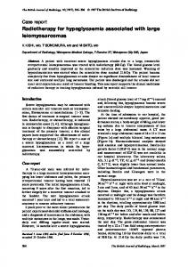
Figure 4 Non-displaced mandibular fractures with a posterior maxillary fracture in a 28-year-old female (Case 5) involved in a motor vehicle accident. This patient was initially diagnosed on CT as having a unifocal symphyseal fracture (a). Detection of a concomitant fracture of the posterior maxillary sinus wall (b), which corresponds to a posterior maxillary fracture Type A, prompted further investigation of the posterior mandible, leading to the identification of an additional non-displaced fracture in the ramus on the coronal CT (c) that would probably have been overlooked. Arrows indicate fracture lines.
5 of 7
Hakansson15 of a directly isolated fracture of the pterygoid plate through the cheek caused by a bicycle handlebar. Instead, these structures are more likely to be disrupted by a displaced neighbouring bone, specifically the mandible. Furthermore, this mechanism of posterior maxillary sinus fracture14 can apply to pterygoid plate fractures, because both fractures are likely to involve the posterior mandible with anteromedial and medial displacement, respectively. This is supported by our present findings that the case group had a significant number of injuries related to a lateral impact. The mandible is more susceptible to a lateral than to a frontal impact.23 When the mandibular vertical unit is fractured by a lateral force, it will be medially displaced towards the posterior maxillary sinus or pterygoid plate and, accordingly, is likely to have a strong chance of fracturing these structures (Figure 5). In comparison with fractures of the posterior maxillary sinus only (Type A), fractures involving the pterygoid plate (Type B) would require more medial displacement of the mandible caused by a higher energy impact and the associated mandibular fractures are likely to be more displaced or comminuted. The branches of the maxillary artery and a part of the pterygoid venous plexus are located around the lateral pterygoid plate.24 The maxillary artery often attaches to the posterior portion of the pterygoid plate in cases with medial travel of the artery to the lateral pterygoid muscle.24 LeFort-type fractures, generally caused by highenergy frontal impact, are occasionally accompanied by life-threatening bleeding caused by vascular injury in the pterygopalatine region and by severe epitaxis.25,26 Bozkurt et al27 reported a case of life-threatening pseudoaneurysm of the maxillary artery secondary to subcondylar fracture with displacement in the oblique and medial directions. Although we did not encounter any patients with active bleeding or acute pseudoaneurysm in the pterygopalatine region confirmed using angiography, these vessels might be susceptible to injury owing to the high lateral energy that fractures the pterygoid plate. The high contrast of soft tissue revealed using MDCT would allow the clear and rapid detection of any relationship between fractured bones and the surrounding changes that cause air collection, bleeding or soft-tissue swelling.10 Although severely displaced mandible fractures can threaten airway patency, this complication may not be realistic in cases with slight or non-displaced fractures in the posterior mandible. However, if the CT scan of these cases displays concomitant posterior maxillary fractures, particularly Type B, a considerable degree of external force may impact the lateral aspect of the mandible. In such a situation, clinicians should carefully evaluate the possibility of a progressively compromised airway caused by swelling of the medially placed parapharyngeal soft tissues (Case 11). In the present study, mandibular fractures in the case group were exclusively produced at two sites by a lateral impact. These fracture sites were predominantly localized dmfr.birjournals.org
Dentomaxillofac Radiol, 43, 20130355
6 of 7
Mandibular fracture patterns with posterior maxillary fractures T Imai et al
Figure 5 Proposed model of posterior maxillary fractures. Lateral force on the posterior mandible (arrows) medially displaces the coronoid process and can induce posterior maxillary fractures simultaneously with transient subluxation or rotation of the temporomandibular joint (A).14 In the setting of mandibular fracture due to high-energy impact or individual structural predisposition, the fragment that includes the coronoid process would gain more mobility for medial displacement to the posterior sinus wall, pterygoid plate or both, with more chances of fracturing these structures (B).
in the vertical unit, particularly in the angle/ramus area as a direct fracture and in the central unit or contralateral vertical unit of the condyle as an indirect fracture (Figure 6). Furthermore, a significant correlation was evident in the posterior mandible between Type A and non-displaced fracture and Type B and displaced fracture, respectively. Because .50% of mandibular fractures display multifocal sites on CT,12,13 a finding of one obvious fracture line should generally prompt the search for another possible subtle fracture with consideration to the direction and the region impacted by the external force. As shown in Figure 4, clinicians who find an isolated posterior maxillary sinus fracture (i.e. Type A) would be encouraged to search for ipsilateral non-displaced fractures in the posterior mandible. The mandibular fracture pattern in the case group is basically consistent with those areas found to be highly stressed in biomechanical studies. A cadaver model of a lateral impact on the mandibular body revealed deformation at the impacted area and posterior mandible, accompanied by a high-stretch deformation at the parasymphysis and condyle of the contralateral side.28 Petzel and Bulles29 demonstrated that the contralateral condyle might be fractured at a more cranial than ipsilateral site when the mandible is impacted by a lateral force. Sites with the highest strain in a laterally explosive model were the posterior mandible on the blast side, the chin and the contralateral condylar neck.30 Our Case 11, with mandibular fractures of the ipsilateral vertical unit, probably reflected the results of direct force only. Among cases for which the impact site is unknown, Case 10 had fractures in the central unit, ipsilateral subcondyle and contralateral condylar neck. Therefore, if an external force were to impact from the anterior aspect, the mechanism of posterior maxillary Dentomaxillofac Radiol, 43, 20130355
dmfr.birjournals.org
Figure 6 Lateral impact on the mandible and associated mandibular fractures. Lateral force (arrows) imposed on the posterior mandible caused a direct fracture (solid lines). An indirect fracture would also occur in the anterior mandible or contralateral condyle (dashed lines). Fractured fragments including the coronoid process would displace medially (arrowheads) and therefore potentially lead to posterior maxillary fractures.
fractures would be difficult to explain. If it originated from the ipsilateral side, the occurrence of these fractures would be reasonable, and the pattern of accompanying condylar fractures could be explained by the concept described above by Petzel and Bulles.29 Simonds et al14 presented a confusing case that does not fit their proposed injury mechanism, namely a posterior sinus wall fracture with only a contralateral condylar fracture. We consider that this fracture pattern could only have resulted from an indirect force. Nevertheless, biomechanical analysis of cadavers focusing on posterior maxillary fractures caused by a lateral external force on the mandible might be warranted. We do not suggest that all posterior maxillary fractures are related to collision with a medially impacted fracture of the mandible. Simonds et at14 suggested a momentary anterior dislocation of the condyle without mandibular fracture, followed by puncturing of the posterior sinus wall. Unlike the unilateral cases presented here, CT scan in patients with traumatic bilateral anterior dislocation of the mandible with impact over the maxillary dental arch revealed bilateral posterior sinus wall fractures, which would bilaterally accommodate the coronoid process.31 This study focused retrospectively on the mandibular fracture pattern on CT with or without posterior maxillary fractures. Several limitations warrant mention. First, not all patients with facial trauma received CT scans in clinical settings. Thus, conventional radiographs might have failed to reveal a non-displaced fracture line in the mandible, and some additional cases might have had posterior maxillary fractures. In the facial trauma cases examined using CT, it is possible to miss mandibular fractures owing to the involvement of clinicians with different levels of expertise and the use of different MDCTs among institutions. Second, the clinical data partially depend on the accuracy of medical records and patient comments. Third, the number of posterior maxillary fractures was small, notwithstanding that our cases were acquired from two independent hospitals with trauma centres.
Mandibular fracture patterns with posterior maxillary fractures T Imai et al
In conclusion, mandibular fractures accompanied by posterior maxillary fractures are not rare on CT. Associated mandibular fractures impacted by direct force would be seen in the posterior region. Symphyseal/parasymphyseal or contralateral condylar neck fractures related to indirect
7 of 7
force would also be expected. Using a systematically routine approach to evaluating CT scans of maxillofacial trauma, the finding of a unilateral posterior maxillary fracture may aid efficient radiological examination of the mandible according to the patterns of the associated fractures.
REFERENCES 1. Papageorge MB, Oreadi D. Radiographic evaluation of facial injuries. In: Fonseca RJ, ed. Oral and maxillofacial trauma. 4th edn. Philadelphia, PA: Saunders; 2012. pp. 232–47. 2. Schuknecht B, Graetz K. Radiologic assessment of maxillofacial, mandibular, and skull base trauma. Eur Radiol 2005; 15: 560–8. doi: 10.1007/s00330-004-2631-7 3. Scarfe WC. Imaging of maxillofacial trauma: evolutions and emerging revolutions. Oral Surg Oral Med Oral Pathol Oral Radiol Endod 2005; 100: S75–96. doi: 10.1016/j.tripleo.2005.05.057 4. Rake PA, Rake SA, Swift JQ, Schubert W. A single reformatted oblique sagittal view as an adjunct to coronal computed tomography for the evaluation of orbital floor fractures. J Oral Maxillofac Surg 2004; 62: 456–9. 5. Tanrikulu R, Erol B. Comparison of computed tomography with conventional radiography for midfacial fractures. Dentomaxillofac Radiol 2001; 30: 141–6. doi: 10.1038/sj/dmfr/4600593 6. Rhea JT, Rao PM, Novelline RA. Helical CT and three-dimensional CT of facial and orbital injury. Radiol Clin North Am 1999; 37: 489–513. 7. Wilson IF, Lokeh A, Benjamin CI, Hilger PA, Hamlar DD, Ondrey FG, et al. Prospective comparison of panoramic tomography (zonography) and helical computed tomography in the diagnosis and operative management of mandibular fractures. Plast Reconstr Surg 2001; 107: 1369–75. 8. Roth FS, Kokoska MS, Awwad EE, Martin DS, Olson GT, Hollier LH, et al. The identification of mandible fractures by helical computed tomography and panorex tomography. J Craniofac Surg 2005; 16: 394–9. 9. Chacon GE, Dawson KH, Myall RW, Beirne OR. A comparative study of 2 imaging techniques for the diagnosis of condylar fractures in children. J Oral Maxillofac Surg 2003; 61: 668–72. doi: 10.1053/joms.2003.50134 10. Kaeppler G, Cornelius CP, Ehrenfeld M, Mast G. Diagnostic efficacy of cone-beam computed tomography for mandibular fractures. Oral Surg Oral Med Oral Pathol Oral Radiol 2013; 116: 98–104. doi: 10.1016/j.oooo.2013.04.004 11. Patel R, Reid RR, Poon CS. Multidetector computed tomography of maxillofacial fractures: the key to high-impact radiological reporting. Semin Ultrasound CT MR 2012; 33: 410–17. doi: 10.1053/j.sult.2012.06.005 12. Ogura I, Kaneda T, Mori S, Sekiya K, Ogawa H, Tsukioka T. Characterization of mandibular fractures using 64-slice multidetector CT. Dentomaxillofac Radiol 2012; 41: 392–5. doi: 10.1259/dmfr/67127210 13. Escott EJ, Branstetter BF. Incidence and characterization of unifocal mandible fractures on CT. AJNR Am J Neuroradiol 2008; 29: 890–4. doi: 10.3174/ajnr.A0973 14. Simonds JS, Whitlow CT, Chen MY, Williams DW 3rd. Isolated fractures of the posterior maxillary sinus: CT appearance and proposed mechanism. AJNR Am J Neuroradiol 2011; 32: 468–70. doi: 10.3174/ajnr.A2337 15. Eriksson L, Hakansson H. Unilateral fracture of the pterygoid process. Report of a case. Oral Surg Oral Med Oral Pathol 1979; 47: 127–30.
16. Matter P. History of the AO and its global effect on operative fracture treatment. Clin Orthop Relat Res 1998; 347: 11–18. 17. Buitrago-Tellez CH, Audige L, Strong B, Gawelin P, Hirsch J, Ehrenfeld M, et al. A comprehensive classification of mandibular fractures: a preliminary agreement validation study. Int J Oral Maxillofac Surg 2008; 37: 1080–8. doi: 10.1016/j.ijom.2008.06.008 18. Ellis E 3rd, Moos KF, el-Attar A. Ten years of mandibular fractures: an analysis of 2,137 cases. Oral Surg Oral Med Oral Pathol 1985; 59: 120–9. 19. Chrcanovic BR, Abreu MH, Freire-Maia B, Souza LN. 1,454 mandibular fractures: a 3-year study in a hospital in Belo Horizonte, Brazil. J Craniomaxillofac Surg 2012; 40: 116–23. doi: 10.1016/j. jcms.2011.03.012 20. Brasileiro BF, Passeri LA. Epidemiological analysis of maxillofacial fractures in Brazil: a 5-year prospective study. Oral Surg Oral Med Oral Pathol Oral Radiol Endod 2006; 102: 28–34. doi: 10.1016/j.tripleo.2005.07.023 21. Gassner R, Tuli T, Hachl O, Rudisch A, Ulmer H. Craniomaxillofacial trauma: a 10 year review of 9,543 cases with 21,067 injuries. J Craniomaxillofac Surg 2003; 31: 51–61. 22. Iida S, Kogo M, Sugiura T, Mima T, Matsuya T. Retrospective analysis of 1502 patients with facial fractures. Int J Oral Maxillofac Surg 2001; 30: 286–90. doi: 10.1054/ijom.2001.0056 23. Nahum AM. The biomechanics of maxillofacial trauma. Clin Plast Surg 1975; 2: 59–64. 24. Lang J. Contents of the masticatory space. In: Lang J (ed.). Clinical anatomy of the masticatory apparatus and peripharyngeal spaces. New York, NY: Thieme Medical Publishers; 1995. pp. 108–40. 25. Dean NR, Ledgard JP, Katsaros J. Massive hemorrhage in facial fracture patients: definition, incidence, and management. Plast Reconstr Surg 2009; 123: 680–90. doi: 10.1097/ PRS.0b013e31819565da 26. Hwang K, Choi HG. Bleeding from posterior superior alveolar artery in Le Fort I fracture. J Craniofac Surg 2009; 20: 1610–12. doi: 10.1097/SCS.0b013e3181b14775 27. Bozkurt M, Kapi E, Karakol P, Yorgancilar E. Sudden rupture of the internal maxillary artery causing pseudoaneurysm (mandibular part) secondary to subcondylar mandible fracture. J Craniofac Surg 2009; 20: 1430–2. doi: 10.1097/SCS.0b013e3181aee442 28. DuChesne A, Unnewehr M, Schmidt PF, Sotonyi P, Brinkmann B, Piffko J, et al. Deformation characteristics of the human mandible in low impact experiments. Int J Legal Med 2003; 117: 257–62. doi: 10.1007/s00414-002-0358-z 29. Petzel JR, Bulles G. Experimental studies of the fracture behaviour of the mandibular condylar process. J Maxillofac Surg 1981; 9: 211–15. 30. Lei T, Xie L, Tu W, Chen Y, Tang Z, Tan Y. Blast injuries to the human mandible: development of a finite element model and a preliminary finite element analysis. Injury 2012; 43: 1850–5. doi: 10.1016/j.injury.2012.07.187 31. Cheng A, Al Hashmi A, Goss AN. Traumatic bilateral anterior dislocation of the mandible with impaction over the maxilla: a case report. J Oral Maxillofac Surg 2009; 67: 673–5. doi: 10.1016/j. joms.2008.06.070
dmfr.birjournals.org
Dentomaxillofac Radiol, 43, 20130355