Research Article
2349
Human melanocortin 1 receptor variants, receptor function and melanocyte response to UV radiation M. Cathy Scott1, Kazumasa Wakamatsu2, Shosuke Ito2, Ana Luisa Kadekaro1, Nobuhiko Kobayashi3, Joanna Groden4, Renny Kavanagh1, Takako Takakuwa5, Victoria Virador6, Vincent J. Hearing6 and Zalfa A. Abdel-Malek1,* 1Department of Dermatology, University of Cincinnati College of Medicine, PO Box 670592, Cincinnati, Ohio 45267-0592, USA 2Fujita Health University School of Health Sciences, Toyoake, Aichi 470-1192, Japan 3Department of Dermatology, Nara Medical University, Kashihara, Nara 634-8522, Japan 4Department of Molecular Genetics, Biochemistry and Microbiology, University of Cincinnati College of Medicine, PO Box 670524,
Cincinnati, Ohio 45267-0524, USA 5POLA Laboratories, 560 Kashio-cho, Totsuka-ku, Yokohama 244-0812, Japan 6Laboratory of Cell Biology, National Cancer Institute, National Institutes of Health, 9000 Rockville Pike, Bethesda, MD 20892, USA *Author for correspondence (e-mail:
[email protected])
Accepted 6 March 2002 Journal of Cell Science 115, 2349-2355 (2002) © The Company of Biologists Ltd
Summary Cutaneous pigmentation is determined by the amounts of eumelanin and pheomelanin synthesized by epidermal melanocytes and is known to protect against sun-induced DNA damage. The synthesis of eumelanin is stimulated by the binding of α-melanotropin (α-melanocyte-stimulating hormone) to the functional melanocortin 1 receptor (MC1R) expressed on melanocytes. The human MC1R gene is highly polymorphic and certain allelic variants of the gene are associated with red hair phenotype, melanoma and non-melanoma skin cancer. The importance of the MC1R gene in determining skin cancer risk led us to examine the impact of specific polymorphisms in this gene on the responses of human melanocytes to α-melanotropin and UV radiation. We compared the ability of human melanocyte cultures, each derived from a single donor, to respond to α-melanotropin with dose-dependent stimulation of cAMP formation, tyrosinase activity and proliferation. In each of those cultures the MC1R gene was
sequenced, and the eumelanin and pheomelanin contents were determined. Human melanocytes homozygous for Arg160Trp, heterozygous for Arg160Trp and Asp294His, or for Arg151Cys and Asp294His substitutions, but not melanocytes homozygous for Val92Met substitution, in the MC1R demonstrated a significantly reduced response to αmelanotropin. Additionally, melanocytes with a nonfunctional MC1R demonstrated a pronounced increase in their sensitivity to the cytotoxic effect of UV radiation compared with melanocytes expressing functional MC1R. We conclude that loss-of-function mutations in the MC1R gene sensitize human melanocytes to the DNA damaging effects of UV radiation, which may increase skin cancer risk.
Key words: Melanocortin 1 receptor, Human melanocytes, MC1R variants
Introduction The significance of human cutaneous pigmentation lies in its protective role against sun-induced DNA damage and photocarcinogenesis (Kaidbey et al., 1979; Pathak et al., 1980; Pathak, 1995). Total melanin content and the relative amounts of eumelanin, the black-brown pigment, and pheomelanin, the red-yellow pigment, synthesized by human epidermal and follicular melanocytes are important determinants of skin and hair color, respectively. Fair skin and red hair, characterized by a low melanin content and a low eumelanin to pheomelanin ratio, is associated with poor tanning ability following sun exposure and with increased risk of skin cancer (Fitzpatrick et al., 1976; Pathak et al., 1980). Melanin-containing granules, or melanosomes, form supranuclear caps in keratinocytes, thus shielding the DNA from excessive exposure to UV radiation (UVR) (Pathak et al., 1971; Kobayashi et al., 1998). In dark skin with a high eumelanin relative to pheomelanin content, melanosomes are found throughout the epidermal layers (Pathak et al., 1971). However, in fair skin, melanosomes are absent from the suprabasal layer, allowing for increased UVR
penetration and DNA damage, and for increased susceptibility to sun-induced genotoxicity and carcinogenesis. Resistance of eumelanin to photodegradation and its ability to scavenge radicals (Menon et al., 1983) suggest that stimulation of eumelanin synthesis by epidermal melanocytes enhance photoprotection. Eumelanin synthesis in melanocytes is stimulated by activation of the α-melanocyte stimulating hormone (α-MSH) receptor, termed the melanocortin 1 receptor (MC1R) (Geschwind et al., 1972; Tamate and Takeuchi, 1984; Hunt et al., 1995). The human MC1R is more polymorphic than several other pigment genes, including tyrosinase, suggesting its importance in determining constitutive pigmentation in humans (Sturm et al., 1998; Rees, 2000). Furthermore, mutations in the gene for proopiomelanocortin, the precursor for melanocortins and other bioactive peptides, result in red hair color in addition to metabolic abnormalities, including adrenal insufficiency and obesity (Krude et al., 1998). This underscores the significance of melanocortins in regulating eumelanin synthesis in humans.
2350
Journal of Cell Science 115 (11)
The MC1R is a G-protein-coupled receptor with seven transmembrane domains (Chhajlani and Wikberg, 1992; Mountjoy et al., 1992). Binding of α-MSH to this receptor activates adenylate cyclase and increases cAMP formation (Suzuki et al., 1996). More than 30 allelic variants of the human MC1R have been identified mainly in northern European populations and in Australia (Valverde et al., 1995; Box et al., 1997; Smith et al., 1998; Rees, 2000). However, the consequences of these variants on the physiological function of the MC1R remain poorly understood. Among the variants so far reported, Arg142His, Arg151Cys, Arg 160Trp and Asp294His are the mutations mostly associated with the red hair phenotype and reduced tanning ability in several populations (Box et al., 1997; Smith et al., 1998; Healy et al., 2000). This supports the notion that α-MSH and its receptor significantly affect the response of melanocytes to UVR (Pawelek et al., 1992; Im et al., 1998). The above four MC1R variants are common in melanoma patients, and increase the risk of melanoma by more than twofold (Palmer et al., 2000). Expression of those variants in heterologous cell cultures reduced the functional coupling of the MC1R to adenylate cyclase (Frändberg et al., 1998; Schiöth et al., 1999). As yet, no studies have shown how allelic variants of MC1R would affect the function of the receptor in human melanocytes, a main physiological target for melanotropins in the skin. Therefore, we sought to analyze the biological consequences of MC1R mutations by investigating the responses of genetically different epidermal melanocyte cultures to α-MSH and UVR. Materials and Methods Melanocyte culture Primary human melanocyte cultures were established from neonatal foreskins with different pigmentation, and maintained as described previously (Abdel-Malek et al., 1993; Abdel-Malek et al., 1995). Obtaining neonatal foreskins for this purpose was approved by the University of Cincinnati Medical Center Institutional Review Board. Bovine pituitary extract contained in the melanocyte growth medium contains high concentrations of melanotropins (Abdel-Malek et al., 1995), thus it was removed from the culture medium 2 to 3 days prior to, and for the duration of, each experiment in which the effects of αMSH were tested. Determination of tyrosinase activity and proliferation To determine the effects of α-MSH on melanocyte proliferation and tyrosinase activity, melanocytes were plated onto 60 mm dishes at a density of 2.5×10⁵ cells. 72 hours later, and every other day thereafter for a total of 6 days, the growth medium was changed and melanocytes were treated with 0 (control), 0.1, 1 or 10 nM α-MSH, or with 1 µM forskolin (n=3 dishes per group) (both were obtained from Sigma Chemical Company, St Louis, Missouri). The tyrosine hydroxylase activity of tyrosinase was determined as described previously (Pomerantz, 1969; Abdel-Malek et al., 1992). The cell number was determined using a Coulter Counter. Each experiment was repeated at least twice for each melanocyte culture. Statistical analysis was carried out using the Student’s t-test to compare the effects of different concentrations of α-MSH on each individual melanocyte culture. Two-way ANOVA was also used to compare the responses to different concentrations of α-MSH of melanocyte cultures that expressed wildtype MC1R and their MC1R mutant counterparts, which had reduced response to α-MSH.
Sequencing of the MC1R gene PCR amplification, sequencing, and restriction fragment length polymorphism (RFLP) analysis of the MC1R gene were carried out as follows. Total RNA was purified from cultured human melanocytes using an RNA Easy Kit (Qiagen, Valencia, CA). The entire coding region of the MC1R was amplified using reverse transcriptase and nested PCR amplification. 3 µg of total RNA was amplified in a PCR reaction containing standard concentrations of reverse transcriptase, Taq DNA polymerase, MgCl2, RNAse inhibitor, dNTPs, buffer and primers, as described previously (Koppula et al., 1997) in a total volume of 50 µl. The complementary strand was synthesized at 43°C for 1 hour, and the MC1R was amplified for 35 cycles (1 minute at 94°C, 1 minute at 60°C, and 2 minutes at 72°C) in an automated thermal cycler (Gene Amp PCR System 9600, Perkin Elmer, Boston, MA). 1-5 µl of the first reaction was amplified in two separate reactions using two sets of M13-adjoined nested primers, first half Nterminal primer+M13up (5′-TGTAAAACGACGGCCAGTCCTGGCAGCACCATGAACTAAGC-3′); first half C-terminal primer+M13rp (5′-CAGGAAACAGCTATGACCTGGTCGTAGTAGGCGATGAAGAGC-3′); second half N-terminal primer+M13up (5′-TGTAAAACGACGGCCAGTCGCTACATCTCCATCTTCTACGCAC3′); and second half C-terminal primer+M13rp (5′CAGGAAACAGCTATGACCCTCTGCCCACACTTAAAGC 3′). The standard concentrations of PCR reagents were added to the first reaction and amplified for 25 cycles (30 seconds at 94°C, 1 minute at 62°C and 1 minute at 72°C). The final reaction yielded a 640 (first half) and a 560 (second half) nucleotide product flanked by the M13 primers. 5 µl of the PCR reaction was electrophoresed on a 2% agarose gel. The remainder was purified in a Centri-Spin Column (Princeton Separations, Adelphia, NJ). The PCR products were sequenced by dye primer chemistry. Briefly, M13 forward and reverse primers were labeled with four fluorescent dyes in four separate basespecific tubes. The products were electrophoresed and read by an automated sequencing machine (Perkin Elmer/Applied Biosystems models 373A or 377). We confirmed some mutations by restriction fragment length polymorphism analysis for variants at codons 151 (HhaI), 160 (SstII) and 294 (TaqI). The PCR products were digested using standard conditions and run on 1-3% agarose gels. Determination of cAMP levels The dose-dependent effect of α-MSH on cAMP formation in human melanocytes was determined using a cyclic AMP radioimmunoassay kit (Dupont-New England Nuclear, Boston, MA), as recommended by the manufacturer and as described previously (Suzuki et al., 1996). Duplicate samples were assayed from each culture well (triplicate wells/group) after the appropriate dilution (1:10 for groups treated with 10 nM α-MSH, and 1:5 for the remaining groups). Each culture was tested twice in two separate experiments. The data were analyzed by two-way ANOVA and Student’s t-test, as described above. Analysis of eumelanin and pheomelanin content and total melanin content Melanocytes were lyophilized, and eumelanin and pheomelanin contents were analyzed using a microassay developed previously (Ito and Fujita, 1985). Duplicate samples of melanocytes deprived of bovine pituitary extract (approximately 1×10⁶ melanocytes/sample) were oxidized by permanganate to pyrrole 2,3,5-tricarboxylic acid (PTCA) and analyzed by HPLC with UV detection to determine eumelanin content. Identical duplicate samples were hydrolyzed with hydriodic acid to aminohydroxyphenylalanine (AHP), and analyzed by HPLC with electrochemical detection to determine pheomelanin content. Variations of PTCA and AHP values were approximately 10% or less when determined on separate occasions in this study. The amount of eumelanin can be obtained by multiplying the amount of PTCA by a conversion factor of 160, while the amount of
MC1R variants and receptor function
2351
Table 1. Determination of MC1R genotype and relative eumelanin and pheomelanin contents in human melanocyte cultures Melanocyte 729-b 747-c 751-b 731-c 754-b 755-c 765-c 777-c 790-c 780-b 753-c 830-c 849-b
MC1R genotype Consensus sequence Consensus sequence Consensus sequence Heterozygous for Arg151Cys Heterozygous for Val92Met Homozygous for Val92Met Heterozygous for Val92Met Heterozygous for Val60Leu Heterozygous for Arg163Gln Heterozygous for Phe196Leu Homozygous for Arg160Trp Compound heterozygous for Arg160Trp and Asp294His Compound heterozygous for Arg151Cys and Asp294His
Eumelanin (µg/106 cells)
Pheomelanin (µg/106 cells)
Eumelanin/ pheomelanin
Total melanin (µg/106 cells)
26.7 1.23 ND ND 34.4 2.58 0.83 1.46 1.25 25.9 0.66 2.30 14.9
4.29 1.43 ND ND 2.49 2.15 0.55 3.01 0.87 3.05 0.86 1.48 3.64
6.23 0.86 ND ND 13.8 1.20 1.51 0.49 1.44 8.49 0.77 1.55 4.09
49.3 14.7 23.8 5.73 41.8 11.7 4.20 14.6 8.81 41.7 3.3 4.60 33.8
The MC1R genotype of 13 melanocyte cultures was determined by sequence analysis of the entire coding region of the MC1R gene, as described in Materials and Methods. The genotypes for NHM 830-c and 849-c were further confirmed by RFLP analysis. The melanocyte cultures are grouped according to whether they are homozygous for the consensus MC1R, heterozygous for a MC1R variant, or homozygous or compound heterozygous for MC1R variants. Cultures were analyzed for eumelanin, pheomelanin and total melanin contents. ND, not determined.
pheomelanin can be obtained by multiplying the amount of AHP by a conversion factor of 10 (Ozeki et al., 1996). Statistical significance of differences was assessed with the Mann-Whitney test. Differences were considered to be significant when P values were less than 0.05. Total melanin content was determined in 0.5-1×10⁶ melanocytes. Cells were harvested, centrifuged, washed twice with PBS, counted and centrifuged. The cell pellets were solubilized in 0.2 M NaOH (1×10⁶ cells/ml) for 1 hour, and melanin content was determined spectrophotometrically by reading the absorbance at 475 nm. Melanin content was calculated using a standard curve generated from the absorbance of known concentrations of synthetic melanin, as described previously (Barker et al., 1995). Response of melanocytes to UVBR The response of melanocytes to UVBR was determined by plating the cells in complete growth medium at a density of 1×10⁵cells/60 mm dish. 72 hours later, melanocytes were irradiated once with 21 mJ/cm2 UVBR as described previously (Barker et al., 1995). The UV source is a bank of six FS20 sun lamps (Westinghouse) with 75% emission in the UVB and 25% emission in the UVA range. The peak emission of the lamps is at 313 nm. Percent cell death was determined on days 2 and 4 after UV irradiation by calculating the number of dead melanocytes that detached and incorporated Trypan blue dye and the number of viable cells that remained attached to the culture dish and excluded Trypan blue, as described before (Barker et al., 1995). The responses to UVR of the cultures with functional MC1R were compared with those of the cultures with reduced response to α-MSH using one-way ANOVA.
Results Characterization of MC1R genotype and eumelanin to pheomelanin ratios We established 35 human melanocyte (NHM) cultures from light-colored foreskins (NHM-c) and eight cultures from dark foreskins (NHM-b), and compared their ability to respond to α-MSH with dose-dependent increases in proliferation and in the activity of tyrosinase, the rate-limiting enzyme in the melanin synthetic pathway. Of those, 33 NHM-c and seven NHM-b demonstrated dose-dependent increases in tyrosinase activity and proliferation in response to α-MSH over the range
of doses tested. The remaining three cultures had no, or minimal response to α-MSH. We sequenced the entire coding region of the MC1R in the three cultures with impaired responsiveness to α-MSH and in ten α-MSH-responsive cultures (four NHM-b and six NHM-c). We analyzed 11 of those 13 cultures for their eumelanin and pheomelanin contents to assess the differences in their constitutive pigmentation. A higher number of NHM-c than NHM-b cultures was included since variation in the MC1R is expected to be higher in individuals with fair skin and red hair phenotype than in individuals with dark skin and hair color (Valverde et al., 1995; Smith et al., 1998; Harding et al., 2000). The sequencing data presented in Table 1 revealed that NHM 753-c was homozygous for Arg160Trp, NHM 830-c was heterozygous for Arg160Trp and Asp294His, and NHM 849-b was heterozygous for Arg151Cys and Asp294His substitutions in MC1R. All three cultures failed to respond to α-MSH with a significant dose-dependent increase in tyrosinase activity (Fig. 1B). NHM 755-c was homozygous for Val92Met substitution, and homozygous for a silent mutation, Thr314Thr, in the MC1R. Four other cultures were heterozygous for Arg151Cys (NHM 731-c), Val60Leu (NHM 777-c), Arg163Gln (NHM 790-c) and Phe196Leu (NHM 780-b) substitutions. Two cultures were heterozygous for Val92Met substitution (NHM 754-b and 765-c). NHM 754-b and 780-b were heterozygous for a silent mutation, Thr314Thr and Thr177Thr, respectively. Only three cultures, NHM 729b, 747-c and 751-b carried the wild-type MC1R genotype. Analysis of eumelanin to pheomelanin content showed that NHM-b cultures consistently expressed higher eumelanin to pheomelanin ratios than NHM-c cultures. The mean (±s.d.) content of eumelanin of NHM-b cultures (25.5±8.02; n=4) was more than tenfold higher (P≤0.01) than that of NHM-c cultures (1.47±0.72; n=7). Also, the mean (±s.d.) content of pheomelanin of NHM-b cultures (3.37±0.77) was significantly higher (P≤0.01) than that of NHM-c (1.48±0.86). As a result, the mean (±s.d.) eumelanin to pheomelanin ratio of NHM-b (8.15±4.17) was sevenfold higher (P≤0.01) than that of NHMc (1.12±0.41).
2352
Journal of Cell Science 115 (11) 1400
A
1000
800
600
400
200
1200
Tyrosinase activity (% control ± s.e.)
cAMP levels (% control ± s.e.)
1200
Control α-MSH 0.1 nM α-MSH 1 nM α-MSH 10 nM
1000
Control α-MSH 0.1 nM α-MSH 1 nM α-MSH 10 nM
B
Forskolin 1 µM 800
600
400
200
0 753-c 830-c 849-b 755-c 765-c 747-c 729-b 751-b
0 753-c 830-c 849-b 755-c 765-c 747-c 729-b 751-b 450
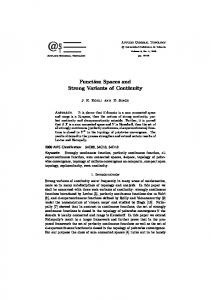
Fig. 1. Dose-responses of various melanocyte cultures to α-MSH. The responses of NHM 753-c, 830-c, 849-b and 755-c, homozygous or compound heterozygous for MC1R variants, and NHM 747-c, 729-c and 751-b, homozygous for the consensus MC1R were compared. The response of NHM 765-c, heterozygous for Val92Met substitution, is also presented, since it is comparable to NHM 753-c and 830-c in the ratio of eumelanin to pheomelanin. The responses to α-MSH were tested as described in the Materials and Methods. The effects of increasing doses of α-MSH on cAMP formation, tyrosinase activity and cell proliferation are presented in A, B and C, respectively. Basal levels of cAMP (Pmole/106 cells) in the cultures tested were as follows: 753-c=2.176±0.134; 830-c=1.451; 849b=1.172±0.0219; 755-c=2.193±0.206; 765-c=1.675±0.137; 747c=1.074±0.098; 729-b=2.007±0.128; 751-b=0.84±0.103. In A and B, each value represents the mean percent of control of six determinations±s.e. In C, each value is the mean percent of control of three determinations±s.e.
Response of melanocytes with known MC1R genotype to α-MSH Melanocyte cultures homozygous for the consensus MC1R or heterozygous for one variant of the MC1R (represented by the data for NHM 765-c) responded to α-MSH with dosedependent increases in cAMP levels, tyrosinase activity and proliferation (Fig. 1). Variations in the magnitude of the response to α-MSH among cultures may be attributed to differential expression of other genes involved in the regulation of pigmentation. NHM 753-c, homozygous for Arg160Trp, 830-c, heterozygous for Arg160Trp and Asp294His, and 849b, heterozygous for Arg151Cys and Asp294His substitutions in MC1R were unresponsive, or 100-times less responsive to α-MSH than melanocytes wild-type or heterozygous for MC1R variants (Fig. 1). Comparing the responses to α-MSH of NHM 753-c, 830-c and 849-b to that of NHM 765-c demonstrated significant differences. NHM 753-c, 830-c and 849-b showed
Cell number (% control ± s.e.)
400 350 300
Control α-MSH 0.1 nM α-MSH 1 nM α-MSH 10 nM
C
Forskolin 1 µM
250 200 150 100 50 0 753-c 830-c 849-b 755-c 765-c 747-c 729-b 751-b R 160W R 160W R 151C V 92M V 92M R 160W D 294H D 294H V 92M +
wild-type MC1R
no significant change in cAMP levels after treatment with αMSH, as determined using Student’s t-test. (Fig. 1A). In contrast, NHM 765-c, heterozygous for Val92Met substitution and with a low eumelanin to pheomelanin ratio, responded to 0.1 or 10 nM α-MSH with significant increases in cAMP levels above control (66% and 4.5-fold, respectively; P≤0.001, as determined by Student’s t-test). NHM 755-c homozygous for the Val92Met substitution responded to 0.1 nM and 10 nM αMSH with a 4-fold and 11-fold increase in cAMP formation (Fig. 1A). Additionally, NHM 753-c, 830-c and 849-b showed no change, while NHM 765-c showed significant increases, in tyrosinase activity in response to 0.1 or 1 nM α-MSH (about 90% or 130% increase, respectively; P