European Cells and Materials Vol. 24 2012 (pages 278-291) J Klein-Nulend et al.
1473-2262 Osteocyte mechanosensing: role of ISSN the cytoskeleton
MECHANICAL LOADING AND HOW IT AFFECTS BONE CELLS: THE ROLE OF THE OSTEOCYTE CYTOSKELETON IN MAINTAINING OUR SKELETON J. Klein-Nulend1,*, R.G. Bacabac2 and A.D. Bakker1 1
Department of Oral Cell Biology, ACTA-University of Amsterdam and VU University Amsterdam, Research Institute MOVE, Amsterdam, The Netherlands 2 Department of Physics, Medical Biophysics Group, University of San Carlos, Cebu City, Philippines
Abstract
Bones adapt to mechanical loading
Lack of physical activity causes bone loss and fractures not only in elderly people, but also in bedridden patients or otherwise inactive youth. This is fast becoming one of the most serious healthcare problems in the world. Osteocytes, cells buried within our bones, stimulate bone formation in the presence of mechanical stimuli, as well as bone degradation in the absence of such stimuli. As yet, we do not fully comprehend how osteocytes sense mechanical stimuli, and only know a fraction of the whole range of molecules that osteocytes subsequently produce to regulate bone formation and degradation in response to mechanical stimuli. This dramatically hampers the design of bone loss prevention strategies. In this review we will focus on the first step in the cascade of events leading to adaptation of bone mass to mechanical loading, i.e., on how osteocytes are able to perceive mechanical stimuli placed on whole bones. We will place particular emphasis on the role of the osteocyte cytoskeleton in mechanosensing. Given the crucial importance of osteocytes in maintaining a proper resistance against bone fracture, greater knowledge of the molecular mechanisms that govern the adaptive response of osteocytes to mechanical stimuli may lead to the development of new strategies towards fracture prevention and enhanced bone healing.
Throughout life bone is constantly remodelled by the coordinated action of bone-resorbing osteoclasts and bone-forming osteoblasts in basic multicellular units. This continuous remodelling likely serves to prevent and remove fatigue-related micro-damage and allows adaptation of the bone mass and structure. The balance between the amount of bone resorption and bone formation determines whether the process of bone remodelling leads to a net gain or loss of bone mass. The number and activity of osteoclasts and osteoblasts are determined by a multitude of factors, such as hormones and cytokines, as well as by locally produced signalling molecules under the influence of mechanical stimuli (Vezerides et al., 2006; You et al., 2008; Onal et al., 2012). The osteocyte is a source of soluble factors not only to target cells on the bone surface but also to target distant organs, such as muscle, kidney, and other tissues (Bonewald, 2011). During physical activity, mechanical forces are exerted on the bones through ground reaction forces and by the contractile activity of muscles (Lanyon et al., 1975; Usui et al., 2003). These physical forces result in a maintenance or gain of bone mass, but also drive adaptation of bone structure. The adaptation of trabecular bone architecture according to the demands of mechanical usage is evident in the vertebrae, where the trabeculae are predominantly oriented in the longitudinal direction, providing the best possible resistance to compression fracture of the vertebrae with a minimal use of material. A classic example of the stimulating effect of mechanical stimuli on bone mass is provided by the bones in the forearm of tennis players. The ulna and radius in the arm that holds the racket are exposed to high impact forces, leading to tiny deformations in the stiff bone matrix and an increase in bone mass of 5 to 10 % compared to the ulna in the contra-lateral arm (Ducher et al., 2004). The deformations that occur in bones as a result of physical forces are expressed as strain, where 1,000 microstrain equals a 0.01 % change in length of the bone compared to its original length. Vigorous exercise induces bone strains up to 1,000 microstrain in humans (Lanyon et al., 1975). By comparison, controlled bouts of whole bone loading resulting in 1,000 to 3,000 microstrain are anabolic in experimental animal models of bone-loading, demonstrating the potential for appropriate physical exercise routines as a means to enhance bone mass (Rubin and Lanyon, 1987; Turner et al., 1994; Reijnders et al., 2007).
Keywords: Mechanical loading; mechanotransduction; fluid shear stress; osteocyte; bone cell; cytoskeleton; inflammatory cytokines; cell mechanics; cell shape; cell stiffness.
*Address for correspondence: J. Klein-Nulend Department of Oral Cell Biology Research Institute MOVE, ACTA-VU University Amsterdam, Gustav Mahlerlaan 3004, 1081 LA Amsterdam, The Netherlands Telephone Number: +31 205980881 FAX Number +31 205980333 E-mail
[email protected]
278
www.ecmjournal.org
J Klein-Nulend et al. In contrast with the increase in bone mass with vigorous physical exercise, we lose bone mass in the absence of mechanical stimuli. Bone mass is rapidly lost under unloading conditions, e.g., during bed rest, hind-limb unloading in mice, or local denervation of muscles (Globus et al., 1986; Vandamme et al., 2012). Interestingly, strains resulting from habitual activity suffice to prevent bone loss compared to complete unloading, even though these habitual strains hardly ever exceed 400 microstrain. Thus, either mechanical loads that lead to tiny strains within the bone matrix are somehow sensed by the cells within bone, which then act to preserve bone mass, or sporadic mechanical stimuli that exceed 2,000 microstrain still occur often enough to prevent bone loss. We have shown that cultured bone cells release nitric oxide (NO) in response to mechanical stimulation in the form of a fluid shear stress, and that the amount of NO released linearly correlates to the rate of the applied fluid shear stress (Bacabac et al., 2004). Since the rate at which a mechanical stimulus is applied is the product of the magnitude (amplitude) and the frequency of the stimulus, this supports the notion that low-magnitude, high-frequency mechanical stimuli are as potent in evoking a response in bone cells as high-amplitude, low-frequency stimuli (Ozcivici et al., 2010). In other words, very small mechanical stimuli may elicit a cellular response only if applied fast enough. The rate dependent-response to stress provides a possible explanation why adaptive bone formation in vivo may occur despite the sporadic occurrence of high-amplitude strains in daily life. The question how daily mechanical loads preserve bone mass is clinically highly relevant, as with our ageing population more and more people suffer from fragile bones. Osteocytes sense mechanical stimuli and direct mechanical adaptation of bone The cells likely responsible for sensing the physical stimuli derived from mechanical forces exerted on bones are the osteocytes, which comprise over 90 % of the bone cells. Osteocytes are stellate cells that are embedded within the calcified bone matrix. They form a large number of cellcell contacts through their long slender cell processes, forming a syncytium capable of rapid transduction of signals (Fig. 1). Osteocytes are highly mechanosensitive, likely more so than periosteal fibroblasts or osteoblasts, and alter the production of a multitude of signalling molecules when triggered by a mechanical stimulus. Mechanically activated osteocytes produce signalling molecules like bone morphogenetic proteins (BMPs), Wnts, prostaglandin E2 (PGE2), and NO, which can modulate the recruitment, differentiation, and activity of osteoblasts and osteoclasts (Robling et al., 2006; Tan et al., 2007; You et al., 2008; Santos et al., 2009 ). Thus, osteocytes are theoretically capable of orchestrating bone adaptation in response to mechanical stimuli. That osteocytes are indeed essential mediators of osteoclastic bone resorption was confirmed in an elegant experiment by Tatsumi et al. (2007). They showed that the loss of bone mass following hind limb unloading of mice was prevented when ~80 % of the
Osteocyte mechanosensing: role of the cytoskeleton osteocytes were ablated. Osteocytes thus seem to stimulate osteoclast activity in the absence of daily mechanical loads, a capability that has been confirmed in in vitro studies (You et al., 2008; Kulkarni et al., 2010). Indeed it has been shown recently by two independent groups that RANKL production by osteocytes determines bone mass in adult mice, demonstrating the importance of osteocytes in the regulation of bone mass (Nakashima et al., 2011; Xiong et al., 2011). Interestingly, the same study demonstrating the requirement of osteocytes for mediating unloadinginduced bone loss also showed that the anabolic response of bone to (re)loading does not require the presence of living osteocytes (Tatsumi et al., 2007). However, this does not eliminate the role of osteocytes in mediating the anabolic response of bone to loading under normal conditions. Fluid flow, strain, and hydrostatic pressure as stimuli for osteocytes If osteocytes are the professional mechanosensing cells of bone, then how do these cells sense whole bone loads? One popular theory entails that matrix strains surrounding the osteocyte cell processes drive a thin layer of extracellular fluid surrounding the osteocyte cell processes to flow across a pressure gradient. This flow of fluid “amplifies” local strains, and is thereby the mechanical signal that is ultimately sensed by the osteocytes. There is ample experimental evidence to support the idea that deformations of the bone matrix drive an interstitial fluid flow. Knothe-Tate et al. have shown experimentally a flow of extra-cellular fluid around the osteocytes as a result of bone tissue strains, by loading of sheep tibiae and following the distribution of tracers through the lacuno-canalicular network (Knothe-Tate et al., 1998; Knothe-Tate et al., 2000). More recently, Price et al. (2011) used fluorescence recovery after photobleaching for imaging fluid displacement synchronised with mechanical loading, to show that the mechanical loading of mouse tibia enhanced fluid transport through the lacuno-canalicular system, demonstrating the correlation of canalicular fluid flow with mechanical load. In addition, several investigators reported that it is not the amount of strain applied to a whole bone that influences bone formation, but the rate at which the strain is applied (Price et al., 2011). Dynamic bone loading, which enhances fluid flow, has also been demonstrated to induce an osteogenic response (Lanyon and Rubin, 1984; Luo et al., 1995; Mosley and Lanyon, 1998). Static loading on the other hand has little effect on lacuno-canalicular fluid flow, and has only a minor effect on bone formation (Lanyon and Rubin, 1984). It has been extensively demonstrated that osteocytes in vitro are sensitive to a flow of fluid when seeded as a monolayer on flat, 2-dimensional (2D) substrates (Klein-Nulend et al., 1995a; Klein-Nulend et al., 1995b; Ajubi et al., 1996; Bakker et al., 2001; Bacabac et al., 2004; Bakker et al., 2009; Litzenberger et al., 2010; Juffer et al., 2012; Kulkarni et al., 2012). One could argue that interstitial fluid is driven to flow within the canaliculi over only osteocyte processes in vivo while a laminar fluid flow over cells seeded on a flat substrate will deform the cell body as well as the cell 279
www.ecmjournal.org
J Klein-Nulend et al.
Osteocyte mechanosensing: role of the cytoskeleton
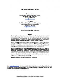
Fig. 1. In osteocytes, all is connected. (A) Macroscopic photograph of a transversely sectioned human femoral head, dipped in black ink to highlight the orientation of the bone trabeculae. Scale bar indicates 5 mm. (B) High magnification osteocytes embedded within their matrix. The osteocytes with their many cell processes are visible (red). Scale bar indicates 5 µm. (C) Schematic drawing of part of an osteocyte cell body with processes (white) residing within the calcified matrix (ECM; beige). The osteocyte cell membrane is surrounded by interstitial fluid and proteoglycans (ECM; brown). Microtubules (blue lines) originate from the centrosome and form a scaffold along which numerous molecules shuttle. Microtubules also form the core of the primary cilium. Osteocyte cell processes contain mostly actin (red lines), cross-linked by fimbrin (not shown) at places where processes bifurcate. The processes contain αvβ3 integrins (blue), possibly located on top of collagen “hillocks”, and protein tethers. These tethers may deform as a result of interstitial fluid flow and subsequently “tug” at the actin cytoskeleton, or pull open stretch-activated ion channels (purple), allowing calcium and other ions to enter the cell and activate a multitude of chemical signalling cascades. The fluid flow may also tug directly at integrins, such as α5β1, present along the cell processes and body and associated with hemi-channels. Activated hemi-channels release signalling molecules such as ATP, triggering a signalling cascade. The mechanical signal may be transduced from sites where integrins are triggered, via the actin cytoskeleton, to distant sites, including the nucleus, which is connected to actin via LINC complexes. Forces applied to actin fibres can likely also open stretch-activated ion channels. Integrins are often clustered together at focal adhesions (yellow), which in osteocytes are predominantly located at places where the cell processes intersect with the cell body. Focal adhesions contain tyrosine kinases, such as focal adhesion kinase, which make focal adhesions a prime location for transducing mechanical signals into a chemical response. Considering that the cytoskeleton interconnects virtually every part of the mechanosensing machinery, one can easily imagine that changes in cytoskeletal properties affect mechanosensing. 280
www.ecmjournal.org
J Klein-Nulend et al. processes, thereby eliciting a response that otherwise would not be provoked (McGarry et al., 2005a; Fritton and Weinbaum, 2009). However, it has been shown recently using a Stokesian fluid stimulus probe that electrical signalling is provoked at much lower forces when applied to a cell process of osteocytes compared to the cell body, thus suggesting that in vitro fluid flow experiments provide a valid confirmation of the sensitivity of osteocytes to fluid flow (Wu et al., 2011). In addition to responding to strain-driven fluid flow, it is also possible that osteocytes respond to matrix strains directly. MC3T3-E1 osteoblasts increase their production of signalling molecules in response to substrate deformations as low as 3,400 microstrain (Robinson et al., 2006), and osteocytes are more mechanosensitive than osteoblasts (Klein-Nulend et al., 1995a). The relative flat and spread shape of isolated bone cells in 2D culture may greatly hamper their sensitivity to a mechanical stimulus (Bacabac et al., 2008), and strains that are not able to elicit a response in bone cells adhered to a flat and stiff surface may be perfectly able to elicit a response in cells in their natural 3-dimensional (3D) conformation. Thus, direct sensation of bone strains may already suffice to activate mechanosensitive bone cells in vivo. It has also been proposed that matrix strains can be locally amplified up to 3-fold by the inhomogeneities in the matrix that are formed by the osteocyte lacunae (Bonivtch et al., 2007). Matrix strains around the osteocyte cell bodies may thus exceed whole bone strains, especially at points where cell processes intersect with the cell body, and could be sufficient to directly activate the osteocytes (Bonivtch et al., 2007). In this regard the observation by Vatsa et al. (2008a), that molecules involved in cellular mechanotransduction such as F-actin and paxillin are concentrated at these intersections, is highly interesting (Fig. 1). Bones can be considered as a material containing large (vasculature) and small (canaliculi) interconnected fluidfilled pores. The permeability of the lacuno-canalicular porosity is several orders of magnitude lower than that of the vascular porosity (Gardinier et al., 2010). Since the lacuno-canalicular porosity has a low permeability, rapidly placed load on bone causes strains that first pressurise the interstitial fluid around the osteocytes, and then drive fluid flow causing dissipation of the build-up of hydraulic pressure (Wang et al., 1999). The magnitude of pressure experienced by osteocytes in vivo may reach up to 5 MPa according to recent calculations (Gardinier et al., 2010). Cyclic hydraulic pressures of 68 kPa can modulate signalling molecule production in cells of the mouse MLO-Y4 osteocyte cell line, and a pressure of 13 kPa was sufficient to stimulate prostaglandin production by primary osteocytes isolated from chicken calvariae (Klein-Nulend et al., 1995a; Liu et al., 2010). This suggests that, besides substrate strain and fluid shear stress, the loading-induced hydraulic pressure could potentially serve as a mechanical stimulus for osteocytes. Whatever the mechanical load-derived stimulus is that activates the osteocytes, the question remains which osteocyte feature enables the perception of the physical stimulus and subsequent
Osteocyte mechanosensing: role of the cytoskeleton transduction into a chemical signal. This question is not necessarily restricted to osteocytes, as eukaryotic cells in general are sensitive to mechanical stress. Cellular features enabling mechanotransduction in osteocytes A multitude of sensory elements exist that allow cells to detect mechanical stimuli. Mechanosensing is enabled by force-induced conformational changes in cellular structures, such as stretch-activated ion channels, integrin complexes, and cell-cell adhesions. The conformational changes enable the influx and efflux of ions or the activation of signalling cascades, resulting in altered cell shape and altered activity and production of proteins (Hoffman et al., 2011). The cytoskeleton, which can be considered a composite gel-like material (of actin, microtubules, intermediate filaments and their crosslinkers) is the scaffold determining cellular shape and stiffness (Sugawara et al., 2008). Molecules like integrins anchor to the extracellular matrix (ECM) and mechanically link the exterior of the cell to the cytoskeleton, forming trans-membrane complex structures. These complexes, often clustered in so-called focal adhesions, are therefore prime suspects as mechanotransducers. With respect to osteocyte mechanosensing, the focal adhesion kinase inhibitor-14 has been shown to abolish fluid flow-induced stabilisation of β-catenin and consequent activation of the Wnt/β-catenin pathway in osteocytes, suggesting that focal adhesions and integrins play an important role in osteocyte mechanosensing (Santos et al., 2010). Indeed, β1-integrins on osteocytes in vivo have been shown to mediate specific aspects of mechanotransduction, confirming the importance of integrins for mediating mechanical stimuli in osteocytes (Litzenberger et al., 2010). Interfering with β1-integrin signalling in vitro reduced the upregulation of cyclooxygenase-2 normally observed after mechanical stimulation of osteocytes, but did not affect mechanically induced intracellular calcium mobilisation (Litzenberger et al., 2010). In addition, α5β1 integrins interact directly with connexin 43 (Cx43), and this interaction is required for mechanical stimulationinduced opening of Cx43-containing hemi-channels (Batra et al., 2012). Cx43-containing hemi-channels are readily expressed on osteocytes and affect the response of osteocytes to mechanical loading in vivo (Zhang et al., 2011). Direct mechanical perturbation of α5β1 integrins leads to the opening of the Cx43-containing hemi-channels (Batra et al., 2012). Integrin attachments thus likely serve as the mechanotransducing units that potentiate the opening of hemichannels (Burra et al., 2010). Other integrins that may mediate osteocyte mechanotransduction are avb3 integrins. Although avb3 integrins are not essential for the attachment of osteocytes to ECM in vitro (Aarden et al., 1996), they may play a role in osteocyte physiology, since it has been shown recently that avb3 integrins mediate signalling via DMP-1, a molecule that is almost exclusively produced by osteocytes (Wu et al., 2011). Interestingly, in vivo avb3 integrins are present along the
281
www.ecmjournal.org
J Klein-Nulend et al. osteocyte processes which are thought to be the main sites of mechanotransduction in osteocytes (McNamara et al., 2009). The glycocalyx of the osteocyte dendritic process is required for the formation of strong integrin attachments (Burra et al., 2010; Burra et al., 2011), demonstrating that the role of integrins should be considered in the context of the complex structures in which they reside before we can fully comprehend how mechanical stimuli are transduced to osteocytes. It should also be noted that as a consequence of osteocytes expressing their own specific set of integrins and the selectivity of integrins for substrates, the nature of the ECM likely affects the mechanoresponse of osteocytes. Although stretch-activated ion channels have long been suspected to play a role in osteocyte mechanotransduction, it is currently unclear which molecules act as stretchactivated ion channels in osteocytes. The osteocyte response to mechanical loading can clearly be inhibited by gadolinium chloride, which is a non-specific blocker of TRP channels (Ajubi et al., 1999; Bakker et al., 2009). However, which member of the extensive family of TRP channels is responsible for transducing mechanical signals in the osteocyte has not been elucidated. It is unlikely that TRPV6 is a candidate as it is only present at low levels in murine osteoblasts and osteocytes and plays a minor functional role in calcium uptake by osteoblasts (Little et al., 2011). To understand how the cellular features mentioned above act as a mechanotransduction complex, one needs to regard the cellular context of these features. It would be difficult, if not impossible, for forces acting on a cell to induce conformational changes in cellular molecules that are freely floating in a semi-liquid cell membrane. However, structures that are anchored to neighbouring cells, the ECM or the glycocalyx, as well as to the cytoskeleton, form a direct mechanical link between the extracellular environment and the intracellular compartment (Fig. 1). Such structures are in an excellent position to sense mechanical forces. Integrins are coupled to the cytoskeleton via molecules such as vinculin, talin, and α-actinin. Though non-trivial, one may imagine three non-linear springs in a series representing the mechanical link between the ECM, the transmembrane proteins (including the focal adhesions), and the cytoskeleton (Fig. 2), to conceptualise the transfer of forces between these protein structure clusters (Fig. 3). Note, however, that each protein network is expected to become stiffer in response to applied forces (Storm et al., 2005), which could support an amplified force transfer. Hence, the importance of anchoring mechanotransduction complexes that connect the ECM to the cytoskeleton predicts that the osteocyte cytoskeleton plays a key role in osteocyte mechanotransduction. Structure of the osteocyte cytoskeleton The cytoskeleton is a scaffold made out of protein components that provide mechanical structure to cells. The viscoelastic properties of the cytoskeletal structure provide cells with resistance to shear or compression, enable cell
Osteocyte mechanosensing: role of the cytoskeleton
Fig. 2. Spring-series conceptualisation of mechanical linkage between the cytoskeleton (CSK), focal adhesion complexes (FAC), and the extracellular matrix (ECM), with respective non-linear spring constants, KCSK , KFAC , KECM .
migration, enable transport of intracellular molecules, determine the mechanical properties of the cells, and allow for mechanosensing. Eukaryotic cells contain three main kinds of cytoskeletal structures, each built up out of their own proteins: actin, intermediate filaments, and microtubules. The distribution of each of these structures seems to change when osteoblasts differentiate into osteocytes. In parallel, the stiffness of osteoblasts decreases during their differentiation towards osteocytes (Sugawara et al., 2008). Microtubules are limited in distribution to the proximal region of osteocyte processes but extend the entire length of cell processes of MC3T3-E1 osteoblasts grown in 3D in collagen gels (Murshid et al., 2007). Microtubules are essential for the integrity and formation of osteoblast cell processes grown in 3D, but processes of primary osteocytes in 3D are dependent on actin (Murshid et al., 2007). Actin filaments are also crucial for maintaining the shape of primary chicken osteocytes when grown on flat substrates (Tanaka-Kamioka et al., 1998). Not surprisingly, osteocytes also contain a set of actin-bundling proteins distinctive from that in osteoblasts. Fimbrin and a-actinin are predominantly present in the processes of osteocytes, with especially strong signals of fimbrin at the sites of bifurcation of the processes (Kamioka et al., 2004). Compared to osteoblasts, osteocytes also contain high amounts of villin, which is present within the osteocyte cytoplasm but not within the processes. Osteoblasts immunostained with anti-spectrin show punctate signals on their cytoplasmic membranes, whereas spectrin is co-localised with actin from the distal portion of the cytoplasmic processes to the cell centre (Kamioka et al., 2004). The typical morphology of the osteocyte, determined by its cytoskeleton, was originally thought to be imposed on differentiating osteoblasts during their incorporation in the bone matrix. Osteocytes have to remain in contact with other cells and ultimately with the bone surface to ensure access to oxygen and nutrients. Culture
282
www.ecmjournal.org
J Klein-Nulend et al.
Osteocyte mechanosensing: role of the cytoskeleton
Fig. 3. Protein network clusters across the extracellular matrix, trans-membrane proteins, and the cytoskeleton regions of a spread cell. ECM, extracellular matrix; TMC, transmembrane complex; FAK, focal adhesion kinase.
experiments with isolated chicken osteocytes have shown, however, that although the cells lose their stellate shape in suspension, they re-express this morphology as soon as they settle on a support (Van der Plas and Nijweide, 1992). Apparently, the typical stellate morphology and the need to establish a cellular network are intrinsic characteristics of terminal osteocyte differentiation. Although the stellate shape of osteocytes seems innate, by no means does this shape represent a fixed or static feature. Dallas and co-workers (Veno et al., 2006) have shown cell body movement and the extension and retraction of cell processes over time using dynamic imaging of living osteocytes within their lacunae. Calvarial explants from transgenic mice with green fluorescent protein expression targeted to osteocytes revealed that the osteocyte is highly dynamic. Therefore the osteocyte processes, rather than being permanent connections between osteocytes as well as between osteocytes and bone surface cells, may have the capacity to connect, disconnect, and re-connect (Bonewald, 2006; Zang et al., 2006). This phenomenon implies a complex role for intercellular connection in bone, and may indicate an adaptive information transfer facility through the ostecytic syncytium beyond mere force transfer mechanisms. Whereas signalling molecule information is diffusion-limited, intercellular communication provides a more efficient means for information transfer.
Since the osteocyte cell processes are of predominant importance for sensing mechanical stimuli, and the osteocyte processes apparently contain mainly actin, the actin cytoskeleton rather than the microtubule cytoskeleton could be important for osteocyte mechanosensing. However, disruption of both the actin cytoskeleton with cytochalasin B and disruption of the microtubule cytoskeleton with colchicine inhibits mechanical stimulation-mediated release of PGE2 in the mouse osteocyte cell line MLO-Y4 (McGarry et al., 2005b). Besides the cell processes, osteocytes may possess a single primary cilium that contributes to mechanosensing (Malone et al., 2007). The primary cilium is a solitary organelle that emanates from the cell surface of most mammalian cell types, and is a key coordinator of signalling pathways during development and in tissue homeostasis (Berbari et al., 2009). Primary cilia consist of an axoneme of nine doublet microtubules that extends from a basal body. This could explain why disruption of actin as well as of microtubules may upset the osteocyte response to mechanical stimulation. In the mouse osteoblast cell line MC3T3-E1, which has been shown to depend on microtubules rather than actin for their cytoskeletal integrity, inhibition of actin polymerisation did not inhibit intracellular calcium mobilisation or PGE2 release in response to a mechanical stimulus (McGarry et al., 2005b; Malone et al., 2007). In contrast, in calvarial 283
www.ecmjournal.org
J Klein-Nulend et al. osteoblasts derived from chicken, disruption of the actin cytoskeleton did strongly inhibit mechanical loadingstimulated PGE2 release (Ajubi et al., 1996). Cytoskeletal mechanics and stress-response signatures Fluid flow over dendrites in the lacuno-canalicular porosity has been suggested to induce strains in the actin filament bundles of the cytoskeleton that are more than an order of magnitude larger than tissue level strains (You et al., 2001). Using ultrastructural data for the cell process cytoskeleton and the tethering elements that attach the process to the canalicular wall, a 3D model was created for the osteocyte process. Using this model the deformed shape of the tethering elements and the hoop strain on the central actin bundle as a result of loading-induced fluid flow was predicted. It was found that tissue-level strains of >1,000 microstrain at 1 Hz result in a hoop strain of >0.5 % (You et al., 2001). The tethering elements of the osteocyte process can thus act as a strain-amplifier. Tethering filaments appear to be absent in the pericellular space surrounding the cell body, likely due to the wide pericellular space (~1 µm) between the cell membrane and the wall of the lacuna, in contrast to the pericellular space surrounding the cell process (~80 nm) (You et al., 2004). Potentially CD44 serves as the tethering molecule since it is expressed in osteocyte cell processes and has an attachment site for hyaluronan (Noonan et al., 1996). Alternatively, a protein tether involved in transduction of mechanical stimuli has recently been identified in cutaneous mechanoreceptors. This molecule is a protein filament with a length of ~100 nm (Hu et al., 2010). It is possible that the osteocyte tether is similar to this protein tether. More recently, a new theoretical model has been developed that predicts that integrin-based attachment complexes along osteocyte cell processes would dramatically and focally amplify small tissue-level strains (Wang et al., 2007). Using rapid fixation techniques it was observed that osteocyte cell processes seem attached directly at canalicular projections emanating from the canalicular wall, via αvβ3 integrins (McNamara et al., 2009). The theoretical model predicts that the tensile forces acting on these integrins are