meeting meeting report report Cell adhesion and signal transduction in cancer Conference on Cadherins, Catenins and Cancer Walter Birchmeier Max-Delbrück-Center for Molecular Medicine, Berlin, Germany
The Centro Nacional de Investigaciones Oncológicas (CNIO) Cancer Conference on ‘Cadherins, Catenins and Cancer’ was held in Madrid, Spain, between 29 November and 1 December 2004. The conference was organized by A. Cano, H. Clevers, J. Palacios and F. van Roy
Keywords: cancer; development; epithelial-mesenchymal transitions; tumours EMBO reports (2005) 6, 413–417. doi:10.1038/sj.embor.7400408
Introduction Cadherin cell-adhesion molecules and their intracellular binding partners, catenins, were discovered in the 1980s (for a review, see Takeichi, 1995). Interest in these molecules was sparked by the discovery that cadherins and, subsequently, catenins are important in the formation and metastasis of carcinomas (Behrens et al, 1989; Berx et al, 1998; Perl et al, 1998; Polakis, 2000). Twenty years later, interest in cadherins and catenins remains high, as seen at the Centro Nacional de Investigaciones Oncológicas (CNIO) Cancer Conference on ‘Cadherins, Catenins and Cancer’ in Madrid. The meeting covered research on all fronts, from the molecular aspects of cadherin and catenin regulation and signalling, to their role in human carcinogenesis and subsequent clinical applications.
E-cadherin in cancer progression In 1998, germline mutations in the E-cadherin gene were identified in New Zealand Maori families with diffuse-type gastric cancer, which firmly established the importance of E-cadherin in the Max-Delbrück-Center for Molecular Medicine, Robert-Rössle-Strasse 10, 13125 Berlin, Germany Tel: +49 30 9406 3800; Fax: +49 30 9406 2656; E-mail:
[email protected] Submitted 04 March 2005; accepted 25 March 2005 published online 22 April 2005
©2005 EUROPEAN MOLECULAR BIOLOGY ORGANIZATION
progression of carcinomas (Guilford et al, 1998). At the Madrid meeting, F. Carneiro (Porto, Portugal) presented new data, which showed that hereditary diffuse gastric cancer is a dominantly inherited familial cancer syndrome that contributes up to 3% of all gastric cancers. Furthermore, E-cadherin mutations are the causal genetic defect for up to 40% of these cases. About 80% of the mutations that have been identified in the E-cadherin gene result in a truncated protein, whereas the remaining 20% are missense mutations. Germline mutations of E-cadherin are distributed throughout the whole gene, in contrast to somatic mutations in sporadic diffuse gastric cancer, which cluster in exons 7-9 (Berx et al, 1998). Inactivation of the remaining normal allele is required for cancerogenesis, and gene silencing through promoter methylation is found in 50% of cases. These studies prompted the International Gastric Cancer Linkage Consortium to develop clinical criteria and guidelines for the management of carriers of E-cadherin mutations, which include the use of intensive screening and prophylactic gastrectomies (Caldas et al, 1999). Despite the identification of somatic mutations in the E-cadherin gene in human cancers, causal evidence for the involvement of E-cadherin in tumour formation and progression in testable animal models was previously lacking. This has now been elegantly shown by J. Jonkers (Amsterdam, the Netherlands), who introduced a conditional loss-of-function mutation in the E-cadherin gene into mice that carry p53 mutations. Although tissue-specific inactivation of E-cadherin alone did not result in tumour formation, the combined inactivation of E-cadherin and p53 led to the accelerated development of mammary gland and skin tumours. Moreover, loss of E-cadherin induced a phenotypic change from non-invasive to highly invasive mammary gland tumours and a conversion from ductal to lobular carcinomas. The latter in mice resemble their human equivalent in that they show strong stromal involvement, invasion into the surrounding tissue and distant metastases to several organs. By contrast, the group of G. Christofori (Basel, Switzerland) has used the Rip1-Tag2 transgenic mouse model of pancreatic β-cell carcinogenesis to show that the loss of E-cadherin is causally involved in the transition from benign adenoma to malignant, invasive carcinoma (Perl et al, 1998). The group also reported that the loss of another neural cell-adhesion molecule (NCAM) during Rip1Tag2 tumour progression results in the formation of lymph-node metastasis. They showed that the loss of NCAM results in the loss of β1 integrin function, which contributes to loss of tissue integrity, EMBO reports
VOL 6 | NO 5 | 2005 4 1 3
reviews upregulated expression of lymphangiogenic vascular endothelial growth factor-C (VEGFC) and VEGFD, and subsequently increases lymphangiogenesis and the lymphogenic dissemination of tumour cells (Crnic et al, 2004). Hence, a new model was proposed, in which there are two distinct pathways to tumour metastasis: the classical pathway of active tumour cell migration and invasion, which is exemplified by the loss of E-cadherin; and a passive pathway in which the increased presence of lymphatic vessels washes tumour cell clusters out into the lymphatics, where they become trapped in the regional lymph nodes.
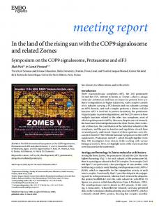
The regulation of E-cadherin expression The invasion of carcinoma cells is associated with the loss-ofexpression of epithelial genes, such as E-cadherin, and the gain-ofexpression of mesenchymal genes (Behrens et al, 1989; Batlle et al, 2000; Cano et al, 2000). This process, which is known as epithelial-mesenchymal transition (EMT), also occurs under strict spatiotemporal control during normal embryonic development (Fig 1; Thiery, 2002). J. Palacios (Madrid, Spain) and G. Berx (Ghent, Belgium) reported that further target genes that are downregulated during EMT include keratins, desmosomal cadherins and components of the basal lamina. By contrast, the upregulated mesenchymal genes include vimentin, secreted protein acidic and rich in cysteine (SPARC), collagens, integrins, protease inhibitors and cytoskeletal components. Altered expression of a selection of these target genes could also be validated in uterine carcinosarcomas and breast cancers. EMT is a crucial event during tumour metastasis and normal embryonic development, as indicated by two further reports at the Madrid meeting. T. Brabletz (Erlangen, Germany) showed that, in colon carcinomas, tumour cells in the central tumour mass and at the invasive fronts differ from one another: the latter undergo EMT, as characterized by a loss of E-cadherin at the membrane and translocation of β-catenin to the nucleus (Brabletz et al, 2001). This is a transient event, given that metastases in the lymph nodes and liver have adherent tumour cells in tubular structures. Genes that become activated at the tumour invasive front include those encoding the transcription factor Slug, laminin-γ2 and, as reported by A. Ben-Ze’ev (Rehovot, Israel), the cell–cell-adhesion molecule L1 and the disintegrin and metalloprotease ADAM10 (Gavert et al, 2005). L1 expression conferred increased cell motility, cell transformation and tumorigenesis in mice. By contrast, E. Dejana (Milan, Italy) showed that EMT is required for normal heart development during embryogenesis. Endocardial cells in the atrioventricular region undergo transforming growth factor-β (TGF-β)dependent EMT and invade the underlying cardiac jelly. This process is accompanied by the activation of β-catenin/T-cell factor (TCF)/lymphocyte-enhancer factor (Lef) transcriptional activity, and is blocked by the absence of β-catenin, which implies an interaction between TGF-β and Wingless (Wnt)-signalling pathways in the induction of endothelial-mesenchymal transformation (Liebner et al, 2004). One of the hallmarks of EMT is the loss of E-cadherin expression. Substantial progress has been made in identifying the mechanisms by which E-cadherin expression is normally regulated. It was reported that different transcription factors—Snail, Slug, δ-crystallin E2-box factor-1/zinc-finger E-box-binding transcriptional repressor (δEF1/ZEB1), Smad-interacting protein-1 (SIP1)/ZEB2 and E12/E47—were responsible for downregulating 4 1 4 EMBO reports VOL 6 | NO 5 | 2005
meeting repor t
E-cadherin expression by binding directly to E-boxes in the E-cadherin promoter (Fig 1; Batlle et al, 2000; Cano et al, 2000). The groups of A. Cano (Madrid, Spain) and A. García de Herreros (Barcelona, Spain) reported new findings on the action of the transcription factors Snail and Slug during EMT. They showed that Snail mediates E-cadherin repression by recruitment— through the amino-terminal Snail/growth-factor independent-1 (SNAG) domain—of a Sin3A corepressor complex that also contains histone deacetylase-1/2 (HDAC1/2), which leads to chromatin inactivation. Activity of the Snail promoter and levels of Snail mRNA are dependent on the extracellular signal-regulated kinase (ERK), mitogen-activated protein (MAP) kinase and phosphatidylinositol 3-kinase (PI3K)/Akt signalling pathways, as well as Snail itself, which therefore functions in an autoregulatory loop (Bachelder et al, 2005).
β-catenin in cancer and embryogenesis It is not only cadherins that have been implicated in tumorigenesis and metastasis; their cytoplasmic binding partners, catenins, are also important in cancer formation and progression. β-catenin has been found to be mutationally activated in several tumours (for example, in 90% of hepatoblastomas, 75% of pilomatricomas and 10% of colon cancers; for reviews, see Bienz & Clevers, 2000; Polakis, 2000). Moreover, β-catenin interacts with the tumour suppressor gene adenomatous polyposis coli (APC), which is mutated in familial and sporadic colon carcinomas. A second armadillo family protein, p120 catenin, which is known to stabilize E-cadherin and thereby prevent its degradation, has also been implicated in cancer progression (for a review, see Reynolds & Roczniak-Ferguson, 2004). A. Reynolds (Nashville, TN, USA) reported the conditional ablation of the p120 catenin gene in mice, using MMTV-Cre or villin-Cre lines. Loss of p120 catenin caused marked E-cadherin deficiency in target tissues, which resulted in notable defects in cell adhesion, polarity and the morphology of epithelial cells. Mammary glands were almost entirely absent, and the animals developed psoriasis-like skin defects that involved massive epidermal proliferation and inflammation. Salivary glands showed hyperplastic growth that was reminiscent of the early stages of tumour progression. These data are consistent with a role of p120 catenin as a tumour modifier and raise the possibility that E-cadherin dysfunction in a subset of human tumours might be associated with p120 downregulation. p120 catenin also binds to the transcription factor Kaiso, which is a BTB/POZ (BR-C, TTK and BAB/Pox virus and zinc finger) family member, and might regulate transcription, similar to β-catenin. P. McCrea (Houston, TX, USA) showed that Kaiso is, indeed, required for gastrulation movements in Xenopus embryos, which are under the control of non-canonical Wnt signalling. Interestingly, p120 catenin relieves Kaiso-mediated repression of the non-canonical Wnt11 (Kim et al, 2004). F. van Roy (Ghent, Belgium) reported on a remarkable nucleocytoplasmic shuttling of Kaiso under the influence of the microenvironment of both normal and tumoral tissues. The identification of the signalling roles of β-catenin in the Wnt signalling pathway provided a breakthrough in the understanding of the roles of cadherin and catenin molecules (Behrens et al, 1996; Bienz & Clevers, 2000; De Robertis et al, 2000; Polakis, 2000). β-catenin was found to have a dual role as a cytoplasmicinteraction partner of cadherins, which is essential for cell adhesions and as a nuclear partner of the TCF/Lef family of ©2005 EUROPEAN MOLECULAR BIOLOGY ORGANIZATION
reviews
meeting repor t
Growth factor
RTK
E-cad
rac
E-cad
β-cat
RTK
α-cat
src Slug/ Snail
BCL9-2 β-cat B
A E-boxes
E-cad
P
LEF-1
EMT
Dedifferentiation
Invasion Metastasis
Fig 1 | Epithelial–mesenchymal transition (EMT) occurs late in tumour progression, but also in normal embryonic development (for example, during gastrulation). Epithelial cells lose the expression of epithelial-specific genes, such as E-cadherin (E-cad), that encode cell–cell-adhesion molecules, and acquire the expression of mesenchymal genes (see text for examples). Carcinomas represent ~85% of human tumours—particularly the ‘big killers’ such as colon, breast and lung cancers—and all originate from epithelial cells. EMT causes cells to lose apical-basal polarity (shown on the left) and gain a fibroblast-like morphology, high motility and invasive properties (shown on the right). When cells are cultured on gels of extracellular matrix proteins, such as collagen (cross-hatched), the fibroblastoid cells can invade the matrix; this process resembles the invasion and metastasis of carcinoma cells. (A) In recent years, transcription factors (such as Snail and Slug) have been identified that control the expression of E-cadherin by binding directly to E-boxes in the gene promoter. Other factors, such as growth factors and their receptors (for example, hepatocyte growth factor (HGF), receptor tyrosine kinases (RTK) and transforming growth factor-β (TGF-β)), the tyrosine kinase src and cytoplasmic G-proteins (such as rac) can also promote EMT indirectly (for a general review on EMT, see Thiery, 2002). (B) β-catenin was found to exert a dual role as an essential cytoplasmic-interaction partner of cadherins, which is essential for cell–cell-adhesion, and as a nuclear partner of the T-cell factor (TCF)/lymphocyte-enhancer factor (Lef) family of transcription factors that regulate genes of the canonical Wnt signalling pathway. The switch of β-catenin from its action in cell adhesion to transcriptional control in the nucleus is controlled by binding to BCL9-2, which is the homologue of a human B-cell oncogene product, and is promoted by tyrosine phosphorylation of β-catenin (Brembeck et al, 2004).
transcription factors that directly regulate gene expression in the canonical Wnt signalling pathway. However, it was not clear how the switch between the roles of β-catenin in cell adhesion and transcription was regulated. B. Gumbiner (Charlottesville, VA, USA) reported that the participation of β-catenin in adhesion and Wnt signalling is dictated by the presence of distinct molecular forms of β-catenin that have different binding properties. A closed form of β-catenin with a folded-back carboxy terminus binds TCFs alone, while an open conformation binds both cadherins and TCFs (Gottardi & Gumbiner, 2004). W. Birchmeier (Berlin, Germany) reported that this switch can be regulated by the binding of β-catenin to BCL9-2 (the homologue of a human B-cell oncogene product, BCL9) or the segment-polarity gene product, legless, in Drosophila. β-Catenin/BCL9-2 binding and transcriptional activation is promoted by tyrosine phosphorylation of β-catenin, which competes with α-catenin binding and cell adhesion (Fig 1; Brembeck et al, 2004). Research has focused on the roles of catenin molecules not only during tumorigenesis, but also during normal embryogenesis. In vertebrate embryogenesis, Wnt/β-catenin signalling participates in dorso-anterior-axis formation, primitive-streak formation and dorsoventral patterning of the mesoderm. During the later stages of development, the Wnt/β-catenin pathway regulates the patterning of the neural tube and brain, limbs, heart, hair, intestine and other organs (for reviews, see De Robertis et al, 2000; Huelsken & Birchmeier, 2001; Moon et al, 2002). R. Kemler (Freiburg, Germany) showed that β-catenin functions in the early ©2005 EUROPEAN MOLECULAR BIOLOGY ORGANIZATION
mammalian telencephalon predominantly as a mediator of cell adhesion. β-catenin, together with N-cadherin, is localized to adhesion junctions at the apical lining of the neuroepithelium. Ablation of β-catenin in the forebrain using the FoxG1-Cre line leads to a disruption of apical adherens junctions and a breakdown of neuroepithelial structures that result in apoptosis. Consequently, β-catenin-mutant embryos have no forebrain or anterior facial structures. The lack of nuclear β-catenin and the absence of TCF/Lef-β-catenin-dependent transcriptional activity in the forebrain of wild-type mice indicates that the canonical Wnt signalling pathway is not crucial in the development of the early telencephalon. During development, the Wnt/β-catenin pathway cooperates with several other signalling pathways, such as the TGF-β/bone morphogenetic protein (BMP), Sonic Hedgehog, Notch and Ras/MAP kinase pathways (Beddington & Robertson, 1999; Jessell, 2000; Capdevila & Izpisua-Belmonte, 2001). E. Fuchs (New York, NY, USA) reported that Wnt/β-catenin and BMP/TGF-β signalling cooperate in the generation of hair follicles. TGF-β2 is necessary to induce the transcription factor Snail, and, by doing so, transiently downregulates E-cadherin expression and activates Ras/MAP kinase signalling (Jamora et al, 2005). H. Clevers (Utrecht, the Netherlands) reported that both the Wnt/β-catenin and Notch pathways are crucial in stem-cell specification of the gut. When the Notch pathway was blocked by an intestine-specific deletion of the transcription factor CSL, all proliferative crypt-precursor cells differentiated towards the goblet-cell lineage. EMBO reports VOL 6 | NO 5 | 2005 4 1 5
reviews Clinical implications Efforts are underway in several laboratories to apply the knowledge that has been gained on cadherin deficiency and activation of Wnt/β-catenin signalling to the diagnosis and treatment of human conditions. At the Madrid meeting, K.-F. Becker (Munich, Germany) showed that it is possible to raise antibodies against the mutant E-cadherin protein that is present in sporadic diffusetype gastric cancers (Gamboa-Dominguez et al, 2005). These could be linked to toxins, thereby providing a way in which to selectively kill cells that bear these mutations in culture. Conversely, in a mouse model, radiolabelled antibodies selectively targeted tumour cells that expressed mutant E-cadherin and prolonged the survival of the treated animals. Such personalized therapy could be applied to the 250,000 new gastric cancers that occur annually worldwide. The group of P. Polakis (San Francisco, CA, USA) has adopted an alterative approach. J. Gonzalez-Sancho (Madrid, Spain) showed that the Wnt pathway inhibitor Dickkopf-1 (DKK1) is a direct β-catenin target and is downregulated in human colon cancer (Niida et al, 2004; Gonzalez-Sancho et al, 2005). Polakis produced a DKK1–Fc–immunoglobulin-G (IgG) fusion protein that is being tested for activity in cancer models that are under the control of Wnts. The purified DKK1 works well in vitro and initial attempts in vivo have focused on the MMTV-Wnt murine mammary gland tumour model. Tumours were transplanted into the cleared fat pads of naive animals and DKK1-Fc was administered daily. A 50% reduction in tumour growth was observed over a 2–3-week period.
Summary and future directions The Madrid meeting reported clear progress in this field. The merging of cell adhesion with concomitant signal transduction has now become accepted knowledge, and the importance of cadherins and catenins in the various stages of tumour formation and progression has also been firmly established. Breakthroughs in the field have often come unexpectedly, such as the discovery of cadherins by mouse embryologists, the identification of β-catenin/armadillo by Drosophila developmental biologists and the uncovering of the β-catenin/APC connection by cancer biologists. Moreover, scientists in this field have worked with organisms ranging from Hydra, which harbours an impressive collection of Wnt-pathway genes (Kusserow et al, 2005), to humans in which several thousands of tumours have been analyzed for β-catenin mutations (Bienz & Clevers, 2000; Polakis, 2000). The Madrid meeting clearly indicated that the results of this basic science are ready to be applied to human cancer patients and several different approaches are now being tested. Certainly, there has never been a feeling of loneliness in the field during the past 20 years, but rather a sense of healthy competition. In the future, we expect the generation and testing of various known, and as yet unknown, molecules that interfere with Wnt signalling and cancer progression (for example, see Dihlmann & von Knebel-Doeberitz, 2005). E-cadherin gene repressors might also provide new targets for anti-invasive therapy. Moreover, target genes of EMT in human cancers might be suitable as new anticancer targets. Finally, the discovery of crosstalk between the cadherin/catenin system and other signalling pathways and adhesion systems might yield new targets for validation in cancer diagnosis and therapy. 4 1 6 EMBO reports VOL 6 | NO 5 | 2005
meeting repor t
ACKNOWLEDGEMENTS I thank the participants of the Madrid meeting for their important contributions to this report, and M. Rosário (Berlin) for helpful discussions and improving the text. Space limitations prevented me from referencing many worthy publications. REFERENCES Bachelder RE, Yoon SO, Franci C, de Herreros AG, Mercurio AM (2005) Glycogen synthase kinase-3 is an endogenous inhibitor of Snail transcription: implications for the epithelial-mesenchymal transition. J Cell Biol 168: 29–33 Batlle E, Sancho E, Franci C, Dominguez D, Monfar M, Baulida J, Garcia De Herreros A (2000) The transcription factor snail is a repressor of E-cadherin gene expression in epithelial tumour cells. Nat Cell Biol 2: 84–89 Beddington RS, Robertson EJ (1999) Axis development and early asymmetry in mammals. Cell 96: 195–209 Behrens J, Mareel MM, van Roy FM, Birchmeier W (1989) Dissecting tumor cell invasion: epithelial cells acquire invasive properties after the loss of uvomorulin-mediated cell–cell adhesion. J Cell Biol 108: 2435–2447 Behrens J, von Kries JP, Kuhl M, Bruhn L, Wedlich D, Grosschedl R, Birchmeier W (1996) Functional interaction of β-catenin with the transcription factor LEF-1. Nature 382: 638–642 Berx G, Becker KF, Hofler H, van Roy F (1998) Mutations of the human E-cadherin (CDH1) gene. Hum Mutat 12: 226–237 Bienz M, Clevers H (2000) Linking colorectal cancer to Wnt signaling. Cell 103: 311–320 Brabletz T, Jung A, Reu S, Porzner M, Hlubek F, Kunz-Schughart LA, Knuechel R, Kirchner T (2001) Variable β-catenin expression in colorectal cancers indicates tumor progression driven by the tumor environment. Proc Natl Acad Sci USA 98: 10356–10361 Brembeck FH, Schwarz-Romond T, Bakkers J, Wilhelm S, Hammerschmidt M, Birchmeier W (2004) Essential role of BCL9-2 in the switch between β-catenin’s adhesive and transcriptional functions. Genes Dev 18: 2225–2230 Caldas C et al (1999) Familial gastric cancer: overview and guidelines for management. J Med Genet 36: 873–880 Cano A, Perez-Moreno MA, Rodrigo I, Locascio A, Blanco MJ, del Barrio MG, Portillo F, Nieto MA (2000) The transcription factor snail controls epithelial-mesenchymal transitions by repressing E-cadherin expression. Nat Cell Biol 2: 76–83 Capdevila J, Izpisua-Belmonte JC (2001) Patterning mechanisms controlling vertebrate limb development. Annu Rev Cell Dev Biol 17: 87–132 Crnic I, Strittmatter K, Cavallaro U, Kopfstein L, Jussila L, Alitalo K, Christofori G (2004) Loss of neural cell adhesion molecule induces tumor metastasis by up-regulating lymphangiogenesis. Cancer Res 64: 8630–8638 De Robertis EM, Larrain J, Oelgeschlager M, Wessely O (2000) The establishment of Spemann’s organizer and patterning of the vertebrate embryo. Nat Rev Genet 1: 171–181 Dihlmann S, von Knebel-Doeberitz M (2005) Wnt/β-catenin-pathway as a molecular target for future anti-cancer therapeutics. Int J Cancer 113: 515–524 Gamboa-Dominguez A et al (2005) E-cadherin expression in sporadic gastric cancer from Mexico: exon 8 and 9 deletions are infrequent events associated with poor survival. Hum Pathol 36: 29–35 Gavert N, Conacci-Sorrell M, Gast D, Schneider A, Altevogt P, Brabletz T, Ben-Ze’ev A (2005) L1, a novel target of β-catenin signaling, transforms cells and is expressed at the invasive front of colon cancers. J Cell Biol 168: 633–642 Gonzalez-Sancho JM, Aguilera O, Garcia JM, Pendas-Franco N, Pena C, Cal S, Garcia de Herreros A, Bonilla F, Munoz A (2005) The Wnt antagonist Dickkopf-1 gene is a downstream target of β-catenin/TCF and is downregulated in human colon cancer. Oncogene 24: 1098–1103 Gottardi CJ, Gumbiner BM (2004) Distinct molecular forms of β-catenin are targeted to adhesive or transcriptional complexes. J Cell Biol 167: 339–349 Guilford P, Hopkins J, Harraway J, McLeod M, McLeod N, Harawira P, Taite H, Scoular R, Miller A, Reeve AE (1998) E-cadherin germline mutations in familial gastric cancer. Nature 392: 402–405 Huelsken J, Birchmeier W (2001) New aspects of Wnt signaling pathways in higher vertebrates. Curr Opin Genet Dev 11: 547–553
©2005 EUROPEAN MOLECULAR BIOLOGY ORGANIZATION
reviews
meeting repor t
Jamora C, Lee P, Kocieniewski P, Azhar M, Hosokawa R, Chai Y, Fuchs E (2005) A signaling pathway involving TGF-β2 and snail in hair follicle morphogenesis. PLoS Biol 3: e11 Jessell TM (2000) Neuronal specification in the spinal cord: inductive signals and transcriptional codes. Nat Rev Genet 1: 20–29 Kim SW, Park JI, Spring CM, Sater AK, Ji H, Otchere AA, Daniel JM, McCrea PD (2004) Non-canonical Wnt signals are modulated by the Kaiso transcriptional repressor and p120-catenin. Nat Cell Biol 6: 1212–1220 Kusserow A et al (2005) Unexpected complexity of the Wnt gene family in a sea anemone. Nature 433: 156–160 Liebner S, Cattelino A, Gallini R, Rudini N, Iurlaro M, Piccolo S, Dejana E (2004) β-catenin is required for endothelial-mesenchymal transformation during heart cushion development in the mouse. J Cell Biol 166: 359–367 Moon RT, Bowerman B, Boutros M, Perrimon N (2002) The promise and perils of Wnt signaling through β-catenin. Science 296: 1644–1646 Niida A, Hiroko T, Kasai M, Furukawa Y, Nakamura Y, Suzuki Y, Sugano S, Akiyama T (2004) DKK1, a negative regulator of Wnt signaling, is a target of the β-catenin/TCF pathway. Oncogene 23: 8520–8526 Perl AK, Wilgenbus P, Dahl U, Semb H, Christofori G (1998) A causal role for E-cadherin in the transition from adenoma to carcinoma. Nature 392: 190–193 Polakis P (2000) Wnt signaling and cancer. Genes Dev 14: 1837–1851 Reynolds AB, Roczniak-Ferguson A (2004) Emerging roles for p120-catenin in cell adhesion and cancer. Oncogene 23: 7947–7956
©2005 EUROPEAN MOLECULAR BIOLOGY ORGANIZATION
Takeichi M (1995) Morphogenetic roles of classic cadherins. Curr Opin Cell Biol 7: 619–627 Thiery JP (2002) Epithelial-mesenchymal transitions in tumour progression. Nat Rev Cancer 2: 442–454
Walter Birchmeier
EMBO reports VOL 6 | NO 5 | 2005 4 1 7